| T H E N I H C A T A L Y S T | S E P T E M B E R – O C T O B E R 2005 |
|
|
|
| P E O P L E |
RECENTLY TENURED
 |
|
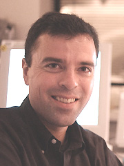
Peter
Bandettini
|
Peter Bandettini received his B.S. in physics from Marquette University in Milwaukee in 1989 and his Ph.D. from the Medical College of Wisconsin in Milwaukee in 1994. There, he contributed to the early development of functional MRI (fMRI). After postdoctoral training at the Massachusetts General Hospital, Boston, from 1994 to 1996 and then serving as an assistant professor of biophysics at the Medical College of Wisconsin, he joined NIMH in 1999 as an investigator in the Laboratory of Brain and Cognition and as the director of the NIH fMRI core facility. Chief of the Section on Functional Imaging Methods, he is also president of the Organization for Human Brain Mapping.
fMRI is a technique that rapidly captures time series images of changes in blood flow, oxygenation, and volume induced by local brain activation. Applications of fMRI are growing rapidly. These range from basic neuroscience to assessment and comparison of clinical populations and presurgical mapping.
Although the technique is widely used and is acknowledged to have high potential impact across neuroscience and medicine, it also has major, but solvable, shortcomings. These include:
![]() The ambiguous and variable relationship between neuronal activity and measured
hemodynamic-based signal changes
The ambiguous and variable relationship between neuronal activity and measured
hemodynamic-based signal changes
![]() Inability to cleanly map transient activity on the order of milliseconds
Inability to cleanly map transient activity on the order of milliseconds
![]() Inability to draw inferences about individual activation maps as they relate
to averaged population brain activation maps
Inability to draw inferences about individual activation maps as they relate
to averaged population brain activation maps
My research, including technology, methodology, interpretation, and research at the interface of applications and development, is aimed at addressing these and other issues in fMRI.
My research group, the Section on Functional Imaging Methods, is a team of physicists, psychologists, engineers, neuroscientists, and computer scientists committed to developing fMRI to its full potential through interrelated advancements in fMRI.
My research has four primary themes. The first is to understand more fully the relationship between measured MRI signal changes and underlying neuronal activity. My main approach to this is the use of multimodal (MEG and EEG) brain activation assessment comparisons, as well as pulse sequence manipulations that allow differential sensitivity to vascular components.
The second theme is to develop methods for increasing spatial and temporal resolution of fMRI through hardware and methodological development. We are collaboratively developing high-sensitivity coils and high-resolution imaging techniques as well as calibration methods that enable us to characterize more fully the hemodynamic response through which fMRI "sees" brain processes.
The third theme is to develop methods for cleanly extracting more subtle spatial and temporal information from the fMRI time series. It has become increasingly evident that the fMRI time series contain significantly more information about brain connectivity and resting-state activity than was previously thought.
What is required—and what my group is working on—are pulse sequence and processing methods to distinguish more robustly the artifacts of physiologic fluctuations from real brain activation fluctuations over space and time.
The fourth theme is to explore new types of functional contrast with fMRI. I am currently working on MRI methods for direct detection and mapping of neuronal activity, bypassing hemodynamic contrast altogether. We have obtained evidence—from in vitro cell cultures and through modeling of neuronal currents in the context of MRI—that NMR signal changes associated with microscopic magnetic field changes from coherent neuronal activity are detectable.
 |
|
Orna
Cohen-Fix
|
Orna Cohen-Fix received her Ph.D. in 1994 from the Weizmann Institute in Rehovot, Israel, and did postdoctoral work at the Carnegie Institution in Baltimore. She joined NIH in 1998 as a tenure- track investigator in the Laboratory of Molecular and Cellular Biology, NIDDK, and is currently a senior investigator in that lab.
Ever since I was a kid I wanted to be a scientist. On weekends my father, who is a microbiologist, would take me to his lab and let me transfer water with a pipet from one test tube to another. We called it an experiment, and I thought science was amazing.
These days my experiments are somewhat more sophisticated, but the fascination remains the same. My lab studies cell-cycle regulation and nuclear architecture in budding yeast.
You’d be hard-pressed to find a yeast researcher who is not infatuated by this model system. With it, one can use a myriad of experimental approaches (for example, genetics, biochemistry, genomics, proteomics, cytology) to examine the biological question at hand. The yeast life cycle is very rapid, it can be manipulated easily, and most processes are evolutionarily conserved, making yeast highly suitable for cell-cycle studies. Sometimes I feel that as yeast researchers, we are limited only by our collective imagination.
I did my graduate thesis on the mechanism of ultraviolet mutagenesis in Escherichia coli, at the Weizmann Institute of Science under the guidance of Zvi Livneh. It was largely a biochemistry project, but during that time I took the "Advanced Bacterial Genetics" course at Cold Spring Harbor Laboratories (CSHL) in Cold Spring Harbor, N.Y. As a graduate student, I didn’t practice much of what I’d learned in the course, but I was still struck by the remarkable power of a genetic approach. Thus, turning to yeast for my postdoctoral training was a logical choice.
In 1994, I started my postdoc in Doug Koshland’s lab at the Carnegie Institution in Baltimore. As part of my research project, I characterized a mitotic inhibitor that plays a key role in ensuring accurate mitotic progression. This inhibitor, called Pds1 in yeast, or securin in higher eukaryotes, binds to a protease called separase, whose activity is necessary for the separation of duplicated chromosomes at the onset of anaphase.
As a postdoc I found that anaphase initiation is brought about by the degradation of Pds1, and that Pds1 is part of a regulatory mechanism—the DNA damage checkpoint pathway—that inhibits mitotic progression in the presence of DNA damage. These studies set the stage for the current research in my lab.
In the Laboratory of Molecular and Cellular Biology, NIDDK, we used Pds1 as a launching pad to explore various processes related to mitosis. Pds1 is at the epicenter of the metaphase-to-anaphase transition.
This transition is irreversible, and cells pay dearly for any mistakes that occur during this process.
Thus, multiple regulatory pathways converge on the metaphase-to-anaphase transition, and on Pds1 in particular, to ensure that chromosome segregation takes place in an accurate fashion.
With Pds1 as a starting point, we began studying some of these regulatory pathways. We elucidated the molecular mechanism by which Pds1 is targeted for degradation at the onset of anaphase. We learned how the nuclear import of separase is regulated by Pds1. We discovered new aspects of the DNA damage checkpoint pathway. We uncovered a novel mechanism that affects nuclear positioning during mitosis.
Through various genetic screens, we identified novel proteins that affect mitotic regulation, and these have now become research subjects in their own right.
More recently we began studying the underlying proteins and structures that affect yeast nuclear architecture. The yeast nucleus is different from its metazoan counterpart in two important ways: (1) There are no nuclear lamins in yeast, and (2) the yeast nuclear envelope does not break down during mitosis.
Nonetheless, intranuclear organization affects fundamental processes, such as DNA replication and transcription, in both yeast and higher eukaryotes. We reasoned that this genetically amenable model organism would yield important insights into nuclear organization in human cells.
This project is at an early stage, but we have already identified domains in the nuclear envelope that differ in their susceptibility to expansion in the presence of elevated phospholipid levels.
An important part of being a scientist is the involvement in activities that affect the scientific community at large. Shortly after I joined the NIH I began teaching the CSHL Yeast Genetics course, a three-week-long intense course that is taught, coincidently, in the same lab as the Bacterial Genetics course I took as a student.
The decision to teach the course was a difficult one, because as a starting tenure-track investigator I feared that being away from the lab for an extended period of time would harm the progress of my own projects, as well as that of my postdocs.
As it turns out, teaching the course was one of the best things I did as a starting scientist, because I had the opportunity to interact closely with dozens of scientists who either participated in the course as students or were invited to the course to give informal seminars.
It was also a humbling experience: Not only did my NIH lab not fall apart in my absence, but also my postdocs discovered that they were fully capable of making important decisions on their own.
By being in constant contact with the outside world, one increases the chances of stumbling across an interesting piece of information. If there’s one thing I learned from teaching the yeast genetics course and other such activities, it is that one never knows what will stimulate the next exciting idea. And the way I see it, having exciting ideas is what being a scientist is all about.
 |
|
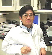
Bin
Gao
|
Bin Gao received his M.D. and Ph.D. from the Wanna Medical College, Wuhu, and the Norman Bethune University of Medical Science, Changchun, China, in 1986 and 1991, respectively. After postdoctoral training at NIH and the Medical College of Virginia (MCV) in Richmond, he became an assistant professor at MCV in 1996. In 2000, he joined the Laboratory of Physiologic Studies at NIAAA. He is now a senior investigator and chief of the Section on Liver Biology.
The primary interests of my laboratory are the molecular pathogenesis of liver diseases and their immunology. In particular, the lab is focused on the study of signal transducers of activators of transcription (STATs) and their roles in liver injury and repair.
Using animal models in examining liver injury, we demonstrated that (1) STAT1, primarily activated by interferons (IFNs), plays a critical role in liver injury and inflammation in T cell–mediated hepatitis and endotoxin-induced liver injury, and that (2) STAT1 is also involved in TLR3 ligand/IFN-g–mediated suppression of liver regeneration.
In the presence of cellular damage, the liver initiates processes to repair itself. Our lab found that, in contrast to STAT1, STAT3 activated by IL-6 and IL-22 in the liver is an active factor in liver repair. Among our findings were that:
![]() IL-6–deficient mice were more susceptible to ethanol-induced liver injury.
They were also more susceptible to concanavalin A–induced T cell hepatitis,
which was prevented by IL-6 treatment.
IL-6–deficient mice were more susceptible to ethanol-induced liver injury.
They were also more susceptible to concanavalin A–induced T cell hepatitis,
which was prevented by IL-6 treatment.
![]() In vivo IL-6 administration ameliorated alcoholic and nonalcoholic fatty liver
and ethanol-induced liver injury.
In vivo IL-6 administration ameliorated alcoholic and nonalcoholic fatty liver
and ethanol-induced liver injury.
![]() In vitro IL-6 treatment prevented mortality associated with alcoholic and nonalcoholic
fatty liver transplants.
In vitro IL-6 treatment prevented mortality associated with alcoholic and nonalcoholic
fatty liver transplants.
Collectively, these findings suggest that IL-6 possesses therapeutic potential for treating alcoholic liver disease and fatty liver transplantation. In fact, we think in vitro application of IL-6 could significantly transform the success rate of liver transplantation.
Investigations into the cellular and molecular mechanisms underlying the hepatoprotective effect of IL-6 in the liver have identified multiple mechanisms.
Our findings show that IL-6 activation of STAT3 can induce anti-apoptotic genes, improve hepatic microcirculation, inhibit NKT cells, inhibit oxidative stress, and ameliorate the accumulation of fatty deposits—powers that combine to prevent further cellular damage and repair existing injuries.
Other studies conducted in our lab show that the T cell–derived cytokine IL-22 is also a survival factor for hepatocytes and plays a protective role in T cell–mediated hepatitis via activation of STAT3.
While investigating the effects of alcohol consumption on STAT1 and STAT3 activation, we found that acute and chronic ethanol consumption attenuates both STAT1 and STAT3 activation, which could be an important mechanism contributing to the pathogenesis of alcoholic liver disease.
So far, our findings suggest that STAT1 and STAT3 play opposite roles in liverinjury and repair. Interestingly, STAT1 and STAT3 also negatively regulate one another through the induction of SOCS (suppressors of cytokine signaling) proteins in the liver.
Because both STAT1 and STAT3 are activated in chronic liver diseases, modulation of the mutual antagonism between STAT1 and STAT3 could offer a novel approach in the treatment of T cell–mediated liver damage in human liver disease.
Related findings from our studies include a determination that ethanol and hepatitis viral proteins synergistically (or additively) produce hepatocyte damage and stimulate inflammatory signals, and that alcohol drinking accelerates concanavalin A–induced T cell hepatitis via dysregulation of NF-kB and STAT3 signaling pathways.
More recently, we also demonstrated that natural killer cells ameliorate liver fibrosis through elimination of activated stellate cells in NKG2D/RAE1- and TRAIL-dependent manners, and IFN-g/STAT1 plays an important role in the activation of natural killer cells.
 |
|
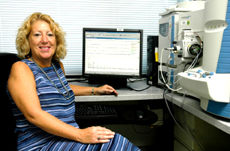
Marilyn
Huestis
|
Marilyn Huestis received her Ph.D. in toxicology in 1992 from the University of Maryland at Baltimore (UMAB) School of Medicine and joined NIDA in 1995 in the Clinical Pharmacology and Therapeutic Research Branch—with more than 20 years of experience in analytical and forensic toxicology. In 1998, she was converted to tenure-track and selected as acting chief of the Chemistry and Drug Metabolism Section. Currently, she is chief of this section and conducts her research at the NIDA facility in Baltimore. She also directs clinical research of toxicology doctoral students from UMAB School of Medicine.
My clinical research program uses chemical and toxicological tools to address drug abuse and addiction. My work focuses on mechanisms of drug action and pathological consequences of human drug exposure.
One area of focus is illicit drug use by pregnant women and the resulting toxicity to their children. Fetuses exposed in utero to excessive amounts of alcohol may suffer from fetal alcohol syndrome. In a similar manner, we believe that fetuses exposed in utero to excessive licit and illicit drugs may experience fetal drug syndromes.
Some consequences are apparent at birth, but subtle cognitive and behavioral abnormalities may not become evident for years.
To date, we do not know the prevalence of illicit drug use during gestation, the most accurate method to identify affected children, or the effects of drugs on developing fetuses. Our studies on the toxicity of prenatal opiate, methamphetamine, and nicotine exposure are helping to provide answers to these important public health problems.
One of the first challenges is accurate differentiation of drug-exposed vs. nonexposed children. I am a co-investigator on the first large-scale study of the developmental consequences of prenatal methamphetamine exposure.
We determined infants to be methamphetamine exposed if the mother self-reported use or if infant meconium, the child’s first fecal material, contained methamphetamine.
Surprisingly, more mothers reported use than were identified by meconium analysis, suggesting a need for additional biomarkers of in utero methamphetamine exposure. We detected 68 percent of neonates whose mothers reported third trimester use, while detection rates were ²10 percent for self-reported use earlier in gestation.
Methamphetamine concentration was significantly correlated with frequency of third-trimester self-reported use. Prior to these data, scientists believed that in utero drug exposure in the last six months of pregnancy might be detected. We are using tandem mass spectrometry to search for unique biomarkers in meconium potentially produced by the fetal liver and kidney.
Research on cannabinoids and the endogenous cannabinoid system is a rapidly developing field of neuroscience due to cannabinoids’ role in mediating important biological processes and the therapeutic potential of cannabinoid agonists and antagonists. Cannabis also is the most commonly abused illicit drug. My research focuses on the neurobiology and pharmacokinetics of cannabinoids.
Rimonabant, the first specific antagonist at CB1 cannabinoid receptors, is a key pharmacological tool for investigating the mechanisms of action of cannabinoids. I led the first investigative team to utilize rimonabant in phase I studies in human cannabis users.
We demonstrated that physiological and psychological effects of smoked cannabis were blocked by pretreatment with oral rimonabant, documenting for the first time in humans that cannabis’ effects were mediated through the CB1 cannabinoid receptor.
We also showed that rimonabant’s blockade of cannabis’ effects was not due to a pharmacokinetic interaction between rimonabant and D9-tetrahydrocannabinol, the primary psychoactive component of cannabis.
We extended these findings in a second clinical study comparing cannabinoid receptor blockade from single and multiple oral doses of rimonabant. We reasoned that the long half-life of rimonabant could lead to drug accumulation and antagonism at lower multiple doses.
Tachycardia associated with active cannabis was significantly lower for the 40-mg 15-day group and the single 90-mg-dose rimonabant groups. Peak subjective effects also were reduced.
Rimonabant holds great potential to treat disorders of the endogenous cannabinoid system and perhaps to treat cannabis and other illicit drug dependence.
Phase III trials of rimonabant have now documented its usefulness in the treatment of obesity and tobacco dependence. If it is approved for two such common indications, it could be prescribed to a large segment of the population, including cannabis users. For this reason, we plan to study the phenomenon of antagonist-elicited withdrawal. These studies of withdrawal may also shed light on retention vs. relapse of cannabis users in drug treatment programs.
 |
|
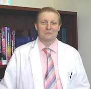
Karel
Pacak
|
Karel Pacak completed his medical training at Charles University, Prague, the Czech Republic, graduating in 1984. He completed a residency program in internal medicine at the same institution and also earned a Ph.D. in 1993 and a D.Sc. in 1999. In 1990, he joined NINDS as a postdoctoral fellow in the Clinical Neuroscience Branch, working with Irwin Kopin and David Goldstein. In 1995, he left NIH to start the residency program in internal medicine at the Washington Hospital Center, Washington, D.C., and in 1997 returned to NIH as a clinical associate in the Inter-Institute Endocrinology Training Program. In 2000, he joined the Pediatric and Reproductive Endocrinology Branch, NICHD, as a tenure-track investigator and chief of the Unit on Clinical Neuroendocrinology, working with George Chrousos. He is currently also an adjunct professor in the Department of Medicine at Georgetown University in Washington.
After joining NINDS in 1990, I focused on the role of catecholamines in the regulation of the hypothalamic-pituitary-adrenocortical axis under stress.
Together with my mentors, Drs. Kopin and Goldstein, as well as Richard Kvetnansky (NINDS), and Miklos Palkovits (NIMH), we established a method to sample and measure catecholamines from individual brain nuclei. We also described new feedback mechanisms between catecholamines and glucocorticoids, and mapped novel stressor-specific brain pathways.In 1999, I introduced a patient-oriented Pheochromocytoma Research Program as a new initiative at NICHD. The program emphasizes bedside to bench projects and multidisciplinary and multi-institutional collaborations, with three main objectives:
![]() To develop and test novel methods and criteria to diagnose and localize pheochromocytoma
in a cost-effective manner
To develop and test novel methods and criteria to diagnose and localize pheochromocytoma
in a cost-effective manner
![]() To develop improved treatments for malignant pheochromocytoma
To develop improved treatments for malignant pheochromocytoma
![]() To identify molecular and genetic mechanisms of pheochromocytoma tumorigenesis
and clinical manifestations of disease
To identify molecular and genetic mechanisms of pheochromocytoma tumorigenesis
and clinical manifestations of disease
Following from earlier collaborative work with Harry Keiser (NHLBI) and Graeme Eisenhofer (NINDS), we initiated large, prospective, multicenter cohort studies to introduce a novel biochemical test—the measurement of plasma-free metanephrines—in the biochemical diagnosis of pheochromocytoma.
Later, we introduced a novel clonidine-suppression test, coupled with the measurement of plasma-free normetanephrine, for diagnosis of this tumor and established new reference ranges for plasma-free metanephrines in adult patients and children. Recommendations of my group and collaborators about the use of plasma-free metanephrines were endorsed by a panel of experts convened for an NIH Consensus Development Conference.
With other colleagues (Goldstein, Jorge Carrasquillo, Clara Chen, CC Nuclear Medicine Department), we introduced a new nuclear imaging method, [18F]-6-fluoro-dopamine positron emission tomography scanning, for diagnostic localization of pheochromocytoma. This approach proved to be a superior imaging method—especially for metastatic tumors—compared with conventional nuclear imaging approaches such as 131I-MIBG.
This is an important advance because correct detection of disease extent often determines the most appropriate therapeutic plan and future evaluation. This new imaging method has substantially changed the imaging algorithm and clinical approach, and many patients have now profited from the correct localization of their tumors.
In the area of treatment, together with Goldstein and James Reynolds (CC Nuclear Medicine Department), we have initiated a new clinical protocol: using pretreatments with certain agents to increase the efficacy of 131I-MIBG therapy in metastatic pheochromocytoma.
We also introduced radiofrequency ablation as a novel treatment of metastases. Frederieke Brouwers from my lab, in collaboration with Tito Fojo (NCI), introduced the use of depsipeptide and trichostatin A (histone deacetylase inhibitors) to increase expression of norepinephrine transporter system in pheochromocytoma cells.
This important development could open the door to a new clinical trial to test whether these compounds may increase entry of 131I-MIBG into pheochromocytoma cells.
Recently, Shoichiro Ohta from my lab, in collaboration with John Morris, Jeff Green, and Mones Abu-Asab (NCI), established a new mouse model of metastatic pheochromocytoma. Ohta and Wai-Yee Chan (NICHD) also uncovered how metastasis-suppression genes might be involved in tumorigenesis of metastatic pheochromocytoma.
In concurrent collaborations with investigators from the Urologic Oncology Branch of NCI (Marston Linehan and McClellan Walther) and Zheng Ping Zhuang, Irina Lubensky, and Alexander Vortmeyer (NINDS), we described mechanisms linking underlying mutations in multiple endocrine neoplasia type 2A (MEN 2A) and von Hippel–Lindau (VHL) syndrome to different pathways of tumorigenesis, biochemical phenotypes, and clinical presentations of pheochromocytomas in the two syndromes. We also described the distinct histopathologic phenotypes of MEN 2A and VHL pheochromocytomas.
This work was extended to collaborations with Abdel Elkahloun (NHGRI) and Peter Munson (CIT), focusing on new approaches to classify various types of pheochromocytomas by gene expression profiling. The work aims to provide much-needed approaches for distinguishing tumors with benign or malignant characteristics and behavior and to identify new molecular and genetic markers for diagnosis and targets for treatment.
We also established the International Working Group on Endocrine Hypertension under the auspices of the European Society of Hypertension and set up a new international consortium to foster collaborative research on the tumor (the Pheochromocytoma Research Support Organization). We are now organizing the 1st International Symposium on Pheochromocytoma, to be held in Bethesda, October 20–23, 2005.
 |
|
Xin
Wei Wang
|
Xin Wei Wang received his Ph.D. from New York University in 1991. He did postdoctoral studies on cancer genetics and molecular carcinogenesis with Michael Newman at the Roche Institute of Molecular Biologyin Nutley, N.J., and with Curtis Harris at NCI. In 1998, he became a tenure-track investigator in the Laboratory of Human Carcinogenesis, NCI, where he is currently a senior investigator and head of the Liver Carcinogenesis Section. He is also an adjunct associate professor at the University of Maryland School of Medicine, Baltimore.
Since the beginning of my undergraduate studies at Shanghai First Medical College, I have been fascinated with the complexity of multicellular organisms, especially their development of fine controls for maintaining homeostasis. The study of cancer is an ideal approach to dissect the underlying mechanisms of homeostasis and its breakdown.
The current paradigm suggests that a tumor evolves through a multistage process, with selection and clonal expansion of an initiating cell undergoing a series of changes.
In the last six years, my laboratory has been investigating the genetic and biochemical pathways involved in human liver carcinogenesis—an excellent model to explore the multistage process of human neoplasms of unknown underlying etiology.
The overall goals of our research are to learn how cancer cells initiate and metastasize and to identify biomarkers that are helpful for early diagnosis and molecular targets for therapeutic intervention.
A key step in our research is identifying molecular fingerprints to elucidate the molecular mechanisms of human cancer and to provide molecular diagnostic tools to guide individualized care for patients with cancer.
Toward this end, we use state-of-the-art gene expression profiling technologies to identify molecular signatures at initiation and progression of hepatocellular carcinoma, particularly those attributable to hepatitis B and C viruses.
For example, by comparing liver samples from chronic liver disease patients with varying degrees of risk for developing hepatocellular carcinoma, we have identified unique fingerprints that may be useful in diagnosing patients with early onset of liver cancer.
Comparing hepatocellular carcinoma with or without accompanying metastasis led us to identify a molecular signature that can be used to predict which liver cancer patients have the potential to develop metastases or recurrence.
In addition, we are examining the role of the liver microenvironment in metastasis and recurrence by focusing particularly on the functions of immune cells and the inflammatory process in liver cancer progression. We identified several potential therapeutic targets that may be useful in eliminating liver cancer cells or metastatic progression.
Because viruses have been invaluable tools for discovering key pathways for human carcinogenesis, a second research emphasis in my laboratory is exploring the interaction between cellular targets and viral hepatitis–encoded proteins. Our initial study focused on molecular aspects of HBx, a viral oncoprotein encoded by hepatitis B virus.
We discovered that HBx contains a functional nuclear export signal motif using the Ran/Crm1 complex, a component essential in nucleocytoplasmic transport of many cellular proteins. Interestingly, this viral protein not only uses but also disrupts Ran/Crm1-dependent activities, presumably to prevent host antiviral response.
The finding implicates the Ran/Crm1 complex in the molecular pathogenesis of HBV.
Our approach also allowed us to uncover a novel role of the Ran/Crm1 complex in regulating cellular proteins that control centrosome duplication and mitotic spindle assembly. Recently, we revealed nucleophosmin as a novel substrate for Ran/Crm1 that negatively regulates unnecessary centrosome duplication.
These results led us to generate a new hypothesis: that the Ran/Crm1 complex serves as the centrosome duplication checkpoint by providing a "loading dock" mechanism to control cellular homeostasis.
Disruption of this complex may result in genomic instability, which may be an early step in viral-mediated hepatocarcinogenesis. Currently, we are exploring other potential partners associated with this complex that may regulate spindle assembly.
Exploration of the roles of these genes and molecular interactions in liver cancer initiation and metastasis has been extremely rewarding, because the findings may be useful in patient management and also challenge current theories of tumor evolution.
Gene expression profiling
and exploration of viral-host molecular interactions have expanded our knowledge
of the global changes that occur in liver cancer and provide numerous insights
into the molecular mechanisms of this disease.
![]()