
| T H E N I H C A T A L Y S T | J A N U A R Y – F E B R U A R Y 2004 |
|
|
|
PATHOLOGY SERVICE UNCOVERS HIDDEN TALENTSOF GENETICALLY ENGINEERED MICE |
by Celia Hooper |
|
The rest of the world may be in perpetual need of a good 10-cent cigar, but NIH scientists have been calling for something else for the past decade: a good, inexpensive, nose-to-tail pathology scan of the mouse models they are churning out via genetic engineering.
NIH’s Division of Veterinary Resources Pathology Service is now offering a service intended to do just that.
Led by veterinary pathologists Michael Eckhaus and Georgina Miller, the group has been piloting a package of phenotyping services that methodically work through a mouse model, generating data on blood cells, serum chemistries, and organ weights, followed by careful necropsy, microscopic evaluation of 40+ organs (see box), and statistical analysis of the data.
| PHENOTYPING REPERTOIRE | |
|
Hematology Blood Smear CBC and Differential
Serum Chemistries Glucose |
Body/Organ Weights Body Necropsy/Tissue Collection Skin |
The cost for this comprehensive analysis is $450 per mouse, approximately half what a small number of outside contractors charge for similar services. Mice between 10 and 14 weeks of age are preferred, but any age can be examined, including embryos (see "fetal urogenital tract").
The phenotyping team anesthetizes the mice, draws a small blood sample for the hematological and serum analyses, and then sacrifices the mice while they are still under anesthesia. Hematology and a panel of serum chemistries are run, using in-house equipment optimized for small sample size.
The team performs a detailed necropsy, collects the tissues, and weighs selected organs. Fixed tissues are sent to a contract histology lab, where they are embedded in paraffin, sectioned, stained, and then returned to the pathologist for microscopic examination
Due to the small size of embryos, blood work and organ weights are not performed on them. Embryos are embedded in paraffin, and step sections are cut and stained. Depending on the embryo size, 50 to 100 sections are examined microscopically, allowing evaluation of all organs in situ.
It takes three to four weeks to complete a study. The pathologists deliver to the investigator an Excel spreadsheet with raw data from the blood chemistry, hematology, and organ weights; statistical analyses (if numbers permit); a list of gross and microscopic diagnoses; and a report on the significance of the findings.
Investigators are also provided the paraffin blocks and slides of the tissues analyzed and digital photographs of gross and microscopic lesions.
Miller recently told NIH’s Scientific Directors that so far, in the 18 months of the pilot service, the program has completed 52 projects from 12 different ICs, turning up potential genotype-associated changes in about half of the models.
Some of these differences have come as a big surprise to the investigators who’d engineered the mice. She cites as an example a mouse that was created by Richard Proia, chief of NIDDK’s Genetics of Development and Disease Branch.
 |
 |
|
Slides
courtesy of Veterinary Resources Pathology Service
|
|
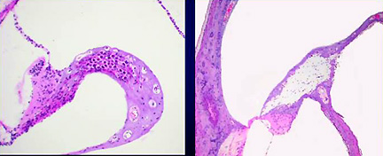
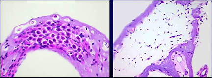
Cross-section of inner ear (close-ups below) from mouse with wild-type G-protein receptors for sphingolipids ((left) and from G-protein receptor mutant (right), which results in degeneration of the spiral ganglia of the inner ear and deafness |
Proia says his group made the mice as they "were looking for functions of G-protein–coupled receptors for sphingolipids."
Initially, things weren’t looking very good because the mutant mice bore no overt phenotype. "Anytime that happens you’re a little disappointed," Proia says.
Under those circumstances, the phenotyping service—free at the time—was hard to resist. "The results came back indicating from the pathology that the mice had degeneration of the spiral ganglia of the inner ear, suggesting they should be deaf," Proia recalls.
"We tested our colony and the mice were, in fact, deaf." He says the result was "totally unexpected" and points to "a new function for these receptors. We are currently working on this with collaborators in IDCD."
Pursuing this unexpected lead will mean looking at the protein expression patterns in the ear and sorting out the mechanism that causes the cells to degenerate. Proia notes that the ear problems could be secondary to some other functions of the gene.
The phenotyping service won’t provide the details that come from careful molecular dissection of a pathology, but Proia credits it with opening a new door in his research. He also gives the service high marks for its timeliness and responsiveness.
So, for all interested
NIH investigators, the mic is open and the spotlight’s on in Bldg. 28A’s
theatre, awaiting the chance to expose the hidden talents of the next murine
American Idol. ![]()
|
CONTACTS Investigators
interested in the phenotyping service should contact either Michael
Eckhaus (301-496-4465)or Georgina Miller
(301-496-4465)for more details. |