| T H E N I H C A T A L Y S T | J A N U A R Y – F E B R U A R Y 2002 |
|
|
|
Translational ResearchGRIFF
RODGERS:
|
text and photo by Fran Pollner |
 |
|
Griff
Rodgers
|
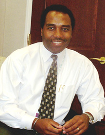
Speaking of the work he does in the lab and in the clinic, Griff Rodgers uses phrases like "reactivating genes" and "reversing ontogeny." This is not hyperbole. These are essential strategies in the business of deciphering and destabilizing sickle cell disease (SCD)—a prime focus for Rodgers since his arrival at NIH in 1982.
Now NIDDK deputy director and chief of the Molecular and Clinical Hematology Branch, Rodgers’ research over the past two decades has advanced the understanding and management of SCD and related thalassemias. His basic and clinical studies established that hydroxyurea, a cancer chemotherapy agent, reactivated the dormant fetal hemoglobin gene in SCD patients, producing a normal red blood cell population and substantial relief from the crippling clinical manifestations of the disease. In 1998, hydroxyurea became the first—and thus far only—drug to gain FDA approval for use in SCD.
Today, he and his team are involved in determining the molecular basis of hydroxyurea’s effect.
The methods used in this pursuit are providing a bridge from pharmaceutical to stem cell and genetic approaches to hereditary blood disorders—and to the broader sphere of "regenerative" medicine with hematopoietic stem cells as source material—an outgrowth of SCD research he laughingly calls an "aside."
Counting the Shoulders
Rodgers cannot describe his sickle cell research without enumerating the achievements of others that informed his own visions. "When I arrived on the scene," he says, "a number of factors had converged to allow for the discovery of the value of hydroxyurea."
There was already a body of epidemiological literature that clearly suggested that high levels of fetal hemoglobin—conferred, like sickle cell itself, through mutation—were protective against the clinical manifestation of SCD.
The work of a dermatology professor at Rodgers’ medical school who used a chemotherapeutic agent to establish what appeared to be normal skin structure and function in patients with psoriasis invited the notion that a drug that decreased the rate of cell proliferation could be of benefit in nonmalignant diseases that were nonetheless characterized by rapid cell turnover—such as SCD.
A small but statistically significant and reproducible rise in fetal hemoglobin levels observed in cancer patients on chemotherapy suggested that fetal hemoglobin levels could indeed be manipulated pharmacologically.
Studies in rhesus macaques, which resemble humans in having fetal and adult hemoglobin systems, demonstrated proof of concept that the fetal hemoglobin gene could be turned back on by chemotherapeutic agents (in this case, 5-azacytidine).
Clinical studies at the University of Illinois and at the NIH Clinical Center (CC) showed that the same drug could do the same thing in SCD patients.
But the potential adverse effects of 5-azacytidine would preclude its long-term use. So it was in the testing of hydroxyurea as a less toxic and more effective alternative to 5-azacytidine that Rodgers took his place in the research chain of events, he says.
In this endeavor, he gives the greatest credit to his CC patients, who were "most generous with their time," spending three to four months in the CC and enabling the researchers to ensure compliance and serially check blood counts and serum hydroxyurea levels. The 70 percent response rate, reported in 1990(1), established hydroxyurea as an agent to be pursued.
Expanding the Search
While hydroxyurea testing moved to extramural venues for the large, definitive clinical trials that would serve as the basis for FDA approval, Rodgers and his colleagues looked for reasons for the variable response to hydroxyurea and ways to enhance it in those who responded with only modest fetal hemoglobin increases.
They could discern no way to predict response to hydroxyurea, but they succeeded in improving response with the addition of growth factors to the regimen. First in primates and then in CC patients, erythropoietin (EPO) proved to be the best of the growth factors tested in augmenting fetal hemoglobin response. Fetal hemoglobin levels in modest responders to hydroxyurea alone—in the 2–8 percent range—rose to 20 percent with the addition of EPO. The findings were reported in 1993(2).
The drawback, Rodgers says, is the prohibitive cost of EPO at the doses given to change the kinetics of red cell maturation—about an order of magnitude higher than those required to restore normal hemoglobin levels in dialysis patients. "Again, we have good proof of concept, but not a practical approach," he says.
The minimum effective dose of EPO is the subject of an impending pharmaceutical company trial to be carried out at several U.S. universities. Rodgers will sit on the study’s independent data safety and monitoring board.
Differential Displays
Meanwhile, the team has begun to unravel the molecular mechanisms of hydroxyurea’s actions, using a liquid culture system in which they can "take a white cell component of blood and in three weeks watch it grow into red blood cells–in the presence or absence of hydroxyurea." They have found that the fetal hemoglobin levels in these cells mirror those obtained in vivo in SCD patients—in "a couple of dozen" patients thus far, not yet enough upon which to base treatment decisions.
"We are, however, confident that the underlying basis of what causes fetal hemoglobin to be induced relates to the molecular biology of the cells as they differentiate—and this is different in different groups of people," Rodgers notes, observing that this finding could have relevance to emerging hematopoietic stem cell therapies for cancer and other diseases.
Using differential display techniques, Rodgers and his colleagues take blood stem cells, treat them with hydroxyurea, and compare what genes are expressed in the absence and presence of the drug.
"We have cloned four genes differentially expressed in the presence of hydroxyurea," Rodgers notes.
The first, a small GTP-binding protein involved in protein trafficking from the endoplasmic reticulum to the Golgi, induces fetal hemoglobin expression in a human leukemia cell line.
"Either alone or in combination with any of the three other genes we’ve found, this GTP-binding protein might be the basis of hydroxyurea’s effects," Rodgers says. Using flow cytometry, he and his colleagues have observed that the cells of patients treated with hydroxyurea tend to be arrested in S phase. Overexpression of the GTP-binding protein in these cells magnifies the drug’s effects and is associated with larger size, increased fetal hemoglobin, and diminished cell doubling rate. "This gene," he says, "may have yet unimagined effects."
The team is now creating a transgenic mouse model to further characterize the GTP-binding protein and to test the effects of partner proteins on red cell kinetics.
Shifting into Reverse
In parallel with efforts to understand and augment means to express fetal hemoglobin, Rodgers is also set upon saving a particular adult form of hemoglobin from extinction.
Hemoglobin A2, which accounts for perhaps 1–2 percent of adult hemoglobin, acts much the same as fetal hemoglobin in interfering with the sickling process by inhibiting polymerization of the sickle protein. The gene that encodes A2, however, is "evolutionarily on the way to becoming a pseudogene because of mutations in its promoter," Rodgers says.
"We are trying to reverse evolution—to restore these critical pieces of mutated DNA and build a better DNA-binding motif coupled with the normal activation domain to get higher levels of transcription in transgenic models. Ultimately," he says, "we’d like to get this chimeric molecule into the stem cells of sickle cell and thalassemia patients—but that’s a long way off."
In anticipation of developing useful gene-based blood stem cell strategies, Rodgers’ team and collaborators in the CC Department of Transfusion Medicine have begun a project with several local hospitals to collect, store, and conduct research on cord blood from about 100 newborns with SCD. "We are optimistic that this line of investigative inquiry will yield important results relevant to adoptive stem cell therapy," Rodgers says.
Stem Cell Potentials
The techniques developed to study the effects of different agents on red blood cell development have proved "enormously valuable" in studying the question of lineage commitment in general.
"We were able to expand this system to grow adult hematopoietic cells that have the capacity to make not only red blood cells but white blood cell and platelet progenitors as well. . . . What defines lineage commitment? What is it that instructs the stem cell to become a red cell or a white cell or a platelet? The level of cytokines is one influencing factor, but there must be others," Rodgers observes.
Beyond that, animal studies have suggested that hematopoietic stem cells may have the capacity to make muscle, nerve tissue, and possibly bone. "We’re exploring this area of reparative or regenerative medicine using hematopoietic stem cells," he says, noting that his research has involved adult stem cells only.
Using differential display in liquid culture, Rodgers and his colleagues have cloned ten "novel genes associated with lineage commitment, some of which appear to be expressed not only in hematopoietic development but also in gut, pancreas, and renal development—of obvious interest to an institute dedicated to research on the digestive system and kidney diseases.
"These genes,"
Rodgers says, "may give us some clues into the origins of certain types
of cancers and developmental anomalies, and we are currently exploring this,
too." ![]()
References
1. G.P. Rodgers, G.J. Dover, C.T. Noguchi, A.N. Schechter, and A.W. Nienhuis. "Hematologic responses in patients with sickle cell anemia treated with hydroxyurea." N. Engl. J. Med. 322:1037–1045 (1990).
2. G.P. Rodgers, G.J. Dover, N. Uyesaka, C.T. Noguchi, A.N. Schechter, and A.W. Nienhuis. "Erythropoietin augments the fetal hemoglobin response to hydroxyurea in sickle cell patients." N. Engl. J. Med. 328:75–80 (1993).
|
'Lineage
Commitment' on a Personal Level— |
|
From "the beginning"—call it high school—Griff Rodgers says, he wanted to do more than learn about science and be a physician. He wanted also to contribute to the body of scientific knowledge, and he wanted those contributions to have a direct effect on the patients he treated. For him, basic and clinical research and clinical care were indivisible, and he mapped his career path accordingly. In high school, he secured accelerated admission into medical school via a seven-year combined undergraduate/graduate/medical degree program offered by Brown University in Providence, R.I. At Brown, his general desire to embrace basic research that translated into clinical benefit became focused on blood cell disorders. He worked in a lab where he evaluated function in red blood cells that were passed through a membrane oxygenator in animals receiving artificial organs. He also carried out a research project involving aspects of globin gene expression in a human erythroleukemia cell line (K562)—a cell line he would later use extensively in his sickle cell research at NIH. During his first medical school year in 1976, he applied for a Public Health Service scholarship—a three-year scholarship with a one-year payback. Payback could have been working at a PHS hospital or in an underserved area—or doing biomedical research at NIH. For Rodgers, doing research related to patient care at a world-renowned institution was the obvious choice. Acceptance into the NIH training program, however, required more than his choosing it; it was predicated on a positive review of a grant proposal, "much like competing for an extramural grant," he notes. The terms of the PHS scholarship allowed for the completion of a full clinical residency before embarking on training in research at NIH. Rogers completed his residency training, including a chief residency, at Washington University in St. Louis, where his work with hematologists doing sickle cell disease research solidified his own interest. Rodgers interviewed at several different NIH laboratories, seeking a lab that was involved in both basic research related to red cell gene expression and clinical research on sickle cell disease. He chose the NIDDK Laboratory of Chemical Biology, where, under lab chief Alan Schechter, he advanced through the ranks from fellow in 1982 to senior investigator in 1990 before becoming a unit chief and then a section chief and, in 1998, chief of the Molecular and Clinical Hematology Branch, a position he retained after he became NIDDK deputy director last year. Rodgers was a charter
member of the NIH Central
Tenure Committee and the Clinical Center Board of Governors. He currently
sits on the Board of Tutors of the Clinical
Research Training Program, and the NIH-Duke
Masters in Clinical Research Admissions Committee. He’s a member
of the subspeciality board on hematology for the American Board of Internal
Medicine and of a national advisory committee for the Robert Wood Johnson
Foundation. |