| T
H E N I H C A T A L
Y S T |
J A N U A R Y – F
E B R U A R Y 2002 |
|
The
mouse has always had a central place in studies of human health and disease;
but in the age of the mouse genome and transgenic and knockout mouse models,
its standing and ubiquity in basic and clinical research are beyond measure.
Just about everything
else in the mouse, however, is measurable—and visible in all its
aspects, with the right tools.
NIH has now amassed those
tools and centralized them in a state-of-the-art home that reflects the status
of the mouse in biomedical research.
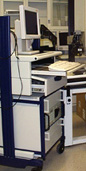
The Mouse
Imaging Facility (MIF), nestled at the end of a labyrinthine series of art-covered
corridors in the B1 level of the Clinical Center, is now ready to accommodate
NIH’s substantial mouse biology community. (It will also be open for general
viewing March 5; see "Open House" below).
| BECKER'S
BRAINCHILD |
Nary
a soul involved in the MIF does not trace the facility’s origins back
to Ted Becker,
former NIDDK investigator and associate director for research services,
who was the driving force behind the idea and creation of what in 1985 was
called the In Vivo NMR Center. Says Becker: "Basically, I brought all
the SDs together and asked for money." That model holds to this day.
 |
The MIF is an NIH-wide
shared resource to which all intramural scientists have access. In 1998, it
was an idea whose time had come, pushed to the fore by NHLBI’s Bob
Balaban and then–CC Radiology’s Nick Bryan. In 1999, it was still
an idea, but it had a director, Alan
Koretsky, recruited by NINDS to help oversee its realization. In 2000 and
2001—the first two years of a three-year pilot project funded by participating
NIH institutes in proportion to their intramural budgets—quarters were
renovated, people were recruited, committees were formed, and equipment dedicated
to the imaging of small animals
was secured.
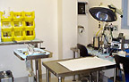
Now investigators
who walk through the door of the MIF will find a cornucopia of modalities optimized
for imaging small rodents at high resolution: ultrasound, computed tomography
(CT), magnetic resonance imaging MRI), positron emission tomography (PET), and
laser Doppler. A luciferase imager is on order.
MRI, however,
is the modality "nearest and dearest" to his heart, says Koretsky,
who also directs the MIF parent facility, the NIH MRI Research Facility (NMRF);
he is also chief of the Laboratory
of Functional and Molecular Imaging, NINDS, which has its own small animal
MRI resources. Koretsky heads two ongoing protocols—one exploring cellular
energy metabolism in the rat liver, heart, and brain and the other imaging brain
function in rats and mice.
These two are among 30 animal studies
in progress in the NMR Center, about one-third of which involve mice and rats.
It was anticipated that some mouse studies begun in the center’s 4.7-tesla
MRI would be moved to the MIF’s new 7-tesla machine.
In a
progress report to the scientific directors late last year, Koretsky announced
that the MIF would indeed be operational
the first month of the new year, and he showcased some examples of MRI studies:
| OPEN
HOUSE |
|
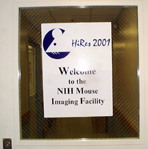
The
Mouse Imaging Facility is throwing open its doors March 5 from 10 a.m. to
4 .m. Take the main elevators in Building 10 to the B1 level, turn away
from the cafeteria, and follow the signs to the door with the welcoming
mouse logo.
|
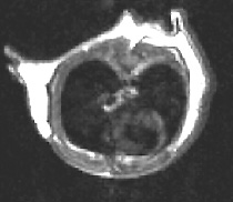
Serial tracking of inducible lung cancer in a mutant mouse model (Galen
Fisher, NHGRI, and Marty
Lizak, MIF; see Figure 1)
Dynamic MRI to assess tumor neovasculature (Steve
Libutti, NCI)
Monitoring genes injected into the rat heart (Jonathan
Sorger and Elliot
McVeigh, NHLBI)
Tracking macrophage infiltration into the ischemic kidney (Robert
Star, NIDDK)
Tracking the course of tagged neural stem cells (Joe
Frank, CC)
The first of these—tracking lung
tumors—Koretsky later observed, "is a good example of imaging that
helps a sophisticated mouse molecular biology study, and it represents something
that we can do routinely. The other MRI studies are in a more developmental
stage.
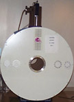
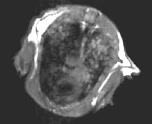
Other MIF modalities being used by NIH
investigators include high-resolution ultrasound, harnessed by NHLBI’s
Cecilia Lo to
elucidate cardiac dynamics and embryo surgery (see Figure
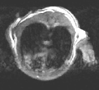
2), and micro-CT, which provides superb contrast between bone, soft tissue,
and fat (see Figure 3). MIF research veterinarian
Brenda Klaunberg
and NIDDK’s Marc
Reitman are developing protocols for routine regional fat determinations
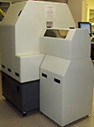
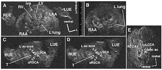
using CT. Also, Koretsky noted, the MIF is helping develop a microPET resource
for NIH—a working prototype (ATLAS) built by the CC’s Michael
Green and his colleagues (see Figure 4). PET
is especially useful, Koretsky said, for detecting specific molecular interactions
in vivo, such as neurotransmitter
receptor distribution. Once the prototype is standardized, it will be moved
into the MIF, he said.
Expert advice on which modality would
best serve any given research objective is part of the MIF package of resources
for investigators new to the field.
Oversight of the MIF comes under the
NMR Center Steering Committee, chaired by Balaban. Balaban established the MIF
subcommittee, which is chaired by King
Li, the new head of the CC radiology department.
In addition to the animal imaging advisory
subcommittee, there are MIF subcommittees to oversee animal safety and to review
research proposals for their technical feasibility and the extent to which they
will require MIF resources, including imaging time, ancillary equipment, and
technician support. MIF structure and procedures can be found at its web
site.
It is not the MIF’s job, however,
to assess the scientific merit of proposed projects or to evaluate issues related
to animal experimentation—those tasks reside within each institute and,
more specifically, with its SD and its animal care and use committee. Before
investigators present a protocol to the MIF, they must have done the legwork
to secure those approvals.
Currently, each institute
supports the MIF with a share proportional to its intramural budget. Once the
pilot phase of the MIF is concluded, the funding formula that has sustained
the NMR Center will go into effect in the MIF:
25 percent of the total budget will continue to be based on intramural budget
and 75 percent will be based n facility usage. For the NMR Center, allocations
for the coming year are estimated on the basis of usage from the previous three
years, but there is a good deal of flexibility based on real-time needs
as they arise. Tracking historical use of the MIF is just beginning.
Koretsky believes investigators
involved in the numerous mouse studies across ampus will find MIF resources
invaluable for analyzing a variety of phenotypes.
Li sees the MIF as a critical
locus in bench-to-bedside research in the postgenomic era. The "challenge
of medical imaging," he says, "is to be able to provide in vivo information
at the molecular level in a spatially and temporally resolved manner. To achieve
this, in vivo experiments in animal models are essential."
 |
 |
 |
|
wild-type
mouse with no lung tumor
|
lung
of mutant mouse model after three months on doxycycline
|
the
same model eight days after doxycycline withdrawal
|
|
Figure
1. MRI of Inducible Lung Tumors:
Mutant K-Ras
transgene whose expression could be controlled with doxycycline caused
tumors in an Ink/Arf knockout background. The tumors regressed
when the expression was switched off.
—Galen
Fisher, NHGRI,
Marty
Lizak, MIF, et al.
Genes and Development, in press
|
 |
|
Figure
2. Ultrasound of a Mouse Embryo:
Ultra-high-frequency (55 MHz) ultrasound imaging of an ex utero, fixed,
tbx1 +/- mouse e13.5d embryo. A. Transverse image showing the heart
within the thorax, flanked by the right (RUE) and left (LUE) forelimbs.
The interventricular septum (ivs) points ventrally, resulting in the ventricular
apex’s appearing mesocardic at this stage. The ventricles are ventral
to the atria. An echo-dense thrombus (starburst) is seen within the left
atrium. The hilum of the left (L) lung can be seen. (LAA = morphologically
left atrial appendage; LV = left ventricle; RAA = morphologically right
atrial appendage; RV = right ventricle; 1 mm calibration is shown.) B.
Transverse image of the heart (same embryo) slightly inferior to that
in A. Pectinate muscle ridges (open arrowheads) within the morphologically
right atrial appendage can now be recognized. The distal aspect of the
left lung can also be seen. C,D. Transverse images at planes more
cephalad than in A. The left aortic (L ao) arch is visualized to the left
of the trachea (T). An aberrant (retro-esophageal) right subclavian artery
(aRSCA) and a normal left subclavian artery (LSCA) can be identified.
E. Sagittal imaging through the left aortic arch reveals the right
(RCCA) and left (LCCA) common carotid arteries and a portion of the descending
(dorsal) aorta (desc ao).
—Cecilia
Lo, NHLBI, and Alvin Chin, Children’s Hospital of Philadelphia
|
 |
 |
 |
|
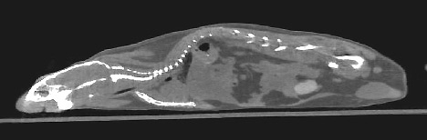
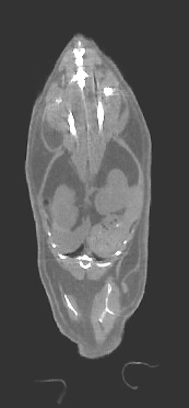
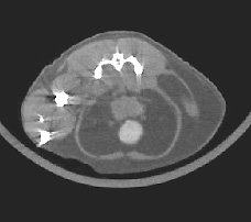
Figure
3. X-ray Computed Tomography (CT):
These images of a mouse were acquired at ~100 micrometer resolution. CT
images are excellent for characterizing bone (brightest intensity), organ
size and distribution (intermediate intensity), and regional fat (lowest
intensity).
—Brenda
Klaunberg and Alan
Olson, MIF
|
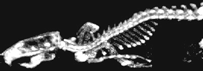 |
Figure
4. Micro-PET:
Whole-body re-projection image of the rat skeleton two hours after intravenous
administration of F-18 fluoride, a bone-seeking PET radiopharmaceutical.
The full 3-D tomographic image set from which this image was synthesized
was obtained with ATLAS, a small animal PET scanner designed and fabricated
at NIH.
—Mike
Green, CC
|
Return to Table of Contents