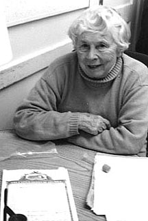
Willadene Zierdt
displays the invention used the world over that earned her a U.S. patent
(certificate shown) and decades of royalties
| T H E N I H C A T A L Y S T | M A R C H – A P R I L 1997 |
|
|
|
TWO NIH INVENTORSAND THEIR JUST DESSERTS |
by Fran Pollner |
The "fecalator" and "laser-capture microdissection" may be worlds apart in complexity and cost, but both sprang from a scientist's need to target and capture the invisible mechanisms of disease so they can be deciphered and disarmed. And both earned patents for the NIH scientists who invented them.
The 17-year patent on the fecalator expired in March 1995, but it still kept its place on the PHS top moneymaking inventions list (see NIH "Inventions" chart) that year, despite the fact that each fecalator costs less than a dollar. Millions of these devices are used the world over to recover parasites from stool specimens."
The patent application for laser capture microdissection was filed in November 1996 and was scheduled to be issued three months later, as this issue of The NIH Catalyst went to press. According to its inventors and the NCI technology development officer who handled the filing, laser-capture microdissection will revolutionize diagnosis and treatment and will become a critical component of every clinical pathology lab in the world. The first-generation, no-frills prototype machine costs about $50,000 and is used to extract, precisely, just the relevant cells from a tissue sample being probed for disease.
|
|
Willadene Zierdt
displays the invention used the world over that earned her a U.S. patent
(certificate shown) and decades of royalties
|
Prospecting for Parasites
Two decades ago, Willadene Zierdt built a better parasite trap; a patent was issued in her name March 28, 1978. She's still getting royalty checks, twice a year, and her latest was the "biggest payment ever"—about $3,000.
Zierdt, who retired in 1993, ran the Clinical Center parasitology lab for 25 of her 35 years there. Her invention, which she dubbed the "fecalator," yielded a hundred-fold increase in parasite recovery from stool samples compared with direct examination methods and afforded better protection from these parasites for laboratory personnel.
The impetus for developing the new apparatus, Zierdt recalls, was a disconnect between specimen yield and the severity of illness in the patient.
"These were sick people, and I knew that we were not recovering all the parasites we should have been recovering," she says. The problem was the use of a nearly century-old method of straining specimens through a gauze-and-funnel apparatus in an attempt to concentrate parasites. "I started sifting through the debris that remained on the gauze and I made dozens of little smears and, sure enough, kept discovering more and more parasites that had been missed." The gauze, Zierdt realized, would have to go.
She puzzled over what sort of nonadherent material she could use to replace the gauze and hot upon stainless steel. She cut out and tested about 20 different stainless steel mesh screens before settling on pore dimensions that let the parasites through while holding back fecal debris. She put the screen between two plastic test tubes that coupled together, then added a small plastic pipe to the top tube where the fecal samples are placed in the device. (The pipe aerates the formalin-washed specimen, enabling the parasites to drip through the mesh screen and concentrate at the bottom of the second plastic tube.) Zierdt taped the pieces of her prototype apparatus together and named it a "fecalator" because it reminded her somewhat of a percolator. Staff in the NIH machine shop glued the pieces of her invention together and made a few more of them. All in all, the fecalator was close to two years in the making.
"I was just trying to help myself do my job. I never dreamed this would become what it became," Zierdt says, recalling that it was the clinical pathology chief who advised her to visit the NIH patent office. After she'd secured her patent, the Department of Commerce requested that she entertain offers from businesses to commercialize the apparatus, and before long, a license was negotiated and the Fecal Parasite Concentratorª was born. Although the patent was issued for the "fecalator, an apparatus and method for concentration of parasite eggs and larvae," the manufacturer did not like her quaint term and renamed the product. The commercial device, manufactured with a screen made of no-stick plastic instead of stainless steel, is disposable. Sales, Zierdt says, have increased each year the product has been on the market. "It's quite a popular item and hasn't been surpassed by anything else. It's sold around the world and is especially useful in developing countries—it's inexpensive and doesn't require electricity," she says.
The fact that it's disposable not only guarantees presumably perpetual sales but also contributes to enhanced safety over the previous apparatus, as does then placement of the screen within the device.
Zierdt's royalties, which she saves for her children, keep coming in, despite the patent expiration—and she still visits the Clinical Center Microbiology Service frequently, often supplying the desserts for celebratory occasions.
 |
|
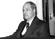
Lance Liotta
|
 |
|
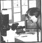
Laser-capture
microdissection: a main attraction on Clinical Research Day
|
Capturing the Disease Process
The driving force behind the invention of laser-capture microdissection was an urgent research need in NCI's Laboratory of Pathology.
"Imagine having the ability to look under a microscope at a disease process, to sample different cellular elements within that disease process, and then place the RNA or DNA from the samples onto a microhybridization system so that thousands of genes can be tracked. . . . We usually diagnose and treat cancer when it's too late. The real opportunity, in the understanding of the genetic basis and somatic progression of cancer, is in looking at premalignant lesions, at the progression from normal epithelium to in situ cancer. We would like to be able to sample each stage of this progression . . . and look at all the genes that are associated with the transition from one step to another. . . . We need a method to microdissect out these cellular elements of interest."
And that method, said Lance Liotta, chief of the NCI Laboratory of Pathology, to a rapt audience on Clinical Research Day, February 10, was on display, right outside the auditorium in one of the hallways filled with posters showcasing clinical research at NIH: laser-capture microdissection.
During his lecture, Liotta showed examples of laser-captured cells: neurofibrillary tangles from the brain tissue of Alzheimer's patients and premalignant lesions of breast carcinoma in situ. "Now," he said, "these individual cellular groupings can be analyzed for RNA, DNA, and protein." he noted, particularly, the utility of microdissection to analyze loss of heterozygosity in premalignant brerast lesions and singled out the extraction of RNA—and with it the ability to develop cDNA libraries and to discover "previously unknown expressed genes that might be associated with that . . . [as] probably the most important advance that can be supported by microdissection." This approach, he said, is being used by Michael Emmert-Buck and David Krizman at NCI to study prostate cancer.
In laser-capture microdissection, described by Emmert-Buck at al. in the Nov. 8, 1996, issue of Science, a thin, transparent, laser-activated film is placed on top of the tissue being studied. Watching via direct microscopy, the researcher procures individual cells by spotlighting them with a laser, inducing the film to stick to just the selected cells. The film with its quarry is removed from the tissue and transferred immediately to appropriate buffers for analysis.
In a booth outside the auditorium, crowds of curious scientists got a chance to see for themselves. Similar excitement was sparked by a demonstration of laser-capture microdissection at the February meeting of the National Cancer Advisory Board when NCRR's Robert Bonner demonstrated the technique there. Bonner heads the instrument-development team for the technique and has produced four generations of laser-capture microdissection prototypes.
"This is probably going to be huge," predicts Gary Colby, senior technology development and patent specialist at the NCI Office of Technology Development. Colby, who is handling tech transfer for laser-capture microdissection, notes that the imminent patent is but one of several anticipated in connection with this invention. Other patents will cover particular aspects of the process and machinery and wide-ranging aspects of its use. The number of inventors on these patents ranges from six to more than a dozen people, including staff in the NCI lab and in NCRR's Biomedical Enginnering and Instrumentation Program, Colby says. For Liotta, the laser-capture microdissection patents are but the latest in a succession of more than 60 patents, dating back to 1973 and including toys, cell lines, genes, proteins, and diagnostic methods.
The first of an anticipated series of laser-capture mirodissection CRADA collaborations with commercial developers has already begun. A demonstration project is being launched at Johns Hopkins, after which the instrumentation will be tested at multiple sites around the country. Patent applications are pending in countries around the world. Prototypes should be available for use at NIH by late summer.
"Every lab is going
to need this," Colby says. ![]()