Mike
Green
| T H E N I H C A T A L Y S T | M A R C H – A P R I L 2002 |
|
|
|
New
Technology
|
by Fran Pollner |
|
|
Mike
Green
|
About 10 years ago, Mike Green recalls, a harmonic convergence of sorts occurred in the positron-emission tomography (PET) community. A small group of investigators had the same idea at the same time—that PET could be used not just to diagnose human disease but also as a basic research tool to probe the physiology and biochemistry of living systems such as the rat and mouse.
Green and his colleagues spent the next decade fashioning the technology to achieve the spatial resolution necessary to image such small subjects. Chief of the Imaging Physics Laboratory in the CC Department of Nuclear Medicine, Green enlisted the aid of the master builders in the ORS precision instrument and electronics shop, the software whizzes at CIT, the electronics wizards at The Thomas Jefferson National Accelerator Facility in Newport News, Va., computer scientists in Spain, and the brainpower of his lab colleagues, most notably staff scientist Jurgen Seidel (see below, "It Takes a Village").
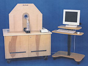 |
With Clinical Center support, a three-year research and development project was launched that culminated last fall in ATLAS (Advanced Technology Laboratory Animal Scanner)—"the most technically sophisticated small animal PET scanner in the world at this moment," says Green, noting that every step of the process to create ATLAS—benchtop experiments, computer simulations, structural designs—is public information. There are probably about a dozen labs worldwide now working on ATLAS-type scanners.
ATLAS is currently housed in Green’s lab, back-to-back with a micro-CT scanner. Spatially registered and superimposed CT and PET images of the same animal will help characterize the biodistribution of the PET tracer and correct the PET image data for radiation attenuation.
PET Digs
A question before the NIH intramural research community is whether ATLAS will take up residence in the newly opened Mouse Imaging Facility (MIF)—the multimodality small-animal imaging suite located in another part of the Clinical Center (see "Mighty Machines for Mini-Models," The NIH Catalyst, January–February 2002).
In Green’s view, the question boils down to whether the unique information obtainable with PET imaging is enough in demand by NIH scientists to warrant its becoming another collectively supported resource within the MIF.
The support required is substantial, Green observes—at the least additional radiochemistry personnel, space, and equipment to produce the theoretically unlimited number of radiopharmaceuticals that investigators might wish to use in small-animal PET imaging.
 |
|
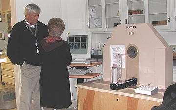
Cameo Appearance: The ATLAS small-animal imaging PET scanner was brought into the Mouse Imaging Facility (MIF) March 5 to participate in the MIF open house (which, by all accounts, was a packed event from beginning to end). In the photo above, Mike Green, ATLAS designer and chief of the Imaging Physics Laboratory in the CC Nuclear Medicine Department, explains the ATLAS modus operandi to an interested scientist: The animal is placed in the scanning imaging bed, which is advanced into the aperture of the scanner; the radiopharmaceutical is injected and its distribution tracked by ATLAS detectors. The data acquired are accumulated within the ATLAS computer (under the shelf upon which the scanner stands). This raw data may be sent via the accompanying software to the Biowulf supercomputer in CIT’s Building 12A to be converted to useful images. The images are ready to be called up in about an hour; meanwhile, another animal can be placed in the scanner. The investigator may also opt for the cruder images obtainable in ATLAS itself in about 5 minutes. At under 1.8-mm spatial resolution, these images are a far cry from the "uselessly blurred" images of small animals obtainable with the 5-7 mm resolution of a human PET scanner. But for the price of a little more time, Biowulf delivers exquisite images at 1.3-mm spatial resolution. |
Bill Eckelman, chief of the CC PET Department, and his staff have been providing the radiopharmaceuticals for the research projects that Green and collaborating investigators from other NIH institutes have been conducting during the decade-long research and development effort leading to the ATLAS machine. This work has taken advantage of the existing cyclotrons and radiochemistry facilities of the CC PET Department in the basement of the Clinical Center.
Radiopharmaceuticals administered to an animal and imaged with ATLAS consist of a positron-emitting isotope attached to a compound whose transport in the body follows a particular biochemical pathway. For example, labeled compounds are available that allow ATLAS to display and quantify the processing of glucose by the brain, the amount and distribution of various receptor types in the brain, gene expression throughout the body, regional hypoxia in tumors, and the movement of various cell types within the body. If the kind and number of such studies were to increase substantially, the additional material and personnel resources mentioned above and some physical modifications to the MIF would be required.
PET Projects
Over the next year or so, demonstration projects will be carried out that will help the community assess whether ATLAS should become a MIF regular. Green has a list of about 10 experiments to be performed with the PET Department and other institute collaborators for this purpose.
One, a "simple experiment," would determine the effect of various anesthetics on the mouse brain distribution of several radiolabeled compounds such as fluorodeoxyglucose (FDG), a popular PET tracer of glucose metabolism in brain, myocardium, and tumors. "This is of particular importance technically," Green notes, because animals must be motionless during imaging and, unless the animal is sacrificed, must be anesthetized during a study.
While live animals must also remain motionless during other forms of imaging, such as MR and CT, anesthetics can potentially distort the "true" time-varying tracer distribution compared to the distribution in the unanesthetized brain. Knowing how much distortion is induced by various anesthetics is important when trying to interpret changes in these distributions.
A study planned by NIMH’s Robert Innis will label amyloid plaques in the mouse brain and track distribution and amount in response to drugs. Fluorinated probes produced for this project will be evaluated. If the probes are effective in labeling and monitoring plaque changes, the payoff would be a treatment model for Alzheimer’s disease, Green notes.
In general, he says, the scope of small-animal PET experiments is limited only by the imagination of the researchers using the modality—and its value is "unequivocal."
PET, he says, provides "extraordinary sensitivity and specificity in visualizing biochemical processes in vivo. One can choose specific biochemical pathways and examine the behavior of those pathways under various conditions."
It’s a better choice for visualizing brain receptors than, for example, MRI, since PET can measure very small tracer amounts of drug that will not alter the pathway under scrutiny.
 |
|
IRP
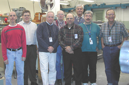
Team ATLAS: (left
to right) Steve
Fellini, CIT; Calvin
Johnson, CIT; Jim
Sullivan, ORS; Jimmie
Powell, ORS; Carroll
Toms, ORS; Mike
Green, CC; Jurgen
Seidel, CC; Burt
Chidakel, ORS (not pictured: Paul
Fitze and Frank
Sharpnack II)
|
Whither Small-Animal PET at NIH
Until now, word-of-mouth has brought investigators to Green’s door to explore the possibility that PET might help solve a particular research question.
Investigators in the radiation oncology branch, for instance, sought a means of assessing hypoxia in tumors so they could determine the effect of drugs or other treatments on tumor hypoxia. Fluorine-18–labeled misonidazole, a compound avidly taken up by hypoxic cells, was used as a probe of hypoxia levels.
PET was also harnessed in the service of cell trafficking in a collaborative study published earlier this year and involving NCI researchers Edit Olasz and Steve Katz, the PET Department’s Eckelman and Lixin Lang, and Nuclear Medicine’s Green and Seidel.
The researchers labeled and followed dendritic cells through the rat lymphatic system, an exercise with implications for research in immunologic processes.
The extent of small-animal PET’s distribution through NIH remains to be seen. PET capabilities, Green observes, do not contribute to achieving one of the main objectives behind the MIF, which was "conceived as a high-throughput lab." PET lends itself to hypothesis-driven experiments, he says, not to such endeavors as mass screening for mutations—a major impetus for the establishment of the MIF.
"The user community
[at NIH for ATLAS] has not been identified," Green says, "but it soon
will be." ![]()
| It Takes a Village | ||
| A collaboration of the following NIH and other entities produced ATLAS. | ||
|
Imaging Physics Laboratory, Nuclear Medicine Department, Clinical Center, NIH (physics, engineering and custom electronics design, assembly, testing and system integration) Michael V. Green (chief) Mechanical Instrument Design and Fabrication Section, SEIB, ORS, NIH (design, mechanical fabrication and assembly) James Sullivan (chief)
|
Computer and Electronics Section, SEIB, ORS, NIH (electronics layout and fabrication) Burt Chidakel Division of Computational Bioscience, Center for Information Technology, NIH (image reconstruction software development, high performance computing) Calvin Johnson (staff scientist) Hospital Universitario Gregorio Maranon, Madrid, Spain (user interface, image registration software) Manuel Desco (chief)
|
Thomas Jefferson National Accelerator Facility, Newport News, VA (custom electronics design, board fabrication and testing) Fernando Barbosa (staff engineer) A and D Precision Co. (data acquisition system with custom modifications) Val Zavarzin (nuclear physicist)
|