| T H E N I H C A T A L Y S T | M A Y - J U N E 1 9 9 8 |
| PEOPLE | |
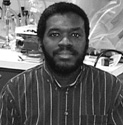
Kuni Iwasa received his Ph.D. in physics from Nagoya (Japan) University in 1974 and continued his work in chemical physics at Dalhousie University (Nova Scotia), Indiana University (Bloomington), University of Ljubljana (Slovenia), and Rice University (Houston). He began his work in membrane biophysics after arriving at NIH in 1979 as a senior staff fellow. He worked at NIMH and NINDS before joining NIDCD in 1991, where he currently heads the Section on Biophysics in the Laboratory of Cellular Biology.
The ear is a mechanoreceptor organ that converts sounds into electrical signals. It is not as simple as a microphone because it also splits signals into their component frequencies and attenuates larger signals. Perhaps for this reason, the ear has a number of cells with intriguing properties.
The outer hair cell is one such cell. It is a cylinder-shaped mechanoreceptor whose cell body shortens and elongates (as much as 5 percent of its length) very quickly, as the membrane potential goes up and down in response to pushes and pulls at its sensory hairs.
Recent studies have revealed that this cell uses electrical energy available at the plasma membrane. By contrast, most biological motility is based on chemical energy, particularly that of ATP. Direct use of electrical energy, which enables fast responses, is suited to the ear, which is sensitive to frequencies up to 20 kHz. The outer hair cell is a key factor in the fine-tuning capacity and wide dynamic range of the mammalian ear.
The mechanism that enables this cell to perform its biological role is not, however, as clearly understood as the role of the cell itself. My goal is to clarify the motility mechanism of the outer hair cell and its biological role.
The lateral membrane of the cell has charges that flip-flop across the membrane, analogous to gating charges of voltage-gated ion channels. These charges are the biophysical basis for voltage sensitivity of the cell, enabling it to use electrical energy.
Charge transfers across the hair-cell membrane are very large (equivalent to several million electrons per cell) and coincide with the cell motility.
If, indeed, such charge transfers provide energy used for length changes, one would expect the system could be run in reverse: The charges should be affected by externally applied tension. I found that is exactly the case.
This experiment also demonstrates that the membrane motor of the hair cell has at least two states that differ in their charges and membrane areas, in contrast to the opened and closed states of ion channels. Detailed knowledge of the motor was obtained by analyzing the charge transfers across the cell membrane induced by changes in voltage and tension. This analysis gives the number of motor units, the differences in charge, and the membrane area per motor unit in its two states.
Combining these data on the motor with data on the passive mechanical properties of the cell membrane, I have constructed a self-consistent biophysical model of the outer hair cell. The model can explain most existing observations on hair-cell motility and predicts forces that can be generated. We have experimentally confirmed the predicted value of 0.1 nN/mV for force production. I plan to use this model for further clarification of the motile mechanism.
To test whether the model can predict kinetic behavior as well as static properties, my group is trying to determine the relaxation time of transitions between motor states by measuring the frequency dependence of the membrane capacitance and the noise spectrum of membrane currents. This project is, in part, designed to address the question of how fast the cell can respond to voltage stimuli.
Perhaps the most intriguing question is which molecular elements contribute to the motility. One approach is to analyze whether the present model can explain the behavior of the cell treated by chemical reagents specifically targeting the cytoskeleton, the membrane, or the motor.
So far my group has shown that softening of the cytoskeleton reduces both cell stiffness and force production in a manner consistent with the model. Another approach to the molecular identity of the motor is via molecular biological methods.
My model predicts that the key element of the motor has a membrane-spanning domain and a charge transferable across the membrane. If candidates for the motor protein can be selectively expressed in a host cell, the electrophysiological techniques that we have developed can then be used to identify the role of individual elements in the motility. One important question is whether the membrane protein needs links to cytoskeletal proteins to function as a motor as the model predicts.

|
Roland Arvel Owens received his Ph.D. in biology from Johns Hopkins University in Baltimore in 1985. He came to NIH as a National Research Service Award Fellow in the Laboratory of Developmental Pharmacology in NICHD. In 1988 he moved to the Laboratory of Molecular and Cellular Biology in NIDDK, where he is now a senior investigator in the Molecular Biology Section.
My group studies the rep gene and Rep proteins of adeno-associated virus type 2 (AAV). AAV is a nonpathogenic human parvo-virus that is being developed as a vector for human gene therapy. AAV requires coinfection with a helper virus, usually an adenovirus or herpesvirus, for efficient productive infection.
It is therefore also a good model system for the study of virus-virus interactions. In the absence of helper virus, the DNA of AAV integrates into the host genome with a strong preference for a 2-kb region of human chromosome 19 (the only example of site-specific integration in a mammalian virus system).
The rep gene of AAV encodes four overlapping proteins involved in AAV replication, gene regulation, and preferential integration. The Rep68 and Rep78 proteins bind specifically to the AAV inverted terminal repeat (ITR) origins of DNA replication and have ATP-dependent, strand-specific endonuclease activity at specific sites within the terminal repeats. Rep68 and Rep78 also have ATP-dependent DNA helicase and DNA-RNA helicase activities, negatively and positively regulate AAV and heterologous gene expression, and can inhibit the production of HIV-1. Rep proteins can inhibit cell division and oncogenic transformation by adenovirus E1A plus an activated ras oncogene.
My group was the first to identify a specific DNA motif within the AAV ITRs that is recognized and bound by Rep78 and Rep68. It is an imperfect repeating ([GCTC]/[GAGC]) motif. We identified a similar Rep recognition sequence (RRS) within the chromosome 19 preferred integration locus and demonstrated that Rep78 or Rep68 can mediate the formation of a complex between the AAV ITRs and the chromosome 19 integration locus. This result led directly to the current model for AAV preferential integration.
We also demonstrated the involvement of an RRS in the regulation of AAV promoters by Rep proteins and have identified more than 20 RRSs within the human genome. Many of these sequences are within genes associated with cell proliferation or DNA repair, such as c-sis, gadd45, and brca1.
We suspect that there has been selection for the Rep proteins to regulate the expression of cellular proteins important for the AAV life cycle. Our working hypothesis is that the inhibition of cell division by Rep proteins is a consequence of this regulation.
We wish to understand better the role of the rep gene and gene products in the AAV life cycle and in AAV's interactions with its host cells and helper viruses. We will use this knowledge to aid in the exploitation of AAV as a gene-therapy vector.
We also wish to understand and exploit the antioncogenic and antiviral properties of the Rep proteins. Over the years, we have developed a unique set of mutant Rep proteins containing subtle mutations throughout the amino acid sequence. We plan to use these mutants, and others we are creating, to identify various functional domains and motifs required for the many activities of the Rep proteins.
Toward these ends, we will test our mutants for the ability to block cell division, interact with various cellular and viral proteins, and regulate the expression of key viral and cellular genes. We will also characterize further the endonuclease activity. This strategy will allow us to explore further the interrelationships between Rep protein functions.
THE 'INTER'NATIONAL INSTITUTES OF HEALTHThe NIH intramural program has been the destination of foreign scientists for five decades, primarily through the NIH Visiting Program. In the 1950s, fewer than 100 foreign postdoctoral fellows and more senior researchers were attached to intramural laboratories. Today, they number more than 2,000, or one-fifth of the intramural community. The program is one of several international programs administered by the Fogarty International Center (FIC) in collaboration with foreign governments and international organizations, some of which offer reciprocal opportunities for NIH intramural scientists. Japan: Give and Take In a program aimed at promoting Japanese-American scientific exchange, the Japan Society for the Promotion of Science (JSPS) supports Japanese fellows at NIH and visits (from one week to two years) by U.S. researchers to Japanese laboratories. NCI's Susanna Rybak, who has pioneered novel therapeutics involving members of the pancreatic RNase A superfamily, was one of last year's recipients. Notice of the fellowship inspired her to contact Masakazu Ueda at the Keio University School of Medicine in Tokyo, whose related work she'd followed in the literature. NIEHS' Sharon Bryant credited her two-month stay in Japan with yielding a "groundbreaking role in my research, expand[ing] my perspectives and [teaching] me a lot about my own culture." Her project in the lab of Yoshio Okada's group at Kobe-Gakuin University involved the structural analysis of newly synthesized ligands for the d-opioid receptor using two-dimensional 'H-NMR spectroscopy. Bryant had met Okada at a peptide symposium, and their labs had collaborated in the study of peptides synthesized by Okada's group before the research visit. Pan American FellowshipNIH also takes part in two Pan American Fellowship programs, one with Mexico and one with Chile. Under a 1996 agreement signed by NIH and the National Council of Science and Technology of Mexico, 14 Mexican postdoctoral scientists have received fellowships to work in NIH intramural and extramural laboratories. In 1997-98, five fellows were placed in intramural labs for research experience in neuropharmacology, cytogenetics, immunotoxicology, cellular biology, and biochemistry. A new agreement between NIH and the government of Chile will bring as many as five Chilean investigators to intramural laboratories. U.S-Russian Collaboration NIH researchers also have benefited from a program developed by the U.S. Civilian Research and Development Foundation (CRDF), a private nonprofit organization authorized by Congress and established by the National Science Foundation in 1995 to facilitate scientific and technological cooperation between the United States and the countries of the former Soviet Union. Financier-philanthropist George Soros provided initial funds of $5 million as an unrestricted gift to the U.S. government. These were matched by another $5 million from the Department of Defense (DoD). In 1996, NIH contributed $1 million for an NIH/CRDF Biomedical and Behavioral Sciences competitive grants program. With additional funds from NSF, DoD, and the Ukraine government, 41 grants were announced in September 1997. CRDF has awarded an additional five grants, supported by funds provided directly from IC budgets. Recently, with DoD funding, CRDF awarded three more grants. NICHD's Andreas Chrambach, in collaboration with Valery Chestkov of the Medical Genetics Center in Moscow, received a two-year CRDF grant to detect and isolate preapoptotic and early apoptotic cells (lymphocytes) by free-flow electrophoresis. In a mutually beneficial arrangement, the study is being conducted in the Moscow lab, where there is the manpower that Chrambach's group lacks-with NIH's electrophoretic instrumentation, which the Moscow group lacks.
-Irene Edwards, FIC
Deadlines
The deadline for the next round of JSPS fellowships is July 10. Contact Kathleen Michels in FIC's Division of International Training and Research (phone: 496-1653; fax: 402-0779; e-mail: <JSPS@nih.gov>). Applications for the NIH-Chile Pan American Fellowship are due on June 15. Contact Jahna Stanton, FIC Division of International Relations (phone: 496-4784; fax: 480-3414; e-mail: <js264e@nih.gov>). |