| T H E N I H C A T A L Y S T | J A N U A R Y - F E B R U A R Y 1 9 9 8 |
S e m i n a r H i g h l i g h t s
CONTEMPLATING TRIPLEX DNA AS EXPLOSIVE GENE THERAPY
|
|
By Ronald Neumann, M.D., Chief, Nuclear Medicine, Clinical Center. Neumann presented this November 12, 1997, at the Stone House at a joint DOE/NIH workshop to assess isotope-based medical research in the post-genome era. These Seminar Highlights were prepared by Celia Hooper.
Abstract
Triplex-forming oligonucleotides (TFOs) labeled with radionuclides that are Auger electron emitters could prove to be ideal vehicles for delivering radioactive decay energy to specific DNA sequences, causing local DNA breaks and subsequent inactivation of genes containing the target sequences. In a DNA triplex, the TFO, a short oligonucleotide generally 15-20 base pairs (bp) in length, occupies the major groove in a DNA double helix. Hoogsteen bonds are formed with the purines of the Watson-Crick base pairs in a sequence-specific fashion. In general, stable triplexes can be formed between polypurine-polypyrimidine duplexes and polypurine or polypyrimidine TFOs. Such sequences are widespread in eukaryotic genomes and are often found in regulatory regions. We have shown that TFOs can serve as suitable vehicles to deliver iodine-125 (125I) to a specific sequence in a DNA target (1).
The radiodecay of certain radionuclides produces a cascade of low-energy electrons, named after Pierre Auger, who first described this process in 1929. For example, radiodecay of 125I results in the emission of approximately 20 electrons of varying energy. Most of these Auger electrons have initial energies of less than 1 keV and a maximum range of only a few nanometers. The radiodecay of incorporated 125I from a TFO in a triplex structure with a targeted sequence in duplex DNA produces strand breaks located within 10 bp of the decay site with an efficiency close to one break per decay (2). Therapeutic applications of Auger electron emitters depend on developing methods for radionuclide delivery to the intranuclear genome of target cells, for example, cancer cells or perhaps even virally infected cells (3).
Sequence-Specific DNA Breaks | |
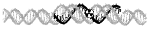 electron emitter binds in the major groove of the target duplex sequence. 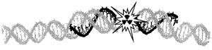 produces DNA strand breaks within five nucleotides from the decay site. | |
The promise of TFOs carrying Auger electron-emitting radionucleotides may be gene-specific radiation therapy if the complexities of triplex formation in vivo can be resolved.
Questions
Q: What was your starting point in this research, and how have your questions evolved?
A: My initial puzzle was how to position the Auger electron-emitting radionuclides sequence specifically and in close proximity to genomic DNA. Fortunately, I went to a lecture in Masur by a visiting Russian DNA chemist, Maxim Frank-Kamenetskii, where I first learned of triplex-forming oligonucleotides, short DNA fragments that incorporate themselves into the major groove of duplex DNA in a sequence-specific fashion. Ironically, the existence of triplex nucleic-acid structures was first demonstrated in 1957, here on campus by Gary Felsenfeld, David Davies, and Alex Rich.
Our first task was to demonstrate triplex binding by radiolabeled TFOs. My colleague, Igor Panyutin, devised a plasmid model system in which we showed DNA double-strand breaks could be produced by TFOs carrying 125I. Next, we measured the frequency and distribution of the breaks to show the small (± 5 bp) zone of DNA damaged by Auger electrons emitted during 125I decay. We then asked gene therapists how best to deliver DNA to specific cell nuclei. We chose cationic liposomes for the in vitro plasmid-model experiments, and demonstrated delivery into cultured cell nuclei by autoradiography and confocal fluorescence microscopy, effected by our visiting fellow, Olga Sedelnikova, in collaboration with Alain Thierry, who was then in Robert Gallo's lab. By attaching chemical linkers to the TFOs, other Auger-emitting radionuclides can now be used. This is work by Valeri Karamyshev, a visiting fellow, and our collaborators at Epoch Pharmaceuticals, Bothell, Washington.
Once formed, the triplexes are quite stable. One that we studied has a melting temperature of 65 C at conditions close to physiological. Double-strand breaks produced by decay of 125I incorporated into genomic DNA are highly radiotoxic and hardly reparable. Their repair usually results in deletion of large (>100 kbp) fragments of DNA.
Q: Which findings have been most surprising to you or to other scientists?
A: Our biggest and most pleasant surprise was finding that 125I-TFOs that do not form triplexes with genomic sequences, yet are present in the nucleus, caused very little radiation damage. These nonbound oligos are nearly 1/300 as toxic to the cell as 125I-5-iododeoxy-uridine, a precursor of DNA synthesis that is incorporated into genomic DNA. This gives us some hope that we can produce breaks in targeted gene sequences without causing excessive nonspecific radiation damage. The half-life of phosphodiester TFO in cell culture is hours and even shorter in vivo. For this reason, we have to freeze the cells after TFO delivery to accumulate DNA breaks and are now developing labeling procedures for phosphoramidate TFOs that are considerably more stable in vivo. Of course, for the therapeutic application we will need radioisotopes with a shorter half-life than 125I.
Q: What were the greatest stumbling blocks, and what new observations, techniques, reagents, or insights helped you get past them?
A: Our current experimental focus is to demonstrate that TFO-mediated Auger breaks in a tumor model have therapeutic benefit. We hypothesize that manipulation of the genomic DNA-151nucleosome complex may affect triplex formation, and we would like to better understand those manipulations to improve the ability of TFOs to find their targets in vivo and to increase the specificity of such targeting.
Q: In which areas do you see this research having the greatest use for clinical scientists? In which areas of basic research will it be most illuminating?
A: Clinically, treating cancers containing amplified genes or viral-derived "foreign" sequences may be the best application of this technique should it be proven to work in vivo. Our chances of hitting a target gene increase if the gene is amplified, as happens in some cancers.
For basic scientists, this method may be useful to probe nucleic acid-protein complexes because the Auger electron damage is so focal and is distance-related. Igor Panyutin and collaborators in NCI and NIDDK laboratories took this approach recently when they analyzed decay-induced DNA breaks to successfully examine nucleic acid conformations.
Q: How are you following up on this work?
A: Beyond the specific cancer genes we're studying now, we would like to explore the application of our techniques in other genetic diseases and are thus in search of collaborators with amplified-gene models of disease in which to test other radiolabeled TFOs for gene-specific radiotherapy.
References
1. I.G. Panyutin and R.D. Neumann, "Sequence specific DNA breaks produced by triplex-directed decay of iodine-125," Acta Oncol. 35: 817-823, (1996).
2. I.G. Panyutin and R.D. Neumann, "Radioprobing of DNA: distribution of DNA breaks produced by decay of 125I incorporated into a triplex-forming oligonucleotide correlates with geometry of the triplex," Nucleic Acids Res. 25: 883-887 (1997).
3. O. Sedelnikova, I.G.Panyutin, A. Thierry, and R.D. Neumann, "Delivery of triplex-forming oligonucleotides into cells: Comparison of different liposome compositions." Gene-therapy poster session, NIH Research Festival (1997).