DECONSTRUCTING DEVELOPMENT
The advent of mammalian cloning extends key observations John Gurdon made more than 30 years ago regarding the frog Xenopus laevis (2). Gurdon used a strategy for nuclear transplantation developed earlier by Robert Briggs and Thomas King (3) to demonstrate unequivocally that nuclei from tadpole intestinal epithelial cells could direct the development of fertile adult frogs. Over the subsequent two decades, these experiments were repeated in ever-increasing detail by a dedicated group of investigators until the pluripotency of adult cell nuclei was definitively established in 1986 (4, 5).
This work laid a firm scientific foundation for mammalian cloning. Nevertheless, despite much effort, no single transplanted adult frog nucleus has ever yielded a cell that grew into another adult frog. The work by Wilmut and colleagues shattered a barrier and revealed that cells of mature higher animals are not just pluripotent, but totipotent. Their use of an adult cell derived from sheep mammary epithelium as a donor in the nuclear transplantation experiment that gave rise to Dolly indicates that adult nuclei can also become totipotent.
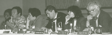 National Bioethics Advisory Commission members, left to right: Rhetaugh Dumas, vice provost for health affairs, University of Michigan; Lawrence Miike,director, Honolulu State Department of Health; R. Alta Charo, professor of law, University of Wisconsin; Arturo Britto, assistant professor of clinical pediatrics, University of Miami; Carol Greider, senior staff scientist, Cold Spring Harbor Laboratory; Eric Cassell, professor of public health, Cornell Medical College. [Other members are Chairman Harold Shapiro, Patricia Backlar, Alexander Capron, James Childress, David Cox, Ezekiel Emanuel, Laurie Flynn, Steven Holtzman, Bette Kramer, Bernard Lo, Thomas Murray, and Diane Scott-Jones. |
In the wake of this revelation, developmental biologists are asking: What does the re-acquisition of totipotency imply for the molecular mechanisms that establish cell fate? Early embryogenesis requires the totipotent egg nucleus to cleave during repeated cell division, generating daughter cells that progressively acquire all of the separate cellular identities that exist in the tissues of an organism. This requires the precisely staged association of transcription factors and specialized chromosomal proteins with the regulatory elements of genes. As development proceeds, an increasing number of cells exists in the embryo, and the regulatory nucleoprotein complexes that establish cell lineages or identities become more elaborate and resistant to physical and biochemical perturbation. This functional specialization of chromatin and chromosomes also becomes more difficult to reverse when an embryonic cell nucleus is transplanted into an enucleated egg. As a general rule, the more differentiated the cell from which a donor nucleus is taken, the more unlikely it is that correct development will proceed.
Nevertheless, persistent attempts to break the rule have led to dividends in amphibia, where nuclei taken from adult keratinocytes or reticulocytes have, in a few notable instances, been shown to support the development of all the cell types found in a tadpole (4, 5). These findings demonstrate that the regulatory nucleoprotein complexes that control the specific patterns of gene activity in highly differentiated keratinocytes or reticulocytes can be disassembled.
The first experiments that examined specific gene activity within somatic nuclei that had been transplanted into an egg revealed that nucleoli disappeared and active ribosomal RNA genes were inactivated (6). The disappearance of the nucleoli - which represent the compartmentalization of rRNA synthesis to a specific chromosomal structure - and the inhibition of rRNA transcription clearly demonstrated that egg cytoplasm has the capacity to influence nuclear function. As development of the embryo containing the transplanted nucleus proceeds, the ribosomal genes are reactivated and nucleoli reappear.
Researchers were better able to understand the influence of cytoplasm on nuclear function after Merriam and Barry demonstrated that there is considerable movement of proteins from the egg cytoplasm to the somatic nucleus following transplantation (7, 8). This movement is concomitant with nuclear swelling and with a significant reduction in the amount of transcriptionally inactive heterochromatin within the nucleus. Subsequent work examined the association of specific components of the transcriptional machinery with individual genes. Both the regulatory nucleoprotein complexes that activated transcription and those that repressed transcription were found to be unstable in an egg environment (9, 10). This was in marked contrast to their stability in the nuclei of differentiated cells (11). Molecular chaperones that are stored in the egg have been shown to have a causal role in directing the loss of chromosomal proteins specific to somatic nuclei and thus the remodeling of somatic nuclei following exposure to egg cytoplasm (12). However, this is only part of the process. The transplanted somatic nucleus progressively acquires the proteins and modifications normally associated with the embryonic chromosome. Many of these specialized modifications are also characteristic of the transformed cell nuclei found in many tumors.
|
The
experiments are simple and powerful. They answer a crucial biological
question...by clearly demonstrating the reversibility of determinative
mechanisms.
|
The orchestrated exchange of somatic nuclear proteins for egg cytoplasmic components takes time, and it is the failure to effect the restructuring of chromatin, chromosomes, and nuclei before cell division that most probably leads to chromosomal damage and the developmental abnormalities apparent in many nuclear-transplant embryos. In this regard, the mammalian cloning experiments provide additional insight (1). These investigators made use of adult somatic cells that they synchronized in G0 - a quiescent state within the cell cycle. This state of quiescence is normally achieved by starving cells for serum, causing cells in G1 to leave the cell cycle. This exit can be reversed by adding back serum in culture or, evidently, by transplanting a G0 cell nucleus into the egg. This might facilitate the remodeling of chromatin by attuning the nuclear and cytoplasmic cell cycles just before entry into S phase. DNA replication itself will further facilitate the disruption of regulatory nucleoprotein complexes (13). Therefore, using the strategy of Wilmut and colleagues (1), an embryonic chromosomal structure might be established in a transplanted nucleus before cell division occurs, thereby preventing chromosomal damage. If this hypothesis is true, then simple manipulations of somatic cell nuclei that would facilitate nuclear remodeling, such as pre-incubation with the molecular chaperone nucleoplasmin, would greatly facilitate the efficiency of animal cloning.
The original interpretation of nuclear transplantation experiments in amphibia suggested that the genetic material is not irreversibly altered as development proceeds. This concept had a major impact on developmental science at the time. However, we now know that this is not universally true, since cell-type-specific rearrangement of immunoglobulin genes and generalized loss of telomeric sequences occur in conjunction with differentiation and aging. Moreover, specific patterns of cytosine methylation and demethylation correlate with gene activity and repression in particular cells. The mammary epithelial cell nucleus used by Wilmut did not undergo VDJ recombination to define the antibody repertoire; however, loss of telomeric sequences presumably has occurred. Hence, the aging of mammalian clones may differ from that in animals derived from the fusion of gametes. As for DNA methylation, it must either be reversible or unimportant for the establishment of differential states of gene activity - the stuff for future studies.
A final technical point is that much of early embryonic development is driven through the activity of proteins and messenger RNAs stored in the egg. Masked maternal mRNA is translationally silent until fertilization (or nuclear transplantation). Recruitment to the translational machinery then initiates the determinative events that restrict the fates of embryonic cells. The same molecular chaperones that facilitate the remodeling of somatic nuclei following transplantation also facilitate the unmasking of maternal mRNA (14, 15). Importantly, the maternal determinants will differ from egg to egg. Thus, a true clone from a nuclear-transplant embryo is not achievable in the sense that monozygotic identical twins are clonal.
Recognition of the success of mammalian cloning will surely have a major impact on many aspects of basic developmental biology. The experiments are simple and powerful. They answer a crucial biological question regarding development by clearly demonstrating the reversibility of determinative mechanisms. Differential gene activation is what drives development, not the irreversible alteration of the genetic material itself. The dramatic affirmation of this conclusion should stimulate experiments that seek to define the developmental ground state of totipotency. Molecular understanding of how specific patterns of gene activity can be reversed in the egg surely will have general relevance for human disease.
1. I. Wilmut, A.E. Schnieke, J. McWhir, A.J. Kind, and K.H.S Campbell. "Viable offspring derived from fetal and adult mammalian cells." Nature 385: 810-813 (1997).
2. J.B. Gurdon and V. Ueblinger. "`Fertile' intestine nuclei." Nature 210: 1240 (1966).
3. R. Briggs and T.J. King. "Transplantation of living nuclei from blastula cells in enucleated frog's eggs." Proc. Natl. Acad. Sci. USA 38: 455 (1952).
4. J.B. Gurdon, R.A. Laskey, and D.R. Reeves. "The developmental capacity of nuclei
transplanted from keratinized skin cells of adult frogs." J. Embryol. Exp. Morphol. 34: 93 (1975).
5. M.A. Di Berardino, N.H. Orr, and R.G. McKinnell. "Feeding tadpoles cloned from Rana erythrocyte nuclei." Proc. Natl. Acad. Sci. USA 83: 8231-8234 (1986).
6. J.B. Gurdon and D.D. Brown. "Cytoplasmic regulation of RNA synthesis and nucleolus formation in developing embryos of Xenopus laevis. J. Mol. Biol. 12: 27-35 (1965).
7. R.W. Merriam. "Movement of cytoplasmic proteins into nuclei induced to enlarge and initiate DNA or RNA synthesis." J. Cell Sci. 5: 333-349 (1969).
8. J.M. Barry and R.W. Merriam. "Swelling of hen erythrocyte nuclei in cytoplasm from Xenopus eggs." Exp. Cell Res. 71: 90-96 (1972).
9. A.P. Wolffe and D.D. Brown. "Differential 5S RNA gene expression in vitro." Cell 51: 733- 740 (1987).
10. A.P. Wolffe. "Transcriptional activation of Xenopus class III genes in chromatin isolated from sperm and somatic nuclei." Nucl. Acids Res. 17: 767 (1989).
11. M.S. Schlissel and D.D. Brown. "The transcriptional regulation of Xenopus 5S RNA genes in chromatin: the roles of active stable transcription complexes and histone H1." Cell 37: 903-911 (1984).
12. S. Dimitrov and A.P. Wolffe. "Remodeling somatic nuclei in Xenopus laevis egg extracts: molecular mechanisms for the selective release of histones H1 and H1° from chromatin and the acquisition of transcriptional competence." EMBO J. 15: 5897-5906 (1996).
13. A.P. Wolffe and D.D. Brown. "DNA replication in vitro erases a Xenopus 5S RNA gene transcription complex." Cell 47: 217-227 (1986).
14. S. Tafuri and A.P. Wolffe. "Selective recruitment of masked maternal mRNA from mRNPs containing FRGY2/mRNP4." J. Biol. Chem. 268: 24255-24261 (1993).
15. F. Meric, K. Matsumoto, and A.P. Wolffe. "Regulated unmasking of in vivo synthesized maternal mRNA at oocyte maturation." J. Biol. Chem. 272 (in press).
Return to Table of Contents