HALL
EFFECT IMAGING MAY GIVE
MEDICINE A NEW SENSE
Those physicists have done it again. Doctors had barely heard of gamma radiation, beta decay, and nuclear magnetic resonance before they found themselves applying the concepts in X-rays, positron emission tomography, and magnetic resonance imaging of the human body. The next addition to the list of obscure physics topics to sweep clinicians off their feet could well be the Hall effect, which may some day allow painless diagnoses of tumors, kidney function, and fetal health in the womb, along with major improvements in conventional ultrasound imaging.
Han Wen, of the Lab of Cardiac Energetics, NHLBI, is the inventor of Hall effect imaging (HEI), and he got the idea partly from artifacts his group and others observed in the electrocardiograms (EKGs) taken of patients while they were simultaneously undergoing magnetic resonance imaging (MRI).
![[ Hall Effect Schematic ]](images/schematic.gif) |
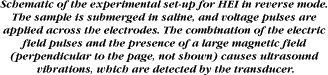
Wen realized that the effect was closely related to the blood's electrical conductivity, a property that happened to be of interest in a variety of body tissues because of its other effects on certain MRI data. He reasoned that the Hall effect could be used to map conductivity in the body - as long as some motion of the tissue could be generated that would play the role of blood flow through the heart in the EKGs. The motion also had to be spatially confined so that signals originating from different locations in the body could be distinguished. Fitting these requirements, ultrasound proved to be a good source of motion.
In ultrasound imaging, pulses of high-frequency sound are sent into the body, and the time of arrival of the echoes indicates the distances to the various sound-reflecting tissues. Because the sound penetration and reflection are mainly determined by tissue density, ultrasound is essentially a density probe. In HEI, the ultrasound pulses are applied to tissue within a magnetic field and jiggle it just enough to generate a Hall voltage, which is detected with electrodes; thus, HEI measures the electrical conductivity of the tissues, rather than their density. Although this was the original concept, Wen discovered in the lab that the HEI signal's noise level could be drastically reduced by running it in "reverse mode," that is, by using the electrodes to apply voltage pulses and measuring the resulting ultrasound signal. In the reverse mode, the combined effects of the voltage pulse and the magnetic field on the tissue's charges cause an ultrasound vibration. Either way, the output measures tissue conductivity.
The beauty of Wen's technique is that it should be able to give high-resolution pictures of tissue conductivity, which, unlike density, varies quite a bit from one tissue to another. That should yield images with far better contrast than conventional ultrasound and permit a new kind of tissue characterization based on conductivity. Wen cites an example from intravascular ultrasound imaging, where a "bulge" might be seen on the wall of a blood vessel. "It's very hard to tell whether that bulge is just a bulge of the muscle lining of the artery, or [whether] it's actually a fatty plaque. Now, potentially, you could use this technique [to identify the nature of the bulge], because it's conductivity-sensitive. There's a big difference in conductivity between fat and [muscle]."
![[ Sample Hall Effect Image: Bacon ]](images/hall-fx_.gif) |
Wen and Balaban can imagine other possible applications that might allow patients to avoid invasive diagnostic procedures. A kidney that isn't properly concentrating electrolytes in the urine, for example, ought to have a clearly different conductivity from a healthy kidney, so HEI could save the trouble of catheterizing the individual kidneys for diagnosis. The cerebrospinal fluid in a developing fetus is sometimes tested for signs of proper development, and, according to Wen, "the conductivity is one of the standard test parameters. And if you can do that noninvasively, it's going to be much less painful for the mother and for the baby."
Although quite promising, the HEI technology has not yet been tested on an animal. The most complicated sample so far was a piece of bacon. "I thought bacon was just too bizarre," Wen recalls. But Balaban explained that bacon is animal tissue with interlaced fat and muscle, which ought to have distinctly contrasting conductivities. After Balaban purchased the sample at a Bethesda grocery store, Wen observed the expected result: the layers of the bacon showed up much more clearly in the HEI image than with conventional ultrasound.
Before imaging an animal, a few engineering problems must be solved, the largest of which is to design a new, nonmetal ultrasound detector. Balaban explains that it's needed to defeat the largest source of noise in the current system. "If you put any metal in the magnet and it [vibrates] at ultrasound frequencies, it generates a Hall voltage, and that's an interference. Han and I have suffered through that in these initial studies. It's a new class of [sound] detectors that we have to come up with." Fortunately, they have found collaborators at the Naval Research Lab, in Washington, who are already experts on such detectors, which rely on interferometry and coiled fiber optics to give high sensitivity without the use of any metal parts. The main problem is to adapt these detectors for use at ultrasound frequencies.
Some other challenges, which appear less difficult, include designing a good way to deliver electric pulses to the body and adapting conventional ultrasound electronics and data processing. But despite these hurdles, Balaban is optimistic. He foresees experiments on humans within a year and a clinical device three to five years after that. "The reason I'm that positive about it is because basically we know a lot about ultrasound as a clinical tool already. What we're doing is adding the magnetic field, which is also now a commercial device." And Wen points out that expensive MRI magnets aren't needed for HEI; fairly cheap ones will suffice - magnets "that they use in junkyards to pull up cars and things like that. That's good enough for us." He adds that an HEI magnet could be much smaller than one from a junkyard.
Balaban stresses that the best applications of this technology may not yet have been imagined. Conductivity has not been observed so directly in the past, and new HEI data may reveal new physiological information and new directions for basic research. He draws a parallel with MRI, which yielded much unexpected information after researchers began experimenting with it. "We're going to look around a little bit with this new technology. We have a few ideas, but the real thing is now to explore the body with this new 'sense' and really see what comes out of it."
New Center To Tackle Inherited DiseaseBorn of the joint efforts of seven NIH institutes and one center, a new Center for Inherited Disease Research (CIDR) has materialized at the Bayview, Md., campus of the Johns Hopkins School of Medicine. Its main purpose in life is the identification of the genetic loci and allelic variants that play important roles in multifactorial human disease, including, but not limited to, cardiovascular and pulmonary disease, cancer, psychiatric disorders, hearing and language disorders, neurological disease, diabetes, and autoimmune diseases. To achieve that end, CIDR will utilize high-throughput genotyping in support of relevant research involving human populations and families and, possibly, pertinent animal models. Access to CIDR is open to all investigators on a competitive basis. Intramural scientists should get approval of their scientific directors first. CIDR will carry out genome-wide genotyping scans on samples provided by principal investigators whose proposals have been accepted. A variety of different mapping approaches may be supported by genotyping within CIDR, including affected-pedigree-member methods, transmission-disequilibrium testing, and linkage analysis in pedigrees. Investigators may also consult with CIDR researchers about study design and statistical analysis. Once CIDR has completed its studies, the data and results of the analyses will be returned to the principal investigator for further research. Proposals from extramural investigators will undergo the customary NIH review for scientific merit. Additionally, all proposals, whether of intramural or extramural origin, will be examined by a chartered CIDR Access Advisory Committee for compliance with criteria, including suitability of the project for the high-throughput genotyping capabilities of CIDR, feasibility of study design for detecting genetic contribution to disease, the likely impact of the study on biomedical research and, for intramural studies, the scientific merit of the proposal. A Board of Governors, the policy-setting body for CIDR, will review the recommendations of the Access Advisory Committee, determine what resources are available, and then advise the center director regarding when the most highly rated projects can be initiated. The board will be made up of the directors of the eight participating institutes and centers (or their designees). This scrutiny by the CIDR Access Advisory Committee is not expected to lengthen the review process beyond what is normally required for extramural grant submission and review. CIDR's lead agent and manager is the National Center for Human Genome Research; its seven other sires are the National Cancer Institute, the National Institute of Child Health and Human Development, the National Institute on Deafness and Communication Disorders, the National Institute on Drug Abuse, the National Institute of Environmental Health Sciences, the National Institute of Mental Health, and the National Institute of Neurological Disorders and Stroke. A description of CIDR will soon be available at the NCHGR homepage on the World Wide Web at <http://www.nchgr.nih.gov/home.html>. If you want more information about CIDR or are interested in using its services and facilities, contact Jerry Roberts, scientific review administrator and chief of staff, CIDR Board of Governors, in the NCHGR Office of Scientific Review, 496-0838. |