NIH Catalyst - Leader of the Pack: Dideoxy Fingerprinting

LEADER OF THE PACK: DIDEOXY FINGERPRINTING
FOR FINDING UNKNOWN MUTATIONS IN GENES

by Zsolt Orban, M.D., NIDDK;
A. Lee. Burns, Ph.D., NIDDK;
Michael Emmert-Buck, M.D., Ph.D., NCI;
and Lance A. Liotta, M.D., Ph.D., NCI


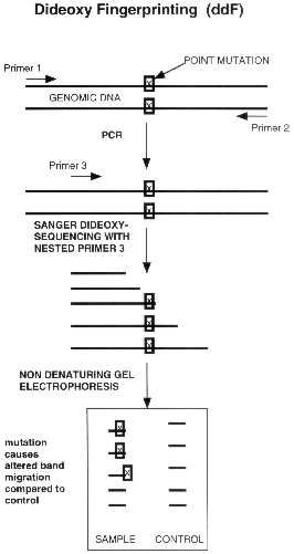
ddf schematic

Figure 1. Schematic
diagram of ddF

Explosive progress in understanding the genetic basis of human disease and drug resistance in microorganisms has left researchers urgently in need of a method for routinely screening specific genes for mutations. A wide variety of different types of mutations may underlie the pathogenicity caused by changes in any given human or microbial gene so the exact mutations in the causative gene cannot be predicted ahead of time in most cases. Even investigators who are attempting to link a newly identified gene with a specific hereditary syndrome would be well served by having a simple way to check for the existence of any mutations before they begin the laborious process of sequencing the entire gene. Consequently, whether the complete gene is known or not, investigators would like to have a foolproof "mutation test." The new dideoxy fingerprinting (ddF) technique reviewed in this Hot Methods Clinic appears to have the sensitivity and ease of use sufficient to put it ahead of a small pack of other techniques in fulfilling this important research need.

Direct genomic sequencing, the gold standard for mutation analysis, is labor-intensive, expensive, and time-consuming. Several shortcut mutation-screening methods have therefore been developed. These include heteroduplex analysis, chemical mismatch cleavage, and denaturing gradient gel electrophoresis (1). Although faster and cheaper than direct genomic sequencing, these methods are technically difficult, labor-intensive, prone to false negatives or false positives, insensitive, and may require relatively large amounts of input template DNA.

The most widely used system for detecting point mutations has been single-strand conformation polymorphism analysis (SSCP) (2). SSCP starts with PCR amplification of target DNA. The amplified and partially denatured strands are then separated on a nondenaturing polyacrylamide gel. A mutation in the DNA strand generally causes a change in the three dimensional conformation, and the altered DNA migrates to a different point on the gel compared with the wild-type control. SSCP is simple to perform; unfortunately, however, it can miss many mutations (4), such as single-base substitutions of cytosine (C) to thymidine (T), that do not alter the 3-D shape enough to significantly change the migrational properties of the strand. In addition, SSCP will provide no information about the approximate location of the mutation within the screened segment.
The Method and How it Works

To overcome the drawbacks of SSCP, Sarkar et al. proposed ddF (3), which is a hybrid between direct genomic dideoxy-sequencing and SSCP analysis. The concept is simple: a standard Sanger sequencing reaction using only one dideoxy terminator is electrophoresed through a nondenaturing gel. In addition to detecting mobility shifts in the fragments containing the mutation, this method also picks up mutations that lead to a gain or loss of dideoxy termination bands (see Fig. 1). Thus, ddF is less influenced than SSCP by the nature of the mutation. ddF can detect mutations with close to 100% sensitivity up to 250 base pairs (bp) from the 51 end of the nested primer, and bi-directional ddF can screen a 500- to 550-bp region of DNA. By comparison, SSCP allows detection of between 70% and 95% of mutations for small PCR segments of 200 bp or less, and its sensitivity decreases rapidly with increasing size of the PCR product (3).

The superior performance of ddF was demonstrated by Teresa Felmlee and associates at the Mayo Clinic in Rochester, Minn., who compared it with SSCP on blinded samples of drug resistant Mycobacterium tuberculosis. They found that prolonged electrophoresis time was required for discrimination of SSCP differences in strand migration compared with ddF. They also noted that some C-to-T transition mutations that were correctly identified by ddF could not be picked up by SSCP. Other researchers report that ddF detected 100% of p53 mutations in breast cancer (6) and all 84 different mutations, including all 12 possible types of base substitutions, in the human blood-clotting factor IX gene (7).

As we cross into the new millennium and the Human Genome Project approaches the identification of all human genes, the medical diagnostic lab will be transformed (8). While we wait for genetic testing to be miniaturized on a chip (8), methods such as ddF, or its modification, bi-directional ddF (7), will likely play a key role in finding mutations in hereditary-syndrome genes, in detecting drug resistant microorganisms, and in determining risk for acquired diseases. As a research tool, ddF will undoubtedly serve as a prominent mutation screening test that will aid in the linkage of newly identified genes to specific diseases.

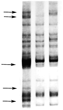
Figure 2. Example of a positive screen. Arrows denote alterations in the band migration pattern. Lane 1: Mutation Present. Lane 2: Control. Lane 3: Mutation Present (different than in sample 1). Mutants were provided as a courtesy of Mary Grace, NCHGR


Protocol

Mention of a specific commercial product or company does not constitute an endorsement.

  1. To prepare the mutation-detection-enhancement (MDE) gel, swirl the following in an empty plastic gel-bottle: 35 mL of 2x MDE stock (manufactured by FMC Bioproducts); 4.2 mL of 10x Tris-boric acid-EDTA buffer; and sufficient distilled water to bring the total up to 70 mL. Mix in 280 uL of 10% ammonium persulfate and 28 uL of tetramethyl-ethylene diamine (TEMED). Pour a 0.4-mm-thick sequencing-type gel, pressing a saw-tooth comb into the gel to create the wells. This comb gives results superior to those obtained with a shark-tooth comb. Store gel in a cold room (8 šC) and allow to set there for a minimum of 2 to 3 h. before removing the comb.

  2. Prepare a stop solution with final concentrations of the following: 7 mol urea/L, 50% formamide, 3 mmol EDTA/L, and 0.5% bromphenol-blue-xylene cyanol.

  3. Generate the amplicon to be analyzed by standard polymerase-chain reaction (PCR) using a proof-reading DNA polymerase. Store at -20 šC until used for fingerprinting.

  4. The next step is to perform dideoxy fingerprinting on the amplicon with a nested, end-labeled primer, using your preferred T4 kinase labeling method. Design this primer so that its melting temperature is somewhat higher than that of the two primers used in the first PCR reaction. For a typical dideoxy fingerprinting reaction, mix the following: 6.0 uL distilled water, 2 uL of 5x Taq buffer; 0.1 uL of 2.5 mmol NTP/L; 0.2 uL of 10 mmol ddGTP/L; 2 pmol (1uL) of end-labeled primer; one unit of Taq polymerase; and 0.5 uL of the template DNA from the PCR reaction in step 3. Perform a typical cycle-sequencing reaction (30 to 40 cycles).

  5. Add 50 uL of the stop solution, prepared in step 2 above, to the completed 10 uL of ddF reaction product. Incubate the samples at 90 šC for 5 min. and quick-chill on ice. Load a 3 uL aliquot onto the gel. Run gel at 20 Watts constant power until the xylene cyanol migrates two-thirds of the way down the gel. Subject the gel to autoradiography.


Trouble-Shooting Tips

These tips are adapted from Q. Liu, J. Feng, and S.S. Sommer (7).

  1. Accurate pipeting is critical.

  2. Use sawtooth combs yielding 32 or 64 wells for each gel.

  3. Quick-chill the samples after boiling by immersing in ice water. This will reduce fuzzy bands.

  4. Cool the gel or run at low temperature (8 šC in cold room).

  5. For the fingerprinting, choose a dideoxy nucleotide that has a uniform spacing of termination segments, especially near the top of the gel.

  6. Control experimental conditions closely. The fingerprint obtained is highly reproducible if run under identical conditions (e.g.. temperature, wattage, and gel-preparation). Different running conditions, such as different wattage, can lead to altered sensitivity in picking up mutations and can produce different band-migration patterns.

  7. Never score a sample as negative unless it is directly next to a control sample. To avoid false negatives, make sure the entire region of interest is represented in the gel.

  8. Do not score a sample as positive if the intensity of the signal fades out as the segment gets larger. This pattern could be due to a poor termination reaction.

  9. Assume that any segment of the SSCP migration that is clearly different from a normal segment in the flanking lane in the gel contains a mutation.


Contacts

Zsolt Orban, NIDDK
Phone: 402-7834

Michael Emmert-Buck and Zhengping Zhaung, NCI
Phone: 496-2912

A. Lee. Burns, NIDDK
Phone: 496-4616

Steve Sommer, Mayo Clinic
Sommer offers a kit of protocols and reprints relating to ddF.
Phone: 507 284-4597, Fax: 507 284-3383


References

  1. M. Grompe. "The rapid detection of unknown mutations in nucleic acids." Nature Genet. 5, 111-17 (1993).

  2. M. Orita, H. Iwahana, H. Kanazawa, K. Hayashi, and T. Sekiya. "Detection of polymorphisms of human DNA by gel electrophoresis as single-strand conformation polymorphisms." Proc. Natl. Acad. Sci. USA 86, 2766-70 (1994).

  3. G. Sarkar, H.S. Yoon, and S.S. Sommer. "Dideoxyfingerprinting (ddF): a rapid and efficient screen for the presence of mutations." Genomics 13, 441-43 (1992).

  4. Q. Liu and S.S. Sommer. "Parameters affecting the sensitivities of dideoxy fingerprinting and SSCP." In: PCR Methods and Applications, Vol. 4. Cold Spring Harbor, NY: Cold Spring Harbor Laboratory Press, 97-108 (1994).

  5. T. Felmlee, Q. Liu, C. Whelen, D. Williams, S. Sommer, and D. Persing. "Genotypic detection of Mycobacterium tuberculosis rifampin resistance: comparison of single-stranded conformation polymorphism and dideoxy fingerprinting." Jour. Clin. Microbiol. 33, 1617-23 (1995).

  6. H. Blaszyk, A. Hartmann, J.J. Schroeder, R.M. McGovern, S.S. Sommer, and J.S. Kovach. "Rapid and efficient screening for p53 gene mutations by dideoxyfingerprinting." Biotechniques 18, 256-60 (1995).

  7. Q. Liu, J. Feng, and S.S. Sommer. "Bi-directional dideoxy fingerprinting (Bi-ddF): a rapid method for quantitative detection of mutations in genomic regions of 300-600 bp." Hum. Mol. Genet. (in press).

  8. R. Nowak. "Entering the postgenome era." Science 270, 368-71 (1995).


Return to the Table of Contents