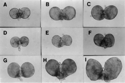
Fruit flies have served as an invaluable model for deconstructing the genetics of development. The power of fly genetics was recently recognized by the award of the Nobel Prize in physiology or medicine to Edward Lewis, Christiane Nusslein-Volhard, and Eric Wieschaus for work that identified Drosophila genes required to establish the body plan of the fly. Many of the genes that were identified in flies by these groups have vertebrate homologs that are also required for development. Could fruit flies be as powerful in helping us understand cancer as they have been in development? In this Hot Methods Clinic, we report on new models that extend the power of Drosophila genetics to the field of cancer progression and metastasis.
Current methods for studying metastasis, tumorigenesis, and tumor suppression in vivo generally rely on rodent models. For example, studies of metastasis typically require injection of tumor cells into nude mice, a delicate animal system that requires a large investment of time and upkeep. Comparatively little use has been made of Drosophila, one of the best-defined animal models. Although much smaller than more traditional models for metastasis, fruit flies also have tumors, including tumors that metastasize when transplanted from a larval donor into an adult host. By studying metastasis in Drosophila, it is possible to do experiments in a few bottles that would otherwise consume substantial resources with rodents. Furthermore, one can take advantage of a large body of genetic techniques and knowledge about Drosophila that make it possible to generate mutants, clone genes, and express transgenes with relative ease.
 |
Recent studies in the field of Drosophila tumorigenesis and metastasis have shown that this genetic system can provide an excellent means for studying metastasis, as well as tumorigenesis and tumor suppressors. Mutations in single genes in Drosophila can cause tumorous overgrowth of the larval brain and imaginal discs - groups of cells in the larva that will give rise to adult structures. The overgrowth is accompanied by the loss of capacity of the brain and imaginal disc cells to differentiate. These tumors are not metastatic in the larva, but when transplanted into adult hosts, they cause large primary tumors which can metastasize and invade host organs. Aided by the introduction of a lacZ reporter gene into various tumor mutant backgrounds, we have studied the tumors which form after transplantation. The reporter gene is used to identify the tumor cells after transplantation since none of the adult host cells contain the lacZ reporter. Using the reporter gene to follow tumor cells after transplantation, we have been able to study the metastasis of tumorous brain tissue and imaginal discs from several Drosophila mutants. Tumors can also form in other tissues of Drosophila such as the gonads and hematopoietic organs. Overall, more than 50 tumor suppressor genes have been identified in Drosophila (1). Although many of these genes have not been extensively characterized, they open exciting new avenues of research.
The general approach of our lab and that of Allen Shearn at Johns Hopkins University in Baltimore is to use Drosophila to investigate factors involved in metastasis, as exemplified by our work on the lethal giant larvae mutant. Loss of function of the Drosophila gene lethal giant larvae, which is located on the second chromosome, leads to tumors of the imaginal discs and brain, and death at the late larval stage. The lethal giant larvae mutants have an extended larval period during which cells in the brain and discs continue to proliferate and become tumorous. When this brain or imaginal disc tissue is transplanted into adult hosts - for example, by injecting the tissue into a fly's abdomen - it can grow as a primary tumor and metastasize to adult structures.
In 1995, Mechler and colleagues cloned the lethal giant larvae gene, and several investigators are now beginning to elucidate its function (2). Recent biochemical studies have shown that the Lethal Giant Larvae (LGL) protein is part of a large complex and that a major component of this complex is nonmuscle myosin (3), suggesting that the LGL protein may be involved with the cytoskeleton. Immuno-staining has localized the LGL protein to the cell surface at regions of cell junctions. These results hint that the LGL protein may be part of the signal pathway from the cell surface via the cytoskeleton.
For marking and following the lethal giant larvae mutant cells, we used a construct consisting of the lacZ gene under the control of the armadillo promoter and inserted onto the X chromosome so it can easily be crossed into the lethal giant larvae genetic background. Using this marked lethal giant larvae line, we transplanted brain tissue into adult hosts. In addition to the growth of primary tumors in the abdomen near the site of injection, cells metastasized to other regions of the adult hosts. Sometimes these invasive secondary tumors were very small and would have been impossible to identify had they not been marked to differentiate them from the host cells.
This work opens the door to questions about factors involved in metastasis. For example, research we have done with these Drosophila tumors on differential expression of specific proteins in the tumor cells have indicated that there are similarities between human tumor cells and Drosophila tumor cells in the differential expression of two proteins, Type IV collagenase (4) and Abnormal wing discs (Nm23 in humans) (5), two proteins implicated in the metastasis of human tumor cells. The panoply of drugs, proteins, and environmental factors that may play a role in metastasis and protection from metastasis have barely been tapped for study in fruit flies.
It is likely that by pursuing these genes - or many others in Drosophila - researchers will gain new insight into the basic biochemical mechanisms underlying the spread of cancer. The genetics of fruit flies, which are comparatively simple to analyze, provide a way to identify new tumor-suppressor genes or proteins that mediate the invasive phenotype. The fact that tumors can arise due to the loss of function of a single gene in Drosophila greatly simplifies the study of these tumors, because Drosophila lines that are heterozygous for tumor mutations can be maintained and crossed together to generate homozygous tumorous larvae that would die before reproducing as adults. It is also easy to generate mutations in these lines in addition to the tumor mutations to see whether changes in other genes enhance or suppress any of the properties exhibited by these tumor cells. Identification of genes in which mutations have this kind of effect could be very useful for understanding metastasis. This type of mutation can be generated by P-element-mediated-mutagenesis in which single P elements are mobilized in the genome and insert randomly, causing mutations. Single-P-element mutagenesis can be done on a large scale and allows for the mutated genes to be cloned relatively easily. This type of genetic screen could lead to the identification of a completely new group of genes which mediate metastasis.
Although Drosophila can provide clues to the mechanisms of metastasis, the model has limitations due to fundamental differences between flies and humans. For example, the open circulatory system of the fly means that angiogenic factors that promote tumor growth in vertebrates will not be modeled in the fly. Also, the lack of a highly complex immune system in flies is likely to make Drosophila less relevant for probing immune factors affecting tumorigenesis and metastasis. However, despite these limitations, the many functional similarities between human and Drosophila malignant tumor cells and the ease of genetic manipulations suggest that Drosophila cancer may provide insight into the primordial conserved proteins, or their functional equivalents, necessary for cancer invasion in both Drosophila and in humans.
Researchers interested in obtaining Drosophila stocks should contact the Bloomington Stock Center (e-mail: matthewk@fly.bio.Indiana.edu). Information on basic techniques for working with Drosophila is contained in two books by Michael Ashburner (6,7).
Elisa Woodhouse, Ph.D, NCI
Phone: 496-1843; fax 480-0853
E-mail: elisa@box-e.nih.gov
Lance A. Liotta, M.D., Ph.D, NCI
Phone: 496-3185
E-mail: lance@helix.nih.gov
Allen Shearn, Ph.D., Johns Hopkins
Phone: 410 516-7285
E-mail: bio_cals@jhuvms.hcf.jhu.edu
Susan Haynes, Ph.D, NICHD
Drosophila Interest Group
Phone: 496-7879
E-mail: sh4i@nih.gov