RECENTLY TENURED
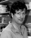
 David Bodine joined the Clinical Hematology Branch (CHB), NHLBI, in
1984 as a postdoc. He remained with the CHB until 1993, when he joined NCHGR as
the Chief of the Hematopoiesis Section. Bodine did his graduate work at Jackson
Laboratory in Bar Harbor, Maine, and received his Ph.D. from the University of
Maine in 1984.
Recent research in my laboratory has focused on pluripotent hematopoietic stem
cells (PHSCs). PHSCs are the ultimate progenitors of all circulating blood
cells and have the ability to self-renew numerous times without losing the
ability to differentiate into cells as diverse as red blood cells and T
lymphocytes. PHSCs are found in the bone marrow, and due to the great
proliferative capacity of their descendants, they are exceedingly rare, less
than one per 100,000 bone marrow cells. We have recently concentrated our
efforts on describing gene expression in PHSCs and devising methods to
introduce genes into PHSCs via retrovirus-mediated gene transfer.
To study gene expression in PHSCs, these rare cells must be greatly enriched.
Our work, using mice as a model system, has shown that murine PHSCs express
high levels of c-kit, the receptor for the hematopoietic growth factor, on
their surface. By combining this observation with techniques to subtract cells
expressing markers of mature blood cells, we were able to use
fluorescence-activated cell sorting to isolate a population of cells highly
enriched in PHSCs. It only takes 100 of these cells to fully reconstitute the
hematopoietic system of a mouse, at least a 1,000-fold enrichment over the
concentration of PHSCs in the original bone marrow cell population. Examination
of messenger RNA purified from those highly enriched PHSCs revealed that c-kit,
stem cell factor (SCF), and the receptors for the hematopoietic growth factors
interleukin-3 (IL-3) and interleukin-6 (IL-6) were all expressed at high
levels. Other work showed that when bone marrow cells were cultured for six
days in SCF, IL-3, and IL-6, the number of PHSCs in the cultures increased two
to threefold.
Efficient retrovirus-mediated gene transfer requires that the target cell
divide, thus allowing the virus to become integrated into the host-cell DNA.
Our finding that PHSC numbers could be increased by culture in SCF, IL-3, and
IL-6 suggested that treatment with these growth factors might significantly
increase the frequency of retrovirus-mediated gene transfer into these cells.
Our results showed that the frequency of gene transfer into mouse PHSCs
cultured without growth factor or in just a single growth factor was
approximately 5%. The frequency of gene-transfer into mouse PHSCs cultured in
all three factors was as high as 75%. These observations were successfully
extended into a rhesus monkey model, and the success of our monkey gene
transfer experiments helped serve as preclinical justification for human
gene-therapy experiments in which retroviruses containing the adenosine
deaminase (ADA) gene were introduced into the bone marrow of patients
with severe combined immunodeficiency syndrome. These human trials were
performed in Europe and the United States in 1993.
In the future, we hope to isolate novel genes that control PHSC division and
differentiation from c-DNA libraries generated from mRNA from highly enriched
populations of PHSCs. In addition, we are continuing our efforts to further
define the factors required for the growth and differentiation of PHSCs both in
vitro and in vivo.
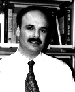
 Mark Boguski received his M.D. and Ph.D. from Washington University
in St. Louis in 1986, and in 1988, he joined the Mathematical Research Branch,
NIDDK, as a medical staff fellow. He became one of the first staff members of
the newly formed National Center for Biotechnology Information (NCBI) in 1989,
where he remains as a researcher in the Computational Biology Branch.
My earlier work at NIH focused on sequence motifs and conserved domains in
proteins involved in signal transduction, particularly those that interact with
and regulate GTPases. Although I continue to study the interrelationships of
sequence, structure, and function in proteins, I have also been working on
information analysis and retrieval problems in genome research. Three years
ago, NCBI's Carolyn Tolstoshev and I founded the database of expressed
sequence tags (dbEST) which is a division of GenBank for cDNA sequence and
mapping data. Now, dbEST contains more than 100,000 sequences, has been queried
by researchers more than 100,000 times, and is currently used nearly 7,000
times per month by intramural and extramural scientists.
We are now collaborating with researchers who are working on genetic and
physical mapping to build a "transcript map" of the human genome. Only a small
fraction of human DNA, probably less than 5%, consists of transcribed coding
sequences. Our goal is to locate all of these transcribed coding sequences,
starting with a comprehensive set of cDNAs, called expressed sequence tags, and
map them back with high resolution onto the chromosomes. Such a map will help
us to understand gene regulation, to pinpoint gene-rich regions for concerted
genomic-sequencing efforts, and to greatly accelerate positional cloning of
genes responsible for genetically complex diseases such as diabetes.
I am also collaborating with Phil Hieter's group at the Johns Hopkins
University of School of Medicine in Baltimore on a project to identify and map
all homologous genes in the yeast and human genomes. In so many instances, such
as the recent studies of cystic fibrosis, neurofibromatosis and familial colon
cancer, yeast biochemistry has shed tremendous light on human pathophysiology.
However, these connections are usually made late in the research process, and
much effort and expense could be saved if the relationships are identified
earlier. Now, however, we're working toward making both the complete sequence
of the Saccharomyces cervisiae genome and a comprehensive sampling of
human coding sequences available within the next 18 months. This creates an
unprecedented opportunity to construct a molecular cross-reference between
yeast and humans and to populate the human-genome map with yeast-gene functions
and phenotypes. This will facilitate the identification of candidate genes for
human diseases and the development of assay systems for studying the functions
of human gene products.
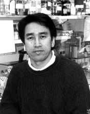
 Seong-Jin Kim received his Ph.D. from the Tsukuba University in Japan
in 1987. He came to NIH in 1987 and is currently a visiting scientist at the
Laboratory of Chemoprevention, Division of Cancer Etiology, NCI.
Our laboratory has been studying the transcriptional and posttranscriptional
regulation of the set of three homologous isoforms of transforming growth
factor-ß (TGF-ß), TGF-ßs 1, 2, and 3. My research program has
focused on the regulation of the TGF-ß gene by etiologic agents
that are involved in disease: tumor-suppressor genes (retinoblastoma gene and
Wilm's tumor gene), oncogenes (jun, fos, src, abl, and ras), and
viruses (human T-lymphocyte virus type 1, human cytomegalovirus, and hepatitis
B virus). Taken together, these studies of gene regulation have delineated the
molecular basis for the observation that the type 1 isoform of TGF-ß is
upregulated by cells in response to injury and pathological processes such as
fibrogenesis and carcinogenesis. In contrast, the type 2 and 3 isoforms of
TGF-ß are regulated principally by developmental cues and hormones.
We have also demonstrated that the protein encoded by the retinoblastoma
susceptibility (Rb) gene can regulate expression of the
TGF-ß1 and -ß2 genes through the Sp1 and activating
transcription factor-2 (ATF-2) binding sites in the TGF-ß1 and
TGF-ß1 promotors, respectively. In the latter case, ATF-2 can form
a complex with the Rb protein with the help of an additional, and
as-yet-unidentified, bridging protein. We are currently trying to clone the
gene that encodes the bridging protein.
I have also been interested in posttranscriptional regulation of TGF-ß
isoforms. It has been suggested that TGF-ß expression is regulated at the
posttranscriptional level by members of the steroid-retinoid superfamily of
nuclear receptors in an isoform-specific manner. Retinoic acid stabilizes
TGF-ß2 mRNA, while the serum cholesterol-lowering drug lovastatin
specifically downregulates the TGF-ß2 mRNA through a posttranscriptional
mechanism. We are attempting to identify the factors controlling the stability
TGF-ß2 mRNA.
Most recently, we have begun to explore mechanisms that regulate the
expression of the TGF-ß receptors in human gastric cancer cell lines that
are resistant to the growth-inhibitory effect of TGF-ß. We have found
alterations in the gene that encodes the type II receptor for
TGF-ß. One of our current goals is to characterize the mechanisms
of transcriptional regulation of the TGF-ß type II receptor gene
because several TGF-ß-resistant cell lines that displayed no
alterations of the TGF-ß type II receptor gene
expressed no detectable TGF-ß type II receptor mRNA.
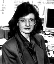
 Lois Travis received her M.D. from the University of Florida
College of Medicine in Gainesville, Fla., in 1980. She received her Sc.D. in
Epidemiology from the Harvard School of Public Health in Boston in 1994 and
joined the Radiation Epidemiology Branch of the Epidemiology and Biostatistics
Program, NCI, in 1989.
One of my major areas of interest is the study of multiple primary cancers,
particularly the evaluation of cancer risk following exposure to ionizing
radiation and/or chemotherapeutic drugs. As long-term-survival rates improve
for many types of cancer patients, it becomes critical to identify the late
consequences of therapy. One of the most serious side effects of cancer
treatment is the induction of new malignancies. Characterization of
therapy-related risks is crucial to enabling the clinician to make an informed
decision regarding treatment, balancing efficacy against acute and chronic
sequelae. In addition, quantification of the late effects of cytotoxic drugs
and radiation therapy provides a unique opportunity for interdisciplinary
studies of carcinogenesis because humans are deliberately treated with measured
amounts of potentially cancer-inducing agents.
When I arrived at NCI, little work had been carried out in the area of
secondary cancers following therapy for non-Hodgkin's lymphoma (NHL). Since
then, in collaboration with investigators worldwide, our group has provided
quantitative estimates of the risk of secondary malignancies among several
populations of NHL patients. In one of my first projects, we identified a
significant excess of solid tumors after NHL, noting that the pattern of risk
increased with time, consistent with the late effects of treatment. In a
subsequent study, we showed that the increased risk of secondary malignancies
persisted for up to two decades after the initial NHL diagnosis. We also
quantified the association between the risk of secondary leukemia and the dose
of various cytotoxic drugs, such prednimustine, chlorambucil, and
cyclophosphamide. We found a dose-response relationship between the cumulative
amount of cyclophosphamide and bladder cancer, and we described the combined
effect of cyclophosphamide and radiotherapy in the induction of bladder cancer.
Now, with the Laboratory of Human Carcinogenesis at NCI, we are collaborating
on molecular studies examining the mutational spectrum of the p53
tumor-suppressor gene in cyclophosphamide-related bladder cancer.
Our group also coordinated an autopsy evaluation of a woman who had been
injected with radioactive Thorotrast (thorium-232) decades previously during
angiography. Thorotrast, once used as a radiologic contrast agent, is not
excreted from the body to any appreciable extent and has induced high rates of
liver angiosarcoma and leukemia. I organized an international workshop that
assembled the clinical and pathologic findings with dosimetric, radiochemical,
autoradiographic, and molecular evaluations for this unique case. Our efforts
enabled correlation of concentrations of radioactive-decay products in various
organs with epidemiologic, histo-pathologic, and molecular observations.
We are now characterizing the risk of cancer after long-term exposure to
radioactive Thorotrast among several large populations. This study may provide
a special opportunity to evaluate the potential effects of low-level radon
exposure. In our investigation, excesses of lung cancer has been noted among
patients exposed to Thorotrast. Since thorium-232 decays into radon, which is
continuously exhaled over the course of the patient's life , it is possible
that this exposure caused the excess lung cancer. Detailed dosimetric studies
are under way to quantify the dose of radon to pulmonary tissues and to relate
dose to lung cancer risk. This study has public-health relevance because indoor
radon is considered the single most important source of radiation exposure and
risks of low-level exposure are poorly understood.
I am also interested in understanding the patterns and determinants of cancer
risk following bone marrow transplantation, an increasingly common procedure
used in the treatment of cancer, and in examining the possible interactions
amoung immunosuppression, total-body radiotherapy, chemotherapy, viral
cofactors, and graft-vs.-host disease. Our recent studies of more than 20,000
recipients of bone marrow transplants identified high rates of secondary
lymphoma, and solid tumors are just now emerging as an important late
consequence. With the Laboratory of Pathology at NCI, we are investigating the
histologic and immunophenotypic characteristics of posttransplant
lymphoproliferative disorders, Epstein-Barr virus status, and host-vs.-donor
origin.
Whenever possible, my research in cancer epidemiology seeks to integrate new
laboratory approaches in efforts to clarify mechanisms of carcinogenesis.
Ultimately, one goal is to develop methods that might predict which cancer
patients are at substantial risk for developing new malignancies and would thus
benefit from targeted screening and preventive measures. Toward this end, we
welcome further collaboration with clinical and laboratory colleagues at NIH.
Table of Contents  David Bodine joined the Clinical Hematology Branch (CHB), NHLBI, in
1984 as a postdoc. He remained with the CHB until 1993, when he joined NCHGR as
the Chief of the Hematopoiesis Section. Bodine did his graduate work at Jackson
Laboratory in Bar Harbor, Maine, and received his Ph.D. from the University of
Maine in 1984.
  Mark Boguski received his M.D. and Ph.D. from Washington University
in St. Louis in 1986, and in 1988, he joined the Mathematical Research Branch,
NIDDK, as a medical staff fellow. He became one of the first staff members of
the newly formed National Center for Biotechnology Information (NCBI) in 1989,
where he remains as a researcher in the Computational Biology Branch.
 Seong-Jin Kim received his Ph.D. from the Tsukuba University in Japan
in 1987. He came to NIH in 1987 and is currently a visiting scientist at the
Laboratory of Chemoprevention, Division of Cancer Etiology, NCI.
 Lois Travis received her M.D. from the University of Florida
College of Medicine in Gainesville, Fla., in 1980. She received her Sc.D. in
Epidemiology from the Harvard School of Public Health in Boston in 1994 and
joined the Radiation Epidemiology Branch of the Epidemiology and Biostatistics
Program, NCI, in 1989.