| T H E N I H C A T A L Y S T | N O V E M B E R – D E C E M B E R 2007 |
|
|
|
Research
Festival
|
by Julie Wallace |
 |
The
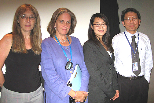
Re-Generation: (left to right) Pamela Robey, NIDCR; Cynthia Dunbar,
NHLBI; Catherine Kuo, NIAMS; and panel chair Rocky Tuan, NIAMS |
Panel chair Rocky Tuan noted that this was the NIH Research Festival’s first dedicated symposium on tissue engineering and regenerative medicine, a reflection that the field is steadily approaching the threshold of clinical application.
Indeed, applying biological and engineering principles to repairing and replacing damaged and destroyed tissues has attracted researchers across NIH; scientists from three institutes described their ongoing research involving adult stem cell–based approaches to tissue regeneration.
Tuan: Creating the Matrix
Adult stem cells and nanomaterials are Tuan’s basic building blocks in his quest to regenerate skeletal tissues. Encouraging results thus far in repairing joint degeneration in rabbits foretell the application of his team’s techniques to the treatment of patients with musculoskeletal diseases such as osteoarthritis.
With the right scaffold and physical and chemical environments, a cartilage micromass could be developed from human mesenchymal stem cells (MSCs) to replace the degraded tissue, said Tuan, chief of the Cartilage Biology and Orthopaedics Branch, NIAMS. The challenge is to generate a cartilage construct of sufficient size to transplant into a human joint, he said.
Tuan’s lab evaluated whether differentiated MSCs could transdifferentiate–change from one differentiated state to another. The team found that osteoblasts, adipocytes, and chondrocytes derived from MSCs could indeed be made to switch identities. This capacity to de-differentiate generated the hypothesis that there might be "stemness" genes that regulate MSC self-renewal and multipotency.
Using microarray analysis, Tuan and his colleagues determined that differentiation genes were upregulated in differentiation and downregulated during de-differentiation; "stemness" genes, on the other hand, were found to support MSC self-renewal and proliferation—"differentiation readiness"—in the undifferentiated state.
A proper scaffold to support these stem cells, Tuan determined, would mimic the native extracellular matrix and be able to fit into a three-dimensional groove. Nanofibers of collagen and other macromolecules are the hallmark of the extracellular matrix of skeletal tissues, Tuan said, explaining how his group used electrospinning to prepare similar nanoscale fibers from a biodegradable polyester and then produced the desired tissue-engineering scaffold.
Their next step was to optimize nutrient supply to the developing cartilage construct to grow in size; the team succeeded in enhancing growth of the engineered cartilage in vitro from a diameter of 1.5 cm a few years ago to around 4 cm today.
Finally, the researchers are using mini-pig and rat models to test the ability of the engineered cartilage construct to repair cartilage defects and integrate into native cartilage. Initial observations have revealed promising signs of tissue repair in six months and four weeks in these respective models, Tuan said.
Robey: Skeletal Cells and Scaffolds
When bone-marrow skeletal stem cells are plated, they are able to form cartilage in vitro, and when they are transplanted, they form bone, marrow, fat, and the stroma that supports blood formation. This multipotentiality suggests broad therapeutic application for dental and craniofacial reconstruction, observed Pamela Robey, chief of the Craniofacial and Skeletal Diseases Branch, NIDCR.
Robey and her team have been characterizing these stem cells and evaluating different scaffold choices for tissue regeneration. The identification of markers for skeletal stem cells will aid the quality of purification of these cells from bone marrow. It will, however, also be necessary to generate large numbers of these cells via ex vivo expansion. The goal, explained Robey, is to trick the stem cells into dividing symmetrically to keep them as multipotent as possible.
In addition to researching ways to isolate and expand the skeletal stem cells, Robey’s group has been testing potential scaffold materials. Currently, there are only a few FDA-approved, commercially available scaffolds. The nature of the scaffold is important, said Robey, observing that only one currently available scaffold (synthetic hydroxyapatite–tricalcium phosphate ceramic particles) supports both bone formation and the stem cell, so that marrow can be formed. The size and shape of the scaffold, which organizes and controls the structure of the bone formation, also matter, she added.
Among research questions requiring attention, Robey said, are determining the number of cells necessary for successful transplantation, identifying an appropriate transplantation method, stimulating incorporation of the transplant into the preexisting tissue, and developing a root structure in order to reconstruct a viable tooth from dental pulp cells.
Kuo: Mechanoactive Tenogenesis
Catherine K. Kuo, a postdoctoral fellow in the NIAMS Cartilage Biology and Orthopaedics Branch, brings her engineering perspective to investigating the potential of MSCs in tissue engineering and regenerative medicine. Kuo is particularly interested in the regeneration of tendons, which transmit forces from the muscle to the bone, and ligaments, which join bone to bone and thus stabilize joint structures.
Nearly half of all skeletal injuries involve tendons and ligaments. Poor healing ability of these tissues and imperfect repair strategies provide an opportunity for regenerative therapies.
There are no known growth factors to induce differentiation of MSCs into tendon/ligament fibroblasts (tenogenesis). These cells are distinct from other musculoskeletal lineages in that mechanical stimulation is the only known factor able to induce tenogenic differentiation of MSCs. Kuo created tendonlike constructs by seeding three-dimensional collagen type I gel scaffolds with MSCs; she cultured the constructs under uniaxial static or dynamic tension.
Kuo monitored tenogenesis via expression of scleraxis, a transcription factor that uniquely marks tendon progenitors during development. To determine whether dynamic mechanical stimulation (cyclic tensile loading) enhanced tenogenesis, Kuo developed engineering devices and tools to place the constructs under either static or dynamic tension.
She observed a similar increase in collagen mRNA in both static and dynamic conditions, but noticed more matrix deposition and persistent scleraxis expression over time with cyclic loading. In addition, expression of matrix metalloproteinases such as collagenase and gelatinase were differentially regulated by cyclic loading, implying that the increased matrix deposition and resulting tenogenic differentiation are regulated by changes in the expression of these genes.
Dunbar: Thwarting Mutagenesis
PI Cynthia Dunbar and her colleagues in the Hematology Branch, NHLBI, have been using gene-transfer techniques in their research on hematopoiesis.
Addressing the safety issues and complications associated with the use of viral transfer vectors, the group has investigated the risks of insertional mutagenesis after retrovirus and lentivirus gene transfer to hematopoietic stem cells (HSCs).
Viral gene-transfer vectors that are used to target HSCs must integrate into the host genome to be effective, but depending on the site of insertion, they can activate adjacent genes, including protooncogenes. These issues did not emerge in studies involving murine models and have come to light during clinical gene-therapy studies, Dunbar said.
In one study, patients with X-linked severe combined immunodeficiency (SCID) had reconstitution of their T-cell immunity and clinical improvement after HSC gene therapy. This optimistic outcome was interrupted three years later by the development of T-cell leukemias in four patients, due to activation of a growth-promoting gene by the inserted gene-therapy vector.
The need for an experimental model more predictive and more closely related to humans prompted Dunbar’s laboratory to turn to the rhesus macaque as a model organism for studies of gene therapy.
Dunbar’s group took advantage of the rhesus macaque model to study the patterns of virus (either murine leukemia virus [MLV] or simian immunodeficiency virus [SIV]) integration sites. The integration sites were determined in circulating granulocytes and lymphocytes of the monkeys up to seven years post-transplantation.
The researchers observed that integration was nonrandom: MLV integrated around transcriptional start sites; SIV integration sites were evenly spaced along the length of a gene.
The researchers also noted that animals receiving MLV-transduced HSCs had marked overrepresentation of cells containing integration events in the Mds1/Evi1 gene complex, suggesting significant in vivo selection for these clones, a very worrisome pattern, Dunbar noted. One macaque developed leukemia due to insertional activation of the BclaA1 locus.
Dunbar
discussed approaches to decrease the risk of insertional mutagenesis, including
alternative vector systems and modification of current vectors to decrease the
likelihood of activation of adjacent genes.
![]()
| Engineering and Physical Sciences SIG Starting Up |
The Working Group on Women in Biomedical Careers subcommittee on Integration of Women into Bioengineering Fields has created the Engineering and Physical Sciences Special Interest Group. The goals of this SIG will be to promote interaction between investigators and laboratories whose research interests involve integrating engineering or physical science with biology, and to educate the NIH community about these approaches. Areas of research interests include tissue engineering and regenerative medicine, biomaterials, nanotechnology, physical regulation in biology, engineering-based enabling technologies, and quantitative approaches based on physical sciences. This
SIG will organize seminars by engineers and physical scientists from inside
and outside NIH and identify mentors available to engineering and physical
science students and fellows at NIH. Particular efforts will be made to
identify outstanding women engineers and physical scientists. E-mail
Catherine Kuo (or call at 301-451-4519) with questions or to join.
|