| T H E N I H C A T A L Y S T | J U L Y – A U G U S T 2007 |
| | |
Recently Tenured
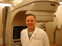
Kevin Camphausen received his M.D. degree from Georgetown University, Washington, D.C., in 1996 and completed a residency in radiation oncology at the Harvard Medical School Joint Center for Radiation Therapy, Boston, where he studied the interaction of angiogenesis inhibitors and radiotherapy in the Judah Folkman lab. He joined NCI in 2001 as a tenure-track investigator and is currently chief of the Radiation Oncology Branch.
Approximately 75 percent of the 1.2 million patients diagnosed with nonskin cancers in North America in 2007 will receive radiotherapy during the course of their disease. Improving the efficacy of radiotherapy would therefore significantly contribute to the field of oncology.
Characterization of the processes regulating cellular radiosensitivity has led to the hypothesis that many of these cellular processes and signaling pathways can be targeted for interruption, thereby predisposing tumor cells to death.
The work in my laboratory and clinic focuses on two major projects: development of novel biomarkers of early failure after radiotherapy, and histone acetylation as a target for tumorcell radiosensitization.
The first focus is the development of a noninvasive biomarker for tumor recurrence after irradiation. An early marker of recurrence would allow prediction of which patients harbor subclinical disease and may benefit from additional therapy, instead of the current practice of delivering treatment to every patient at moderate risk of recurrence.
Because tumor growth depends on angiogenesis, an increase in circulating angiogenic factors could identify patients at risk for persistent or recurrent disease. Although angiogenic factors have been explored as tumor markers, no study has explored their utility as a tumor marker across multiple tumor types or in patients receiving radiation.
Matrix metalloproteinases (MMPs) have been implicated in both primary and metastatic tumor formation. Marsha Moses and colleagues defined the presence of biologically active MMP-2 or MMP-9 in the urine of cancer patients as an independent predictor of organconfined cancer. We developed an in vivo Lewis lung carcinoma (LLC) model to evaluate urinary MMP-2 as a biomarker for disease recurrence after irradiation.
We were able to detect biologically active MMP-2 as an early marker for lung metastases in C57Bl/6 mice that had been implanted with LLC and "cured" with local irradiation. This rise in MMP2 activity was then used as a signal to begin adjuvant angiostatin therapy, which successfully reduced the metastatic burden.
With the knowledge that urinary MMP-2 levels reflect disease status in mice after irradiation, we sought to explore this finding in radiotherapy patients using two biomarkers, MMP-2 and VEGF (vascular endothelial growth factor).
A pilot study (02-C-0064) examining these two urinary biomarkers was opened in early 2002. We recently published early findings from this study. In samples obtained before radiotherapy, there were a greater number of MMPs detectable in the urine of patients with localized cancer than in that of normal controls (1.43 vs. 0.38). Similarly, patients with localized cancer had higher urinary VEGF levels than normal controls (317 vs. 131.6 pg/mg creatinine). Although the absolute values of these biomarkers were not able to predict clinical outcome, comparisons of the MMP or VEGF level on the last day of radiation with levels obtained at one-month follow-up were predictive of disease status at one year.
To validate the utility of these promising markers, patients with the same histology undergoing a homogeneous treatment regimen needed to be studied. Therefore, we initiated a prospective Phase II study (04-C-0200) in patients with glioblastoma multiforme (GBMs) undergoing radiotherapy to determine whether these urinary biomarkers add to the Radiation Therapy Oncology Group Recursive Partition Analysis, which uses clinical parameters to divide patients into subgroups based on historical survival. This study should be mature within the next year.
My second focus is the evaluation of histone deacetylase inhibitors as radiation sensitizers. Histone acetylation plays a critical role in modulating chromatin structure and regulating gene expression. The balance of histone acetylation is determined by the opposing actions of histone acetyltransferase (HAT) and histone deacetylase (HDAC).
In addition to slowing tumor-cell proliferation, the HDAC inhibitors sodium butyrate and trichostatin A have also been shown to enhance tumor-cell radiosensitivity. Their clinical utility, however, is limited by their pharmacologic characteristics.
Although several HDAC inhibitors have recently been developed that possess more favorable pharmacokinetic and toxicity profiles, their ability to increase tumor-cell radiosensitivity cannot be assumed. We have been examining these HDAC inhibitors and have found that several—including MS-275, CI-779, depsipeptide, and valproic acid (VA)—indeed appear to enhance the radiosensitivity of tumor cells.
Because of its potential for clinical application—oral bioavailability, a low toxicity profile, and penetration of the blood-brain barrier—we have conducted further studies with VA.
Our laboratory results suggest that VAinduced HDAC inhibition can lead to an enhancement of radiosensitivity both in vitro and in vivo. Our data further suggest that continuous exposure to VA is necessary to achieve the maximum enhancement.
Based on these findings, I initiated an NCI Phase II trial (06-C-0112) testing this agent as a radiation sensitizer in combination with temozolomide and radiotherapy in patients with newly diagnosed GBM.
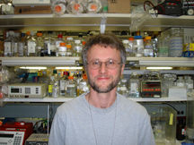
David Clark received his Ph.D. degree from the University of Cambridge, U.K., in 1986. He did his postdoctoral training with Gary Felsenfeld at NIH. In 1995, he joined the Laboratory of Cellular and Developmental Biology, NIDDK, as a tenure-track investigator. In 2003, he moved to the Laboratory of Molecular Growth Regulation, NICHD, where he is now a senior investigator and head of the section on Chromatin and Gene Regulation.
The study of gene regulation is a prerequisite for understanding how cells respond appropriately to a changing environment, how they implement developmental programs, and how a defect in gene regulation can result in carcinogenesis. Gene activation involves the recruitment of a set of factors to a promoter in response to appropriate signals, ultimately resulting in the binding of RNA polymerase II and transcription.
These events must occur in the presence of nucleosomes, in which DNA is coiled around a central octamer of core histone proteins. Nucleosomes are compact structures capable of blocking transcription at every step. To circumvent this chromatin block, eukaryotic cells possess ATPdependent chromatin-remodeling machines and histone-modifying complexes. The former use ATP to alter nucleosome structure and to move nucleosomes along DNA. The latter contain enzymatic activities that modify the histones to alter their DNAbinding properties and to mark them for recognition by other complexes, which have activating or repressive roles (the basis of the histone-code hypothesis).
The current excitement in the chromatin field reflects the realization that chromatin structure is of central importance in gene regulation: The cell has dedicated complex systems to manipulate the repressive properties of chromatin structure to maximum effect. Furthermore, multiple connections between chromatin and disease are apparent.
We have developed a model system to investigate the remodeling and histone modifications that occur on gene activation. We purify native plasmid chromatin containing a model gene in its transcriptionally active or inactive chromatin states from yeast cells and compare their structures. Our studies provide a detailed and surprising picture of the events occurring in the chromatin structure on gene activation.
We discovered that induction results in a remodeled chromatin domain that extends far beyond the promoter to include the entire gene. The formation of this domain requires the transcriptional activator and a remodeling machine. We propose that a highly dynamic chromatin structure is created, facilitating access to the DNA for initiation and elongation factors. Our current studies aim to increase our understanding of the structure and dynamics of remodeled chromatin domains.
In a second, related, project, we are investigating the biological function of the yeast Spt10 protein, which we believed initially to be a co-activator with histone acetyltransferase (HAT) activity that is recruited to promoters by activators. However, we discovered that Spt10p is in fact the activator of the histone genes, which has been sought after for many years.
Spt10p appears to be a rare example of an activator with a sequence-specific DNAbinding domain fused to a HAT domain. Our current aim is to place our observations in their biological context of cell-cycle regulation of the histone genes. In addition, we are investigating the implications of the homology we have observed between the DNA-binding domain of Spt10p and the zinc-finger domain of human foamy retrovirus integrase.
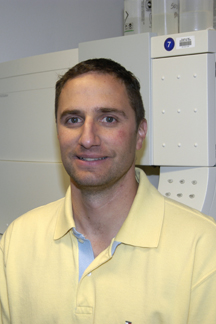
Frank DeLeo received his Ph.D. in microbiology from Montana State University, Bozeman, in 1996 and carried out his postdoctoral training with William Nauseef at the University of Iowa, Iowa City, in the Department of Medicine. He joined the staff at NIAID's Rocky Mountain Laboratories (RML) in Hamilton, Mont., in the fall of 2000 as a tenuretrack investigator in the Laboratory of Human Bacterial Pathogenesis. He is currently a senior investigator and chief of the Pathogen-Host Cell Biology Section.
My laboratory studies the interaction of human polymorphonuclear leukocytes (PMNs, or neutrophils) with pathogenic bacteria. Although most bacteria are killed readily by neutrophils, pathogens such as Staphylococcus aureus have evolved mechanisms to circumvent destruction by these key innate immune cells and thereby cause human infections. Therefore, a better understanding of the bacteria-neutrophil interface at the cell and molecular levels will provide information critical to our understanding, treatment, and control of disease caused by bacterial pathogens.
The Pathogen-Host Cell Biology Section conducts 1) a systematic molecular dissection of steps involved in pathogen-host interaction, with specific emphasis on the interaction of bacterial pathogens with innate host defense (neutrophils), and 2) research investigating mechanisms mediating evasion of innate immunity by pathogens of special interest, such as methicillin-resistant S. aureus (MRSA).
During my first few years at RML, we used genomics strategies to discover a genetic program that links phagocytosis in human neutrophils with apoptosis. This research redirected our thinking about neutrophil function in the innate immune response.
Using similar genomics methodologies, we elucidated a global model of host cell-pathogen interaction, which is broadly applicable to many bacterial pathogens. Collectively, these studies lead to a new paradigm for the resolution of human bacterial infections.
Studies from our laboratory were also among the first to identify complex genetic programs in bacteria that circumvent human innate immunity to promote disease. This work, which comprises a series of studies, identified novel virulence factors in human bacterial pathogens.
Since late 2003, we have focused our research on the emerging problem of community-associated MRSA (CAMRSA), which is epidemic in the United States. CA-MRSA causes disease in otherwise healthy individuals, and these infections can be severe and/or fatal. The molecular basis for the increased incidence and severity of CA-MRSA disease is not known.
Work from our laboratory suggests that enhanced CA-MRSA virulence is linked to (or results from) evasion of killing by neutrophils, which likely underlies the ability of these pathogens to cause disease in otherwise healthy individuals.
An S. aureus leukocyte toxin, PantonValentine leukocidin (PVL), is widely believed to be the cause of the increased severity and incidence of CA-MRSA infections. However, our recent studies indicate PVL does not play a major role in CA-MRSA disease. This unexpected finding will redirect the medical community toward other factors likely responsible for CA-MRSA infections.
Our efforts to understand MRSA pathogenicity are currently focused on identifying the proteins directly responsible for the type and severity of disease caused by these emerging human pathogens.
The long-term objective of this research is to develop and/or promote development of enhanced diagnostics, better prophylactic agents, and new treatments for emerging infectious pathogens such as CA-MRSA.

Aiyi Liu received his Ph.D. degree in statistics in 1997 from the University of Rochester, Rochester, N.Y. He was a postdoctoral research fellow at St. Jude's Children's Research Hospital in Memphis, Tenn., and an assistant professor of biostatistics at Georgetown University Medical Center, Washington, D.C., before joining NIH in 2002 as a tenuretrack investigator in the Biometry and Mathematical Statistics Branch, Division of Epidemiology, Statistics and Prevention Research (DESPR), NICHD. He is now a senior investigator in that branch.
As an intramural investigator at NIH, I conduct independent statistical research and collaborate with investigators within and outside DESPR on design, analysis, and interpretation and publishing of biomedical studies.
Sequential methods is one of my major research areas. Sequential methods, which allow a scientific hypothesis to be tested repeatedly at several time points using accumulated data, are frequently used in designing medical studies, particularly clinical trials, because they increase the ability to reduce sample size and to terminate inferior treatments as early as possible.
Independently or joined by other colleagues, I have published a series of papers addressing various issues in the design and analysis of a sequential trial, with particular focus on statistical inference upon termination of the trial.
It is well know that sequential sampling introduces bias to estimation of treatment effect. I derived some explicit formulas to evaluate the magnitude of such bias and found that the bias can be substantially reduced using a simple segmented estimation by shrinking the estimator toward zero for small treatment effect and subtracting a constant for large treatment effect. For two-stage phase II trials, I discovered that the bias can also be substantially reduced by simply subtracting an empirical estimator of the bias.
Clinical trials are usually designed to test hypotheses for a primary endpoint. However, data on secondary endpoints are also collected and need to be analyzed by taking into account the possibility of early stopping of the trial. I developed a simple bias-corrected confidence interval for the mean of a secondary parameter and showed the interval to have desired coverage probability. For a sequential phase II trial, I obtained several efficient estimators to reduce the bias in estimating a secondary probability. The methods can be used to obtain efficient estimation of the toxicity rate when the response rate is the primary endpoint in a phase II cancer clinical trial.
Power and sample-size calculation are integrated considerations in planning a medical study. Unfortunately, such calculation usually relies on correctly specified values of nuisance parameters, that is, parameters that are not related to the treatment difference but need to be dealt with in the study. The study can be severely underpowered if the specification is far from the truth. This situation calls for sample-size recalculation based on internal data. I investigated this issue for a bivariate phase II cancer trial to evaluate toxicity and response rate simultaneously, and also for studies comparing the accuracy of two diagnostic medical devices. I proposed several approaches to re-estimating the sample size to achieve the needed statistical power.
I have also been exploring statistical issues related to diagnostic devices (or biomarkers), such as receiver operating characteristic (ROC) curves analysis, which plots sensitivity and specificity. I found a linear function to combine several medical devices so that the sensitivity of the resulting diagnosis is maximized over a desired range of high specificity. I also developed several sequential methods to compare the diagnostic accuracy of medical devices.
Recently, I proposed and developed a two-stage testing procedure to evaluate the measurement errors of an instrument in measuring the level of disease-associated biomarkers. This procedure allows the investigator to assess the measurement error at an early stage and make decisions about whether more experiments are needed for further evaluation.
I am also interested in microarray data analysis. In order to extract information from a database with a large number of genes and a relatively small number of samples, I found an efficient way to perform principal components analysis (PCA) via grouping genes into several blocks according to their correlation. Within each block, PCA is conducted and important gene expression features extracted. These selected features are then combined for further PCA. This new procedure, called "block principal components analysis," has the advantage of computational simplicity and is as efficient as ordinary PCA.
I have now begun to examine several new research areas, including sequential and adaptive design methods for studies in diagnostic medicine, sequential ranking and selection of diagnostic biomarkers, and a new model for survival and ordinal data. With the continued excellent support of my branch chief and division director, I look forward to making more contributions to the science of statistics.

Enrique Schisterman received his Ph.D. in epidemiology from the State University of New York at Buffalo in 1999 and completed his postdoctoral training in epidemiological methods at the Harvard School of Public Health in the Department of Epidemiology. In 2002, he came to NIH as a tenure-track investigator in the Division of Epidemiology, Statistics, and Prevention Research, NICHD. He is currently a senior investigator in the division.
My current research interests are twofold: 1) I have a long-standing interest in reproductive and perinatal epidemiology, and 2) I am working on developing analytical tools that are closely tied to etiological questions. In pursuit of the first interest, I am in the midst of conducting two studies.
The Effects of Aspirin on Gestation and Reproduction (EAGeR) trial is a multisite, double-blind, placebocontrolled, randomized trial that will evaluate the effect of daily low-dose aspirin on all phases of reproduction beginning at preconception and continuing throughout pregnancy, including implantation and live births.
We plan to recruit 1,600 women preconception and actively follow them for two menstrual cycles or until becoming pregnant, whichever comes first. Active follow-up entails collection of questionnaire data as well as regular specimen collection. Women who do not become pregnant within the first two cycles will remain in the study under passive follow-up for four cycles or until pregnancy occurs, whichever comes first.
Study volunteers will be followed throughout gestation, and pregnancy outcomes will be recorded. Our main outcome is spontaneous abortion, which we hypothesize will be decreased in the aspirin arm compared with the placebo arm. Aspirin is a primary target of interest because of its anti-inflammatory, vasodilatory, and platelet-aggregation inhibition properties. We expect to be in the field by July 1, 2007.
The second epidemiological study is the BioCycle study, which is a longitudinal assessment of the effects of endogenous hormones (that is, estrogen and progesterone) on biomarkers of oxidative stress and antioxidant status during the menstrual cycle.
Specifically, we measured F28-isoprostanes, TBARS (thiobarbiturate acid reactive substances), and total plasma protein carbonyls as markers of oxidative stress; fat-soluble antioxidant vitamins, watersoluble vitamin C, and the major antioxidant enzymes were measured as markers of antioxidant defense. We assessed oxidative stress, hormones, and antioxidant levels at specific times during the menstrual cycle that represent the days with the greatest hormonal variation. The study recruited and completed follow-up on 259 women, and we are now in the midst of analyzing the data gathered.
In addition to these trials, I have explored and am currently exploring pooledsample designs because they provide practical and cost-effective solutions for investigating oxidative stress as a mediator of reproductive outcomes.
I am also involved in developing new methods for estimating the receiver operating characteristic (ROC) curve and the accompanying Youden index accounting for measurement error, selection bias, and information bias.
Finally, I am interested in methodological research focused on the assessment of biomarkers and exposure data, consideration of the limits of detection, statistical modeling of causal effects, and design studies with pooled biospecimens.
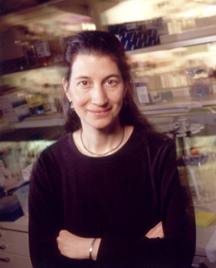
Julie Segre received her Ph.D. degree in genetics from the Massachusetts Institute of Technology, Cambridge, in 1996, working with Eric Lander at the inception of the MIT Genome Center. She was then a Damon Runyon-sponsored postdoctoral fellow in the laboratory of Elaine Fuchs at the University of Chicago, where she developed an interest in skin biology. In 2000, she was awarded a Burroughs-Wellcome Career Award in Biomedical Research and joined NIH as a tenure-track investigator. She is currently a senior investigator and head of the Epithelial Biology Section, Genetics and Molecular Biology Branch, NHGRI.
Combining classical genetics techniques and modern genomic tools, I study gene-environment interactions at the skin surface. I study the formation of the skin barrier during development, which is necessary to prevent the escape of moisture and the entry of infectious agents.
The skin barrier is established in utero at 32 weeks (resulting in an incomplete barrier and increased risk for dehydration and infection in children born preterm—about 2 percent). The skin barrier is maintained throughout life and reestablished after trauma such as abrasion or wounding.
Even when the skin regrows, the ensuing scar at the site of trauma continues to exhibit a mild barrier deficiency compared to uninvolved skin for as along as one year.
We modeled in animals the establishment of the skin barrier during development and the interaction of the pathways regulated by the transcription factors, KLF4, GATA-3, and the glucocorticoid receptor.
We showed that mice with increased gap junctional communication exhibit mild barrier impairment under homeostatic conditions. After wounding, an inability to restore the barrier arrested the wounds in the hyperproliferative state and resulted in immune cell infiltration.
These studies suggest that the signal to restore the homeostatic balance between proliferation and differentiation after regrowth of the skin is restoration of the barrier and that, in its absence, the skin enters a pathologic state resembling psoriasis.
Psoriasis is a common postnatal skin disorder, affecting 2 percent of the population in the United States; its typical age of onset is in the third decade. Wounding is an initiating event in a significant percentage of psoriatic patients and is clinically referred to as the isormorphic phenomenon.
Our future goal is to evaluate the hypothesis that skin microbial diversity (a mix, for instance, of bacteria, fungi, and archae) plays a role in many common dermatological conditions, including atopic dermatitis (eczema). The onset of atopic dermatitis is typically within the first year of life and affects about 15 percent of children in the United States. More than half of patients with severe atopic dermatitis will progress to develop asthma and/or allergic rhinitis (hay fever), disorders with significant morbidity and mortality.
There are two classical explanations for the role microbes play in skin disease: 1) A specific microbe colonizes the skin to disrupt the balance of commensal microflora, and 2) microbes release toxic substances or invade cells to induce an inflammatory response directly.
In fact, there are numerous possibilities of how microbial communities contribute to human health and disease. However, culture-dependent skin sampling methods are incomplete assessments of microbial diversity and thus are insufficient to fully address these basic questions.
We propose to use novel genomic methods to sample skin microflora directly and shed light on the above conjectures in both normal and diseased dermatologic states.
We genomically classify the phylum and genus of the bacteria by sequencing the 16S rRNA genes directly from skin biopsies and will characterize the full bacterial genomes with metagenomic sequencing.
The genetic program to specify, maintain, and heal the skin is complex. However, the skin is an ideal system in which to perform genetic and genomic experiments because it offers easy access to diverse human skin disorders, excellent animal models, and well-developed cell and organotypic culture systems.

Giorgio Trinchieri received his medical degree from the University of Torino, Italy, in 1973. He was a member of the Basel Institute for Immunology (Basel, Switzerland) and an investigator at the Swiss Institute for Experimental Cancer Research (Epalinges sur Lausanne, Switzerland). From 1979 to 1999 he was at Wistar Institute in Philadelphia and became Hilary Koprowski Professor and Chairman of the Immunology Program; he was also Wistar Professor of Medicine at the University of Pennsylvania in Philadelphia. He was director of the Schering Plough Laboratory for Immunological Research in Dardilly, France, and an NIH Fogarty Scholar at the Laboratory for Parasitic Diseases, NIAID, before becoming director of the Cancer and Inflammation Program (CIP) and chief of the Laboratory of Experimental Immunology at NCI in August 2006.
As CIP director, I oversee the operations of two major NCI intramural laboratories, the Laboratory of Experimental Immunology and the Laboratory of Molecular Immunoregulation.
These two laboratories constitute the major immunologic component of the CCR's newly announced inflammation and cancer initiative, which spans the NCI's campuses in Frederick and Bethesda and seeks to partner NCI's expertise in inflammation and immunology with its cutting-edge cancer etiology and carcinogenesis program.
My research interest has always focused on the interplay between inflammation/innate resistance and adaptive immunity and on the role of pro-inflammatory cytokines in the regulation of hematopoiesis, innate resistance, and immunity.
My focus now is on the role of inflammation, innate resistance, and immunity in carcinogenesis, cancer progression, and prevention or destruction of cancer.
Recent studies are shedding a new light on how innate resistance, as an integral part of inflammation, participates in oncogenesis and tumor surveillance.
For a long time, innate resistance was considered a primitive nonspecific form of resistance to infections that was eclipsed by the potent and specific acquired immunity of higher organisms.
More recently, it has been recognized that innate resistance is not only the first line of defense against infections but also sets the stage and is necessary for the development of adaptive immunity.
Advances in cancer biology now reveal that what used to be considered the defensive mechanisms of innate resistance and inflammation are indeed manifestations of tissue homeostasis and control of cellular proliferation that have many pleiotropic effects on carcinogenesis and tumor progression and dissemination.
The interaction of the inflammatory mediators and effector cells with carcinogenesis and tumor progression is complicated and results in effects that either favor or impede tumor progression. In my own group and in collaboration with the other CIP investigators, research on the interface between inflammation, natural resistance, and adaptive immunity in the mouse and in humans will focus on:
1) The molecular and cellular mechanisms regulating the activity of dendritic-cell subsets, particularly the type I interferon-producing plasmacytoid dendritic cells, as well as conventional dendritic cells, and their crossactivating interaction with other inflammatory cell types, NK cells, and T cells;
2) The role of plasmacytoid dendritic cells in the regulation of immune response and tolerance in experimental models of cancer, autoimmunity, and infections;
3) The molecular, structural, and signaling aspects of receptors that recognize pathogens—particularly Toll-like receptors, cytoplasmic NOD and RIGI-like receptors—and other surface receptors and their synergism or antagonism in the regulation of the inflammatory response;
4) The immunosuppressive tumor environment that results in alternate activation of macrophages and myeloid cells and paralysis of dendritic cells, with an emphasis on the role of IL-10 and STAT3 activation;
5) Treatments aimed at reversing the immunosuppressive environment, combined with others to activate innate resistance or modulation of the inflammatory response for antitumor therapy;
6) Using genetic or chemical models of colon and skin carcinogenesis to explore the role of inflammation-related gene products, such as tumor necrosis factor, IL-12, IL-23, IL-27, IL-10, IL-17, IL-22, Toll-like receptors and their signaling molecules.