Leighton
Chan
| T H E N I H C A T A L Y S T | M A R C H – A P R I L 2007 |
|
|
|
| P E O P L E |
RECENTLY
TENURED
|
|
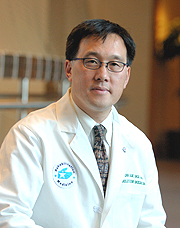
Leighton
Chan
|
Leighton Chan received his M.D. degree from the University of California at Los Angeles in 1990. He did his residency in physical medicine and rehabilitation and was a Robert Wood Johnson Clinical Scholar at the University of Washington in Seattle, where he also received his M.P.H. He spent 10 years on the faculty of the University of Washington while also working at the Center of Medicare and Medicaid Service’s Seattle regional office. In January 2007, he joined NIH as chief of the Rehabilitation Medicine Department at the Clinical Research Center.
If one were to select a single institution to monitor health care in the United States, it would likely be Medicare. Medicare is the largest purchaser of health care in this country, accounting for 20 percent of all health-care dollars spent here. This includes 20 percent of all physician bills, 10 percent of all skilled-nursing facility spending, 32 percent of all inpatient costs, and 70 percent of all inpatient rehab costs. In all, there are 44 million Medicare enrollees whose medical care costs about $220 billion per year.
As a health-services researcher, I have focused my research efforts on using Medicare billing data to monitor secular trends, to measure geographic variation, and to support the evaluation of specific conditions, treatments, and procedures.
Much of my prior work dealt with issues around improving Medicare’s payment policy. For instance, I performed a large cohort study of approximately 300,000 Medicare rehabilitation inpatients and found that certain types of hospitals were incentivized to prolong patients’ hospital stays to maximize profits. This policy cost the federal government more than $200 million over a seven-year period. These results helped galvanize changes in this payment methodology over the past few years.
I have also used Medicare billing data to examine outcomes after specific surgical procedures, most recently bariatric surgery for obesity. Using Medicare billing data, we collected the largest bariatric surgery cohort to date—more than 16,000 patients—and determined that short-term mortality rates were exceptionally high for individuals over 65 years old and for individuals whose surgery took place in hospitals that performed the procedure infrequently. These results helped lead to changes in Medicare’s payment policy for bariatric surgery.
More recently I have become interested in the concept of health-care disparities, in particular the notion that disability—whether it involves mobility or communication or is cognitively based—is a risk factor for poor health care. I have been using a large Medicare survey dataset that is designed so that one can draw conclusions from the entire Medicare population.
Our findings suggest that—after controlling for important and known demographic confounders—individuals with disabilities are indeed at risk for inadequate primary care, poor satisfaction with care, and higher health-care costs. We are in the process of confirming these results through triangulation with other datasets.
In my new position as chief of the CRC’s Rehabilitation Medicine Department, I plan to expand the activities of the department’s Physical Disabilities Research Branch. This branch focuses its efforts on the biomechanics of movement. I plan to continue this important work as well as add areas of focus, including health-services research and possibly cardiopulmonary rehabilitation.
Ultimately,
my hope is to create models of care for individuals with disabilities to maximize
their function and reduce or eliminate health-care disparities.
 |
|
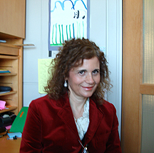
Patrizia
Farci
|
Patrizia Farci received her M.D. degree from the University of Cagliari Medical School, Cagliari, Italy, in 1979 and became a board-certified specialist in infectious diseases and in gastroenterology. She had her postdoctoral training at the Molinette Hospital in Torino under Giorgio Verme and Mario Rizzetto and at the Royal Free Hospital, University Medical School, London, under Sheila Sherlock. In 1989, as a visiting scientist, she joined the NIAID laboratory of Robert Purcell. She returned to Italy in 1994 and in 2000 became full professor of medicine and director of the Liver Unit and of the Laboratory of Molecular Virology, as well as director of the Post-Graduate School of Gastroenterology at the University of Cagliari. Earlier this year she moved to the Laboratory of Infectious Diseases, NIAID, where she is a senior investigator and head of the Hepatic Pathogenesis Unit in the Hepatitis Viruses Section.
Most of my work has been devoted to the study of chronic viral hepatitis, especially hepatitis C. My research has been driven by my natural inclination to merge clinical medicine with basic research.
In the early 1990s, soon after the discovery of HCV, I developed one of the first PCR assays for HCV and used it to define the molecular events that occur during the natural history of HCV infection in humans and chimpanzees, providing evidence that HCV continuously replicates for more than 20 years. I became fascinated by the extremely high propensity of HCV to induce chronic infection, and I focused my research on trying to understand the mechanisms whereby HCV escapes immune recognition.
Among the most significant findings, we showed that HCV does not confer protective immunity in either humans or chimpanzees and that it may cause multiple episodes of acute hepatitis C in the same individual. Because of the major implications of these observations for the development of an effective vaccine against HCV, we investigated why HCV does not confer protective immunity in the host.
We were able to demonstrate for the first time in vivo that HCV does elicit neutralizing antibodies, but their efficacy is isolate-restricted and ineffective against minor viral variants present in the complex quasispecies. We then identified HVR1, the most variable region of the HCV genome, as a major target of neutralizing antibodies.
Capitalizing on these observations, I started to focus my research on understanding the biological implications of HCV genetic heterogeneity (quasispecies) for viral persistence, pathogenesis, therapy, and prevention of hepatitis C. One of the most important observations was the discovery that the early evolution of the viral quasispecies during primary HCV infection predicts the outcome of acute hepatitis C (resolution vs. persistence).
Another major interest has been hepatitis D, the most severe and progressive form of chronic liver disease that leads to cirrhosis in 80 percent of the cases. We demonstrated that high doses of a-interferon are required for the treatment of this disease and that interferon significantly improves the long-term clinical outcome and survival of patients withchronic hepatitis D, providing the basis for current treatment guidelines.
Moreover, our 20-year long-term study convincingly showed the regression of advanced hepatic fibrosis (and probably cirrhosis) after interferon therapy, thus contributing to a paradigm shift in our understanding of chronic liver disease, challenging the dogma that cirrhosis is an irreversible process.
I am now in the process of establishing the Hepatic Pathogenesis Unit within the Hepatitis Viruses Section. I will pursue two major lines of research: the study of the molecular mechanisms underlying the pathogenesis of chronic viral hepatitis and the search for new hepatitis agents.
I will be continuing studies on the mechanisms of liver disease progression, shifting the focus to the molecular level and engaging proteomics, gene arrays, and bioinformatics analyses, powerful new tools to uncover patterns of disease in which hundreds of genes are jointly regulated.
In particular, I am very interested in the molecular mechanisms that determine the pace of progression of liver fibrosis or, conversely, the regression of liver fibrosis. A major question is to understand why the development of cirrhosis may take decades in chronic hepatitis C, but only one to two years in chronic hepatitis D. I will also investigate the relationship between inflammation, fibrosis, and hepatocellular carcinoma (HCC), cirrhosis being the highest predisposing factor for the development of HCC.
Another major area of research will aim at the identification of new etiologic agents responsible for viral hepatitis or other forms of hepatic inflammation. We will attempt transmission of putative etiologic agents to nonhuman primates, especially chimpanzees, as evidenced by perturbation of specific immunologic or metabolic pathways and then will use global discovery techniques for virus identification.
I am very grateful to my first mentor, Angelo Balestrieri, and to many collaborators, especially Robert Purcell and Harvey Alter, and I hope to continue to expand my trans-NIH collaborations, which represent for me the greatest strengths of this unique place.
 |
|
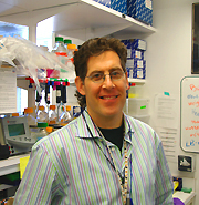
Andy
Golden
|
Andy Golden received his Ph.D. from the State University of New York at Stony Brook in 1990. He completed a postdoctoral fellowship at the California Institute of Technology in Pasadena in the laboratory of Paul Sternberg and in 1995 became a program fellow at the NCI-Frederick. In 2000, he joined the Laboratory of Biochemistry and Genetics, NIDDK, where he is now a senior investigator.
Errors in chromosome segregation during meiosis are responsible for the majority of spontaneous miscarriages in humans and also account for disorders such as Down syndrome. Understanding the regulatory mechanisms of chromosome segregation during meiosis is the primary goal of my laboratory.
We have taken a genetic approach to understanding this process. Using the free-living soil nematode Caenorhabditis elegans, we have isolated mutants that are defective in chromosome segregation during meiosis.
In C. elegans, oocytes are fertilized in prophase of meiosis I. In a screen for temperature-sensitive mutants, we isolated over 30 mutants whose embryos arrest at metaphase of meiosis I. These mutants defined five genes, all of which coded for subunits of the anaphase-promoting complex (APC). This complex is an E3 ubiquitin ligase, which ubiquitinates substrates and targets them for degradation by the 26S proteasome.
Our findings demonstrated that the APC is maternally stored in oocytes to drive the meiotic divisions upon fertilization. Oocytes defective in the APC arrested as one-cell meiotic embryos and failed to develop further.
Spermatocytes defective in the APC also failed to segregate their chromosomes during both meiotic divisions, but surprisingly, still differentiated into spermatids. These sperm were able to fertilize oocytes despite their lack of chromosomes. These embryos undergo a number of divisions before dying.
To further understand the players in meiotic chromosome segregation, we isolated 26 extragenic mutations that restored viability to these APC mutants at the nonpermissive temperature. These 26 mutants defined at least eight different genes. Four of these genes code for known regulators of the APC. One was FZY-1, an APC activator. Three others were orthologs of the spindle assembly checkpoint (SAC).
The SAC functions during mitosis to monitor the assembly of the spindle and the attachment of chromosomes to the spindle microtubules. Defects in either of these processes triggers the SAC to inhibit the APC, thus allowing the cell more time to correct these defects before separating chromosomes at anaphase.
Our findings suggest that the SAC also functions during normal meiotic divisions (in the absence of any obvious spindle damage), perhaps to regulate the timing of the meiotic divisions after fertilization. The identification of the remaining suppressor mutants from this screen is one of our immediate goals.
We have also used a genetic approach to find suppressors of a mutant separase allele. Separase is the protease that cleaves the cohesion molecules that hold metaphase chromosomes together. This mutant has severe defects in meiotic chromosome segregation and cytokinesis.
This strategy has identified only two suppressors, one of which is a conserved, but uncharacterized, phosphatase. We are investigating the mechanism by which a mutation in this phosphatase suppresses the separase phenotypes.
In addition, we are currently working toward the molecular identification of the other suppressor.
Though our studies have focused on the maternal factors in the oocyte that drive the meiotic divisions, we are also curious as to how fertilization triggers the oocyte to resume its meiotic divisions. Sperm are likely the source of this signal, because normal meiotic divisions are not observed in the absence of sperm.
SPE-11 is a protein that is contributed by the sperm that is absolutely essential for meiotic resumption upon fertilization. Mutations in the spe-11 gene result in embryos that fail to complete the meiotic divisions after fertilization and subsequently die during embryonic development.
To understand how paternal SPE-11 triggers meiotic resumption, we have recently begun an extragenic suppressor screen to identify mutants that permit the survival of spe-11 mutants.
Presumably such a screen will identify components of the pathway through which SPE-11 communicates with the oocyte to initiate meiotic resumption. This screen may identify additional paternal factors involved, or at least other maternal factors that receive this sperm signal.
I enjoy the genetic approaches we have taken to understand the regulation of the meiotic divisions in C. elegans. These screens have allowed us to take a naïve approach without making any assumptions about the types of genes for which we should be looking.
In this
way, we have isolated rare gain-of-function alleles of some genes and hypomorphic
alleles of essential genes. These types of alleles allow a more nuanced examination
of the roles of these genes in meiosis and early development.
 |
|
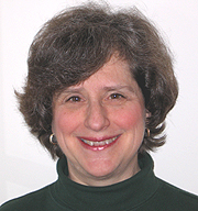
Ellen
Leibenluft
|
Ellen Leibenluft received her M.D. degree from Stanford University, Stanford, calif., in 1978. She did her residency at Georgetown University in Washington, D.C., where she served as director of the psychiatric inpatient unit and day hospital before joining NIMH in 1989. She is currently a senior investigator and chief of the Unit on Bipolar Spectrum Disorders in the Emotion and Development Branch of the Mood and Anxiety Disorders Program, NIMH.
My research has two main goals: to elucidate the pathophysiology of pediatric bipolar disorder (BD) using the principles and techniques of cognitive neuroscience and to ascertain whether children with extremely severe irritability and symptoms of attention deficit hyperactivity disorder (ADHD) are exhibiting a phenotype of pediatric BD.
Our studies of the brain mechanisms mediating BD in youth involve administering standardized behavioral paradigms to assess emotional and cognitive information processing. On paradigms in which patients and control subjects differ behaviorally, we use rapid event-related functional MRI to study between-group differences in neural activation.
Using such techniques, we found that youth with BD have deficits in their ability to identify facial emotion. For example, compared with control subjects, youth with BD rate a neutral face as more hostile, are more afraid of the face, and have increased amygdala activation when rating the hostility on the face or their fear of it (but not when performing a nonemotional rating of a facial feature). Deficits in face labeling and/or increased amygdala activation differentiate youth with BD, not only from controls, but also from those with anxiety, depression, and ADHD.
Recently, we found that deficits in labeling face emotion are also present in unaffected youth at high risk for the disorder, that is, those who have a parent or sibling with BD. Thus, we are now studying whether face emotion–labeling deficits may be an endophenotype of BD, as well as associations between polymorphisms and these behavioral and neuroimaging findings.
We have also found that youth with BD have deficits in their ability to adapt their behavior flexibly in response to changing emotional stimuli. This deficit may be pathophysiologically related to the fact that patients with BD have marked response inflexibility when they are manic or depressed (for example, they are unable to respond appropriately to rewarding stimuli when depressed). Response-flexibility deficits are evident on response-reversal tasks and on tasks in which subjects must inhibit a repetitive response or substitute an alternative one.
We found that patients with BD, compared with control subjects, have decreased activation in the ventrolateral prefrontal cortex and striatum when they try to inhibit a motor response but fail to.
These data indicate that patients with BD may have a deficient striatal error signal and, in concert with the data presented above, implicate amygdala-striatal-prefrontal circuitry in the pathophysiology of BD.
Our second, and parallel, research focus is on youth with extremely severe irritability and ADHD-like symptoms, a syndrome that we have named "severe mood dysregulation" (SMD).
Although youth with SMD are frequently diagnosed with BD in clinical settings, our research indicates that they are at particularly high risk of developing major depression, rather than BD, in early adulthood, and that BD youth are more likely than SMD youth to have a parent with BD. In addition, youth with SMD have less consistent response-flexibility deficits than do those with BD.
When engaging in a frustrating attentional task, both SMD and BD children report more frustration than do control subjects, and both have attentional deficits, as measured by evoked-response potentials. However, the specific deficits differ, with those of the BD youth centering on their inability to focus their attention when frustrated and those of the SMD youth centering on their failure to attend to a stimulus throughout the task.
These data all argue against SMD being a phenotype of BD. However, it is important to note that SMD youth, like those with BD (but unlike those with depression, anxiety, or ADHD) have difficulty labeling facial emotions, indicating that there may be some shared pathophysiological dysfunction between SMD and BD.
We are currently comparing amygdala-striatal-prefrontal activation in SMD, BD, and ADHD youth, as well as in control subjects, during an emotional face-processing task, a motor-inhibition task, and a motor-flexibility task.
The question of whether SMD youth are exhibiting a phenotype of BD is important, because the treatment for BD is very different from that for ADHD or the other psychiatric illnesses for which SMD youth meet diagnostic criteria (such as anxiety disorders). Thus, this line of research should ultimately help to clarify the classification, and thus treatment, of SMD. In addition, research into the pathophysiology of both BD and SMD should provide knowledge that will be relevant to the development of novel treatments for these very impairing illnesses in youth.
 |
|
Clement
McDonald
|
Clement J. McDonald received his M.D. from the University of Illinois in 1965. After his internal medicine residency at Boston City Hospital and Cook County Hospital in Chicago and a two-year fellowship at NIH, he joined the faculty at the Indiana University Medical School and the Regenstrief Institute in Indianapolis, where he developed large-scale medical-record systems. In November 2006, he became the director of the Lister Hill National Center for Biomedical Communications at the National Library of Medicine. He is a member of the Institute of Medicine and a founding fellow of the American Institute for Medical and Biological Engineering.
As an intern at Boston City Hospital, I spent most of my day gathering, organizing, and analyzing patient data, cross-checking new data against the old and what was expected, and adjusting treatments and testing accordingly. The days were consumed by hordes of little data-gathering and -checking tasks—work more like that of a bank clerk summing and double-checking deposits and receipts than that of Sherlock Holmes.
However, back then bank clerks had computer help. Physicians did not and, partly as a result, would sometimes fail to review tasks and therefore overlook an abnormality or a preventive opportunity. Physicians need a computerized medical record to organize and watch their patients’ records—something like a faithful sheepdog that calls attention when it "sees" danger lurking.
When I arrived at Marion County Hospital and the Regenstrief Institute in 1972, I set out to build one such medical record system, one that could accomplish three missions:
![]() Eliminate the logistic problems of the paper record: lost or unavailable, disorganized,
or illegible
Eliminate the logistic problems of the paper record: lost or unavailable, disorganized,
or illegible
![]() Provide reminders to physicians about clinical situations that need action
Provide reminders to physicians about clinical situations that need action
![]() Provide query access across patients for research purposes
Provide query access across patients for research purposes
The project started with 32 patients in a diabetes clinic and then expanded to cover all patients. Early on, we developed a reminder system that could review the patients’ records according to guidelines and remind physicians when it saw that patients needed specific tests, treatments, or immunizations.
In a series of randomized trials beginning in 1973, we showed that such computer reminders increased the appropriate use of such interventions among eligible patients by 15 to 2,000 percent compared with the control state.
The largest of these studies included more than 400 care providers, 1,000 reminder rules, 12,000 patients, and 50,000 visits. In late 1980, we developed one of the first physician order-entry systems and showed in a randomized trial that it significantly reduced costs compared with paper-based ordering.
Along the way, it became obvious that standards were needed for delivering patient data from the computers that produced them (such as pharmacy and laboratory systems) to other computers that needed them (such as medical-record systems). So we developed ASTM 1238, the first standard data structure for interchanging clinical observations between computers.
That structure was adopted by Health Level Seven, an accredited standards-developing organization that focuses on clinical and administrative data. ASTM 1238 is now used worldwide. We also developed a coding system for clinical variables, such as laboratory results and survey questions, called LOINC, which is also used internationally.
The medical record system has grown over the years and has expanded to include all of the Indianapolis hospital systems. It now includes a billion discrete results from 3 million patients and 15 different hospitals, as well as the Indiana State Medicaid and Well Point, and Rx Hub.
It digests HL7 message streams from over 200 distinct computers that carry 180 million HL7 messages per year. It serves direct patient care, public health, and research functions. It also delivers diagnostic study reports and dictations produced by the 15 hospitals to Indianapolis’ physicians—all 3,800 of them. It is now the longest continuously operating electronic medical record system in the world and the best example of a Regional Health Care Network in the United States.
My goals
at NIH are to develop similar informatics tools, systems, and processes, including
data de-identification for preserving patient privacy, text understanding, statistical
analysis, and linkage—tools to improve availability and research utility
of the clinical data contained in electronic medical records and long-term clinical
studies
 |
|
Andrew
Singleton
|
Andrew Singleton received his Ph.D. from the University of Newcastle upon Tyne, U.K., in 1998. His doctoral work focused on genetic causes and contributors to dementia, particularly Alzheimer’s disease and dementia with Lewy bodies. He had postdoctoral training at the Mayo Clinic in Jacksonville, Fla., before joining NIA as an investigator within the newly created Laboratory of Neurogenetics in 2001. He is currently chief of the Molecular Genetics Unit in that lab.
My group focuses on the genetic basis of neurological diseases—particularly the role of genetic variability in Parkinson’s disease, ataxia, dystonia, and stroke—in addition to working on genetic influences on phenotypes of aging.
Driving this research is the belief that this approach is the first step toward understanding the complex clinical and pathological manifestations of disease and aging and that ultimately genetic insights will highlight disease mechanisms and implicate pathways amenable to therapeutic intervention.
Undoubtedly our largest success over the past five years has been in the field of Parkinson’s disease genetics. In 2003, we were the first group to identify a-synuclein locus multiplications as a cause of Parkinson’s disease and Lewy body disorders; in 2004, we identified mutation of the gene LRRK2 as a cause Parkinson’s disease; and in 2005, we showed that a particular mutation in LRRK2, G2019S, is the most common known genetic cause of this disorder, underlying Parkinson’s disease in an estimated 20,000 North Americans.
Each of these findings was a paradigm shift for the Parkinson’s disease field. The first highlighted the role that overexpression of normal wild-type protein can play in diseases of protein deposition. The second finding has provided clues to the underlying biology of Parkinson’s disease. The third has had an impact on the field’s understanding of the clinical and pathological entity of Parkinson’s disease.
My group continues to use familial forms of disease to find monogenic causes of neurological disorders, but most recently we have expanded on this family-based work in order to define the genetic basis of typical sporadic forms of neurological disease, including Parkinson’s disease, amyotrophic lateral sclerosis, and stroke.
In performing this work, we have generated approximately one billion genotypes, about 400 million of which are already in the public domain. It is my belief that this work lays the foundation for future research into the genetic basis of these conditions.
 |
|
Doris
Wu
|
The vertebrate inner ear is a highly intricate organ that is responsible for relaying auditory and vestibular information to the brain. Any malformations of this complex structure during development are likely to result in functional deficits. My research program focuses on:
![]() Identifying the molecular mechanisms underlying inner ear formation using both
the mouse and chick as experimental models
Identifying the molecular mechanisms underlying inner ear formation using both
the mouse and chick as experimental models
![]() Understanding how mutations in specific genes cause human deafness
Understanding how mutations in specific genes cause human deafness
The proper formation of the inner ear depends on signals emanating from surrounding tissues. In our work to identify and characterize such signals, we demonstrated in both mouse and chicken that Sonic hedgehog (Shh), secreted from the ventral neural tube and notochord, is required for the formation of the primary auditory apparatus, the cochlear duct.
The mammalian cochlear duct is a coiled structure that is tonotopically organized, such that the base of the cochlear duct is most sensitive to high-frequency sounds and the apical region to low-frequency sounds.
We showed that apical and basal regions of the cochlear duct depend on different levels of Shh signaling.
The apical, low-frequency region depends on high levels of Shh signaling, whereas the basal, high-frequency region requires lower levels of Shh.
We also discovered that various levels of Shh activate different amounts of Gli transcription activators and repressors that mediate Shh’s functions. In addition, our results provide an explanation as to why mutations in one of the Gli transcription factors, Gli3, cause low-frequency hearing loss in Pallister-Hall syndrome patients.
Proper inner-ear morphogenesis is a dynamic process directed by cellular interactions and proper temporal activation of various molecular pathways within the developing otic epithelium, in response to extrinsic signaling factors from surrounding tissues. Two of the major molecular pathways that we have been studying involve bone morphogenetic proteins (BMP) and fibroblast growth factors (FGF).
The primary sensory cells of the auditory and vestibular systems, referred to as hair cells, are clustered within the various sensory regions of the inner ear. These sensory regions are embedded within complex, highly specialized labyrinths formed by nonsensory tissues. We provided insight into how the formations of sensory and nonsensory regions of the inner ear are coordinated during development.
We demonstrated that three vestibular sensory patches, the presumptive cristae, most likely regulate the formation of their associated nonsensory structures, the semicircular canals, through the actions of secreted molecules such as BMP4 and FGF10.
We are currently investigating whether similar molecular mechanisms are involved in the coordinated development of the sensory and nonsensory components of other auditory and vestibular sensory organs.
 |
|
Keji
Zhao
|
Keji Zhao received his Ph.D. in molecular biology from the University of Geneva in Switzerland in 1996. He did his postdoctoral training with Gerald Crabtree at Stanford University, Palo Alto, Calif., before joining NHLBI in 1999 as a tenure-track investigator. He is currently a principal investigator in the Laboratory of Molecular Immunology, NHLBI.
Eukaryotic DNA forms a complex chromatin structure by interacting with histones and nonhistone proteins in the cell. Our research has focused on understanding how the chromatin structure is regulated and how it regulates gene expression.
Chromatin structure can be modified (or remodeled) by two major mechanisms. One is the activity of ATP-utilizing enzymes exemplified by the yeast SWI/SNF complex, which uses ATP-derived energy to alter the DNA-histone interactions and thereby allows regulatory enzymes to access their target sites in chromatin.
The other mechanism is the post-translational modification of histones—including acetylation, methylation, ubiquitin-ation, and phosphorylation—which may affect gene expression by providing specific recognition platforms for various regulatory enzymes on the chromatin substrate.
The BAF complex is the mammalian homolog of the yeast SWI/SNF complex. The BAF complex remodels nucleosomal structure in vitro, but its function and mechanisms in vivo are not well understood. Our group was the first to identify several BAF target genes, including the cell-cycle inhibitor p21 and interferon-inducible genes, in human cells.
Mutations of the complex have been correlated with various cancers, suggesting it can act as a tumor suppressor. We demonstrated that through its regulation of p21 expression, the BAF complex regulates the activity of the tumor suppressor protein pRb. Furthermore, we found that rapid activation of cellular antiviral activities depends on BAF complex priming of the chromatin structure of interferon-inducible genes.
The nonclassical left-handed zigzag DNA structure (Z-DNA) was discovered three decades ago and, in the absence of known biological function, has been considered "junk" DNA. We recently demonstrated that chromatin remodeling by the BAF complex induces Z-DNA formation at the CSF1 gene promoter.
Both the Z-DNA formation and BAF activity are required for the CSF1 gene activation, indicating that Z-DNA structure plays a role in modifying the chromatin structure.
The availability of genomic sequences of many model organisms has enabled us to monitor the chromatin modification events on a genome-wide scale.
Because the only available technique for high-throughput analysis of chromatin modifications—"ChIP (chromatin immunoprecipitation)-on-chip"—depends on the selected probes on the DNA microarrays, we developed an unbiased genome-wide mapping technique by combining ChIP and serial analysis of gene expression to cover the entire human genome.
Using this technique, we generated the first comprehensive map of major histone modifications in human T cells. Analysis of the histone-modification patterns revealed 46,000 "histone acetylation islands," which represent functional regulatory elements of chromatin and transcription.
Interestingly, the methylation of H3 lysine 4 has been considered an "active" mark, while the methylation of H3 lysine 27 has been considered a "repressive" mark. Our genome-wide analysis, however, revealed that these two apparently opposite marks are co-localized at thousands of genomic loci, suggesting that they may together define the chromatin status.
We have also combined comparative genomics and histone acetylation data to identify functional enhancers of transcription. We found that histone acetylation patterns can predict both conserved and nonconserved enhancers, suggesting a novel method for annotating the functional regulatory elements in the human genome.
The chromatin
modifications—including DNA methylation, histone modification, and ATP-dependent
remodeling—may collectively define epigenomes that contain distinct epigenetic
signals unique for each differentiated cell type and for maintaining the stability
of cellular identity through cell divisions. Our current goal is to define the
epigenomes using the genome-wide techniques and to understand how the epigenetic
signals regulate genome function. ![]()