| T H E N I H C A T A L Y S T | J A N U A R Y – F E B R U A R Y 2007 |
|
|
|
| P E O P L E |
RECENTLY TENURED
 |
|
John
Chiorini
|
John (Jay) Chiorini received his Ph.D. in genetics from the George Washington University in Washington, D.C., in 1993. He completed a postdoctoral fellowship at NHLBI and a PRAT fellowship awarded by NIGMS before joining NHLBI as a senior research fellow in the Molecular Hematology Branch. In 2000, he joined the Gene Therapy and Therapeutics Branch, NIDCR, where he is now a senior investigator.
The development of improved vectors for gene transfer is a critical element for the advancement of the field of gene therapy.
Vectors based on adeno-associated virus (AAV) offer several advantages as a platform for gene transfer—such as stable long-term transfer in vivo into several different cell types—but our understanding of the biology of this virus is limited. My overall research goals are to define the interactions of AAV with its target cell and to develop improved vectors for gene transfer. Our underlying hypothesis is that by understanding these interactions as they apply to the biology of the virus, we can contribute to the development and use of AAV vectors for gene therapy.
AAV is a human parvovirus with a single-stranded DNA genome that contains only two open reading frames that encode either the nonstructural (Rep) proteins or the capsid (VP) proteins.
Our early work focused on the role of the Rep proteins in virus production and regulation of cellular activity; much of our recent work focuses on the capsid genes and the use of recombinant AAV vectors for gene transfer.
The interactions between the capsid of an AAV particle and its host cells that result in transduction are complex and largely not understood.
Our initial work in this area demonstrated that not all AAV serotypes have the same cell tropism. This observation led to the search for and identification of new AAV serotypes and has resulted in improved gene transfer to several target cell types.
However, this process of matching new isolates to those targets where they would be most effective was largely empirical because we lacked understanding of the cellular components necessary for transduction with these new vectors.
We therefore worked to develop a novel bioinformatics-based screening assay that has allowed us to identify the cellular genes responsible for the distinct cell tropism of the different AAV isolates.
The identification of genes critical for transduction with AAV vectors has yielded better matches and, in addition, has enabled us to increase transduction in poorly permissive cells by upregulating the expression of these critical genes.
For example, we have identified the platelet-derived growth factor receptors (PDGFR) as critical for efficient transduction of AAV type 5 (AAV5).
Our further characterization of PDGFR as a receptor for AAV5 has allowed us not only to target the use of the vector to specific cell types in which PDGFR is highly expressed, but also to manipulate the expression level of PDGF and improve transduction.
Currently, natural viral isolates serve as a rich source of vectors for gene transfer. But we envision being able to build on our understanding of the biology of these natural isolates and develop engineered vectors with specific and defined tropisms.
Toward this goal, we have worked to understand gene transfer from the perspective of the virus by developing structural models for different AAV vectors and identifying key regions involved in receptor binding and transduction.
In this way, we can rationally design vectors that incorporate targeting epitopes into the vector capsid. These targeting epitopes will fill the same role as the natural targeting domain in current vector but bind receptors of our design.
 |
|
Philipp
Kaldis
|
Philipp Kaldis received his Ph.D. in 1994 from the Swiss Federal Institute of Technology in Zürich and did postdoctoral work at Yale University in New Haven, Conn. He joined the NCI-Frederick in 2000 and is currently a senior investigator in the Mouse Cancer Genetics Program.
I am interested in how cells divide and multiply. Misregulation of cell growth and proliferation plays an important role in many diseases, including cancer. Detailed understanding of the regulation of cell growth and proliferation is therefore a prerequisite for designing strategies to treat such diseases. Cyclin-dependent kinases (Cdks) are among the central regulators of cell growth and proliferation.
Originally, I was trained as a "hard-core" biochemist and spent lots of time purifying proteins and analyzing enzyme kinetics. But a growing desire to relate my results to in vivo situations inspired an interest in genetics, especially mouse genetics to model cell cycle and cancer as they relate to humans.
Right now, I am trying to dissect the genetic pathways of cell-cycle regulation in the mouse. It will take many years to analyze all the essential pathways that regulate the cell cycle.
Mouse models to investigate in vivo functions of Cdks. Studies of Cdk2 in vitro and in cell lines suggest that Cdk2 controls the transition from G1 (interphase) to S phase, where DNA replication takes place. Inactivation of Cdk2 in mammalian cell lines results in growth arrest in the G1 phase.
To explore Cdk2 functions in the context of a living animal, we have generated Cdk2 knockout mouse models. We have found that these
Cdk2-/- mice are viable but sterile and therefore that Cdk2 seems to be essential for meiosis but not mitosis. These results suggested that other genes can compensate for Cdk2 functions in the mitotic cell cycle.
Among the best candidates were the family members Cdk1, Cdk3, Cdk4, and Cdk6. Cdk3 could be excluded because it is not expressed in the mouse. To investigate the functional overlap, we generated Cdk2-/-Cdk4-/- and Cdk2-/-p27-/- double-knockout mice. Our results indicate that Cdk2-/-Cdk4-/- knockouts die in utero, suggesting that Cdk2 and Cdk4 have overlapping essential functions in controlling the retinoblastoma protein.
The Cdk inhibitor p27Kip1 has been thought to act primarily by controlling its major target, Cdk2. Interestingly, we observed all phenotypes of the single knockouts in the Cdk2-/-p27-/- double-knockout mice. This result suggested that p27 has other targets in addition to Cdk2, one of which we identified as Cdk1.
We used to think that Cdk2 functions in the G1/S phase and Cdk1 only in mitosis, but our results opened the possibility that Cdk1 could also regulate the G1/S transition in addition to its mitosis function. We found that cyclin E (usually a partner of Cdk2) binds to and activates Cdk1 and that this is essential for S phase entry in the absence of Cdk2. Therefore, Cdk1 compensates for the functions of Cdk2 in G1/S.
Our results indicate that Cdk2 and Cdk1 bind the same cyclins and are functioning during the same cell-cycle phases.
These findings raise questions as to why there are two Cdks fulfilling the same functions and why Cdk2 cannot compensate for Cdk1 in mitosis. (Cdk1 seems to be an essential gene in the mouse.) To answer these questions, we will focus our future studies on Cdk1.
In summary, my goal is to investigate the in vivo functions of Cdks during normal development and tumor formation. To achieve our goal, we are generating mouse models that cover pathways that impinge on the functions of Cdks. Our work enhances the basic understanding of cell-cycle regulation and also provides information essential to the design of cancer therapies.
Mouse genetics requires patience and careful long-term planning. I am very grateful to the past and present members of my lab for their contributions. We welcome continuing collaboration with other scientists at NCI, NIH, and elsewhere.
 |
|
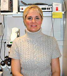
Carole
Parent
|
Carole Parent received her Ph.D. from the University of Illinois at Chicago in 1992. She had her postdoctoral training in the laboratory of Peter Devreotes in the Department of Biological Chemistry of the Johns Hopkins University School of Medicine in Baltimore and in 1996 became an instructor in the same department. In May 2000, she joined the Laboratory of Cellular and Molecular Biology, NCI, where she is now a senior investigator.
Research in my group focuses on uncovering the mechanisms by which chemotactic signals are integrated at the cellular and multicellular levels to regulate directed cell migration.
We use genetic, biochemical, and cell biological approaches to study this clinically relevant topic in two related and complementary model systems, the social amoeba Dictyostelium and human neutrophils.
We are particularly interested in exploring two fundamental questions in chemotaxis:
![]() How do external signals establish and maintain signaling and cellular polarity?
How do external signals establish and maintain signaling and cellular polarity?
![]() How are chemotactic signals relayed to neighboring cells—that is, how do
cells transition from single to group migration?
How are chemotactic signals relayed to neighboring cells—that is, how do
cells transition from single to group migration?
Dictyostelium cells, which are amenable to genetic study, track down and phagocytose bacteria during normal growth. When starved, they enter a developmental program that culminates in the formation of a multicellular organism composed of spores atop a stalk of dead cells.
This impressive transition is driven by chemotaxis. Dictyostelium cells migrate toward secreted adenosine 3’, 5’ cyclic monophosphate (cAMP), which is recognized by a family of G protein–coupled seven-transmembrane receptors.
Thus, in this system, cAMP acts as both an intracellular second messenger and a chemotactic cue. Surface-receptor activation leads to dissociation of G protein into a- and bg-subunits, which then activate a variety of effectors that control signaling cascades leading to cell polarity, a prerequisite for migration.
As Dictyostelium cells chemotax, they align in a head-to-tail fashion and migrate in chains. Our quest to understand how chemoattractants regulate cell polarity and migration led us to discover—thanks in large part to live cell imaging—a novel mechanism of chain migration: Polarized and migrating Dictyostelium cells concentrate their adenylyl cyclase protein at their back, generating and secreting cAMP, which in turn attracts neighboring cells.
Cells lacking adenylyl cyclase activity, or in which the enzyme is active but mislocalized, do not exhibit this remarkable chain migration behavior.
We proposed that this asymmetric distribution provides a compartment from which cAMP is secreted to act locally as a chemoattractant, thereby allowing cells to follow each other and greatly amplifying the chemotactic response.
Indeed, the relaying of chemotactic signals that is exhibited by Dictyostelium cells leads to the recruitment of significantly more cells to aggregates, a potential mechanism of survival of this species.
Using highly sensitive fluorescent microscopy analyses, we also established that adenylyl cyclase is not only highly enriched at the back of chemotaxing Dictyostelium cells but also found on very dynamic vesicles that coalesce at the back of cells.
Photo-bleaching experiments showed that these vesicles are involved in replenishing the plasma membrane, suggesting that they are actively engaged in membrane trafficking and perhaps are part of a regulatory mechanism that controls adenylyl cyclase activity.
This unique chemoattractant amplification mechanism may well be conserved in higher eukaryotes. Migration of cells as groups is common during development and wound repair, as well as in metastatic processes. Yet the mechanism by which this occurs remains obscure.
We are currently expanding our studies to include neutrophils, which share striking behavioral and mechanistic similarities with Dictyostelium cells, and to metastatic cancer cells, which have been shown to migrate in files as they leave tumors and invade other tissues.
Such studies should continue to exert a profound impact on our general understanding of the mechanisms used by various types of cells to attain a specific destination in the context of both normal physiology and disease.
 |
|
Shyamal
Peddada
|
Shyamal Peddada received his Ph.D. in 1983 from the University of Pittsburgh, under the supervision of C. R. Rao. He held various academic positions and was a tenured full professor in the Department of Statistics at the University of Virginia in Charlottesville before joining the Biostatistics Branch, NIEHS, in 2000. He is currently a senior investigator and director of the Statistical Consulting Service in that branch. He is an elected fellow of the American Statistical Association and an elected member of the International Statistical Institute, as wellas a recipient of the American Statistical Association’s Outstanding Statistical Application Award.
I have worked in several different areas of statistical theory, methodology, and applications; one of my major contributions is in the area of statistical inference under order restrictions.
In many applications, a research hypothesis of interest can be formulated in terms of mathematical inequalities (known as order restrictions) among the unknown parameters.
For instance, when conducting dose-response studies, a researcher may hypothesize a particular shape or pattern of mean response with respect to dose, which can be expressed in terms of mathematical inequalities.
As an example, consider a dose-response study with a control, a low-dose, and a high-dose group. The experimenter hypothesizes that the mean response has an increasing pattern (or shape) with dose.
The hypothesis can then be restated in this manner: The mean of the control group is at most as large as that of the low-dose group, which is at most as large as that of the high-dose group. By incorporating the shape of the curve into the statistical analysis, one can greatly enhance the power of a test.
The most commonly used statistical procedures (such as ANOVA and the pairwise t-tests) do not incorporate such information and hence are not powerful in the context of dose-response and time-course experiments, especially when the sample sizes are small.
On the other hand, the order-restricted statistical procedures, such as those developed by my colleagues and me, enjoy substantially greater power over the standard procedures.
Further, results obtained from such analyses have biological implications because they describe the pattern of biological response to treatment over dose and/or time.
An important application of my research program is the analysis of microarray gene-expression data from time-course and/or dose-response experiments.
We have developed user-friendly and freely downloadable JAVA-based computer software called ORIOGEN (order-restricted inference for ordered gene expression) for analyzing microarray gene expression data. This software is available at this website.
Though it was developed specifically for analyzing microarray gene-expression data, ORIOGEN can be applied to data from any time-course or dose-response experiment.
We are now developing new methodologies, including Bayesian techniques, to allow for more complex dependence structures.
These methodologies would
be useful for many different applications, such as analyzing gene-expression
microarray data obtained from dose-response studies with repeated measures,
analyzing data on multiple tissues obtained from the same subject, and associating
different types of correlated data, such as correlating phenotype with genotypes.
![]()