| T
H E N I H C A T A L
Y S T |
J
A N U A R Y – F E B R U A
R Y 2007 |
|
Shedding
Light on Deep Tissue
BIOPHOTONICS:
THE NEW "AGE
OF ENLIGHTENMENT"
|
by Fran Pollner |
 |
|
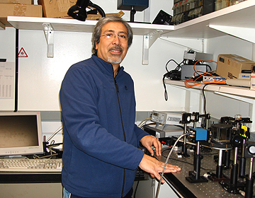
Master
builder:
In addition to constructing equations in his mind, Amir Gandjbakhche builds
countertop prototypes of potential bedside instruments. He notes that
once an experimental technology reaches a point of clinical commercial
potential, his lab moves on to other basic explorations.
|
Sometimes
Dr. McCoy’s hand-held device rendered an instant diagnosis as he waved
it over a fallen crew member of the Star Ship Enterprise; sometimes it
achieved an instant cure.
Such rapid, noninvasive
bedside management is not as much a fantasy as one might think.
Amir
Gandjbakhche calls it his "dream," but it’s a dream that
gets closer to reality, he says, with each advance in optical-imaging research.
"They say the 18th
century was the ‘Age of Enlightenment.’ But, really, it’s the
21st century. It’s optical imaging that’s enlightening us, moving
us from subjective to quantitative diagnosis," says Gandjbakhche, chief
of the Section on Biomedical
Stochastic Physics in the Laboratory
of Integrative & Medical Biophysics, NICHD.
Gandjbakhche and his team
collaborate with other NIH investigators in animal studies and on clinical protocols
that involve noninvasive in vivo optical imaging to characterize the physiologic
and metabolic environment of diseased tissues.
Modeling
Stochastic Processes
The group devises quantitative
theories and designs instrumentation for optical spectroscopy and tomographic
imaging of tissues.
"We are looking at
biological systems with randomness in time and space—that’s a stochastic
prcess," says Gandjbakhche.
Using mathematical models
to localize lesions and track their changing functional status requires analyzing
different optical sources of contrast such as fluorescent labels, absorption,
and/or scattering.
Gandjbakhche also builds
countertop prototypes of the instruments that might be used at the bedside to
characterize the tissues under scrutiny and to monitor response to therapy,
instruments that involve neither ionizing radiation nor surgical biopsy, just
light.
When light enters biological
tissue, it doesn’t go straight in but scatters in many directions, requiring
sophisticated methods such as "random-walk theory" to explain the
path the light takes through the tissue. This stochastic method, developed at
NIH, takes into account the absorption and scattering properties of tissue,
which are wavelength dependent, Gandjbakhche explains, noting that a wealth
of information can be obtained by spectroscopic methods.
"Light traveling
through tissue is a stochastic process. The photons are going everywhere. It’s
beautiful," says Gandjbakhche, pointing to a display on his computer screen.
The creation of the vascular
network is also stochastic. Fellows Franck
Amyot and Alex
Small are modeling the stochastic process of tumor-induced angiogenesis
in collaboration with NCI’s Kevin
Camphausen.
Optical
Imaging Advances at a Glance
|
|
The
attributes that make optical imaging a choice diagnostic and monitoring
modality, says Amir Gandjbakhche, are these:
 Optical imaging uses nonionizing visible and near-infrared light and is
therefore safer than such techniques as X-ray, CT, and PET imaging to
gather information about what’s going on beneath the skin’s
surface.
Optical imaging uses nonionizing visible and near-infrared light and is
therefore safer than such techniques as X-ray, CT, and PET imaging to
gather information about what’s going on beneath the skin’s
surface.
Optical-imaging instruments are portable and can be brought to the patient,
not large and stationary like MRI machinery.
Optical imaging provides functional information and penetrates far deeper
than the 1—2 mm accessible by two-photon or confocal microscopy.
Other kinds of imaging equipment, such as MRI and PET, are between 10
and 20 times as expensive as the tools of optical imaging.
In short, it’s
"portable, safe, cheap, fast," and provides functional information,
says Gandjbakhche.
|
Some
Ongoing Studies:
Tracking Vasculature Responses In
Kaposi’s Sarcoma Patients
Graduate student Abby
Vogel and postdoc Moinuddin
Hassan, with NIBIB’s Paul
Smith, are monitoring the effect of experimental drugs to counter angiogenesis.
For the past five years,
Gandjbakhche’s team has collaborated with Robert
Yarchoan, chief of the HIV
and AIDS Malignancy Branch, NCI, and Richard
Little, senior oncologist, on four clinical protocols involving drug regimens
for patients with Kaposi’s sarcoma (KS), a highly vascular tumor.
"We created quantitative
methods to assess the vascularity of these tumors using three noninvasive imaging
modalities," Gandjbakhche says. "We monitor the results of the drugs
being tested and discuss them with the physicians."
"All three imaging
modalities," he adds, "take less than five minutes."
Each of these modalities—laser
Doppler imaging (LDI), infrared thermal imaging (thermography), and near-infrared
multispectral imaging—provides specific, complementary information.
LDI measures blood flux,
a combination of red blood cell velocity and concentration; spectral imaging
measures changes in blood volume and oxygenated and deoxygenated hemoglobin;
and thermography measures temperature as a reflection of blood flow, providing
confirmation that changes in the vasculature are related to blood flow.
The spectral-imaging component
came about through collaboration with Stavros Demos of the Lawrence Livermore
National Laboratory, Livermore, Calif., in designing a portable spectral-imaging
system, Gandjbakhche notes.
Overall, the KS studies
have thus far demonstrated that the lesions—in addition to being hotter
than normal tissue—also register higher blood volume, deoxyhemoglobin,
and blood velocity. The cytotoxic/anti-angiogenic combination of liposomal doxorubicin
and interleukin-12 is among the agents that have been tested. These imaging
techniques can easily be adapted to a variety of skin diseases.
 |
|
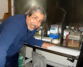
Amir
Gandjbakhche leans into his lab’s home for mice in the mouse-imaging
area in Building 9
|
Fluorescence
Lifetime Imaging In
Mouse Tumor Studies
The applications of fluorescence
imaging are limitless, constrained only by the development of fluorophores sensitive
to the biological targets of interest.
A fluorophore, Gandjbakhche
explains, "is a molecule that has the property to be excited in one wavelength
and emits light in a longer wavelength after a delay called lifetime."
"Any condition in
which receptors on the cell surface play a role, for instance, is a candidate
for fluorescence imaging," says Gandjbakhche. "We need only create
antibodies tagged with fluorophores that bind to a specific receptor of interest."
For the past two years,
the Gandjbakhche lab has assisted in the mouse tumor studies conducted by Jacek
Capala, an investigator in the Radiation
Oncology Branch, NCI.
Postdocs Jason
Riley and Hassan
and staff scientist Victor
Chernomordik, along with NICHD colleagues Hacene
Boukari and Dan
Sackett in the lab of Ralph
Nossal, have developed quantitative methods to characterize the molecular
and functional status of deeply embedded tumors—breast cancer cells expressing
high levels of the HER2 protein.
Gandjbakhche’s team
measures fluorophore lifetime because it varies, for instance, with degree of
oxygenation or pH value.
"The instrinsic optical
properties of the tissues under investigation," he explains, "will
yield different functional properties." Fluorophores are chosen for their
specific sensitivity to the tissue environment.
Localization of the tumor
and quantification of pH was achieved with the use of a fluorophore-Herceptin
(a HER2-specific monoclonal antibody) conjugate. The fluorophore was a pH-sensitive
near-infrared dye called Alexa Fluor 750. The team intends to continue these
studies to investigate an affibody-based molecular probe for imaging HER2 receptors.
"We know exactly
what kinds of antibodies to use; we know that tumor cells tend to be hypoxic
and have lower pHs than the surrounding tissues."
The
IPDC Connection
Gandjbakhche sits on the
steering committee of the Imaging
Probe Development Center, a new NIH core resource, directed by NHLBI’s
Gary Griffiths,
for the production of imaging probes, both known but not commercially available
and novel (see The
NIH Catalyst, January-February 2006).
Gandjbakhche’s proposal
to the IPDC that it manufacture a near-infrared dye for optical-imaging research
(the review of which he did not participate in) was recently approved. "This
dye uses metal chelates to modulate the fluorescence lifetime," Gandjbakhche
says, "and it will increase our ability to detect smaller fluorophore concentrations."
Acknowledging that familiarity
with the concepts, language, and calculations of his research is not widespread,
he observes that the "most important part of this work is its multidisciplinary
nature–it takes physicists, engineers, biologists, physicians, chemists.
. . ."
For
an in-depth look at the Gandjbakhche lab and its collaborative work, visit this
website.
Return
to Table of Contents