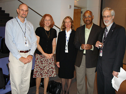
Propspecting
for the best regimens: (left to right)William Copeland, NIEHS;
Lynne Mofenson, NICHD; Kristine Witt, NIEHS; Robert Sills, NIEHS; and
panel chair John Bucher, NIEHS
| T H E N I H C A T A L Y S T | N O V E M B E R – D E C E M B E R 2006 |
|
|
|
Research
Festival
|
by Fran Pollner |
|
|||
|
Propspecting
for the best regimens: (left to right)William Copeland, NIEHS;
Lynne Mofenson, NICHD; Kristine Witt, NIEHS; Robert Sills, NIEHS; and
panel chair John Bucher, NIEHS
|
When it comes to reducing the rate of mother-to-child HIV transmission, the elusive perfect is not the enemy of the good; nonetheless, investigators would like to better define and minimize the risk, however small it may currently be, of genetic damage or mitochondrial dysfunction in fetuses and infants exposed to antiretroviral therapy.
To that end, NIEHS and the National Toxicology Program (NTP) have been examining the mitochondrial and potential carcinogenic effects of zidovudine (AZT), the first FDA-approved anti-HIV agent, and other nucleoside reverse transcriptase inhibitors (NTRIs); and NICHD, in collaboration with NIAID, NIDA, NIMH, NIDCD, and NHLBI, has launched the Pediatric HIV/AIDS Cohort Study (PHACS) to assess the long-term safety of fetal and infant exposure to prophylactic antiretroviral therapy.
According to John Bucher, deputy director of the Environmental Toxicology Program, NIEHS, and chair of the NIH Research Festival symposium on the benefits and risks of antiretroviral therapy in preventing mother-to-child HIV transmission, antiretroviral HIV regimens are among NTP’s top targets of investigation today (cell phone radiation and dietary supplements are two others).
Three NIEHS scientists reported recent findings:
![]() William Copeland,
of the Laboratory of Molecular Genetics,
reported on the propensities of NRTIs to induce disruption of mitochondrial
DNA replication through inhibition of DNA polymerase-g.
William Copeland,
of the Laboratory of Molecular Genetics,
reported on the propensities of NRTIs to induce disruption of mitochondrial
DNA replication through inhibition of DNA polymerase-g.
A new NTP study, he said, establishes mitochondrial DNA damage in mouse-pup hearts from perinatal exposure to two NRTIs—AZT and 3TC. A possible cascade of NRTI-induced oncogenic events starts with the inhibition of thymidine kinase 2 and DNA polymerase-g and potentially culminates in activated protooncogenes and cancer.
![]() Robert Sills,
of the Laboratory of Experimental
Pathology, elaborated on AZT-induced lung tumors in mice after in utero
exposure. Mutations in the K-ras oncogene and P53 tumor-suppressor gene
were among the findings reported in mouse lung tumors.
Robert Sills,
of the Laboratory of Experimental
Pathology, elaborated on AZT-induced lung tumors in mice after in utero
exposure. Mutations in the K-ras oncogene and P53 tumor-suppressor gene
were among the findings reported in mouse lung tumors.
![]() Kristine Witt,
of the Environmental Toxicology Program, noted that NTP studies revealing chromosomal
damage in mouse pups were designed to echo human therapeutic levels of AZT.
Transplacental exposure alone to low-dose AZT (50 mg/kg) is associated with
a 10-fold increase in micronucleated reticulocytes—a standard biomarker
of chromosomal damage—in newborn pups.
Kristine Witt,
of the Environmental Toxicology Program, noted that NTP studies revealing chromosomal
damage in mouse pups were designed to echo human therapeutic levels of AZT.
Transplacental exposure alone to low-dose AZT (50 mg/kg) is associated with
a 10-fold increase in micronucleated reticulocytes—a standard biomarker
of chromosomal damage—in newborn pups.
"These were the findings," she said, "that prompted human studies"—studies in which the frequency of micronucleated reticulocytes in 13 infants exposed prenatally to AZT was 10-fold that found in cord blood of control subjects and in three infants whose HIV-infected mothers had received prenatal antiretroviral therapy that had not included AZT. "Transplacental AZT is genotoxic to erythrocytes," Witt remarked.
Whether findings of this sort have any long-term clinical consequences, however, remains to be determined.
In addition to transplacental exposure to maternal treatment for HIV during pregnancy and at labor and delivery, infants of HIV-infected mothers also receive prophylactic antiretroviral therapy for the first six weeks of life. About 6,000 to 7,000 HIV-infected women give birth annually in the United States.
The most salient consequence of this treatment is that most offspring of HIV-infected mothers are now shielded from the ravages of HIV infection, a resounding public health success story, observed Lynne Mofenson, chief of the Pediatric and Adolescent AIDS Branch, NICHD, and executive secretary of the PHS committee that issues guidelines for HIV/AIDS treatment and prevention of transmission in pregnancy.
Indeed, mother-to-child transmission rates have decreased from 25 percent to 1 percent or less with the use of combination antiretroviral therapy.
Mofenson noted, however, that the long-term clinical effects of in utero exposure to these drugs is unknown, as combination regimens have been used for only 8 to 10 years, and the data on mitochondrial dysfunction and genetic toxicity are concerning.
This concern, she added, informs the recommendation for long-term follow-up of uninfected children born to HIV-infected mothers who receive such drugs during pregnancy.
French studies have suggested that in utero antiretroviral exposure may rarely be associated with development of symptoms (primarily neurologic) of mitochondrial dysfunction in young HIV-exposed but uninfected infants; two deaths in the perinatal period in children with such findings have been reported. Other studies in the United States and Europe have not observed these findings, she said, but large numbers of children need to be followed to detect a rare event.
Additionally, there have been reports suggesting that mild, persistent, but clinically insignificant, hematologic abnormalities may be associated with antiretroviral exposure in HIV-exposed uninfected infants. Similar findings have been reported regarding asymptomatic, mild echocardiographic abnormalities.
Mofenson noted that the
PHACS study will provide systematic follow-up of several thousand antiretroviral-exposed
infants, with a focus on growth, metabolic, cardiac, and neurologic/neurodevelopmental
evaluations. The study, she said, should provide more answers and perhaps clues
to which combination regimens may have the fewest risk of long-term adverse
effects. ![]()