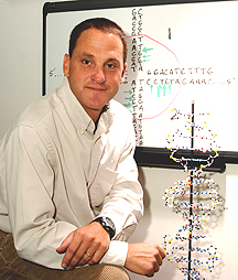
Robert
Brosh
| T H E N I H C A T A L Y S T | S E P T E M B E R – O C T O B E R 2006 |
|
|
|
| P E O P L E |
RECENTLY TENURED
|
|
Robert
Brosh
|
Robert Brosh received his Ph.D. in biology in 1996 from the University of North Carolina at Chapel Hill. He did postdoctoral work in the Laboratory of Molecular Gerontology (LMG), NIA, becoming a tenure-track investigator in that lab in 2000. He is currently a senior investigator in the LMG.
Our research has focused on the mechanisms of human helicases in pathways of nucleic acid metabolism that are important for the maintenance of genomic stability.
We have been particularly interested in the molecular and cellular functions of the WRN helicase, which is defective in the premature aging disorder Werner syndrome. It is our belief that understanding how the WRN helicase and related DNA helicases work to maintain chromosomal stability will lead to greater insights into the processes of cellular senescence, aging, and cancer.
A significant challenge to understanding the basis for the rapid onset of aging phenotypes in Werner syndrome has been defining the mysterious cellular role of the WRN helicase. To address this issue, we developed a model genetic system to explore WRN catalytic functions and protein interactions. This approach enabled us to provide the first in vivo evidence that a conserved noncatalytic domain of WRN helicase plays a critical role in facilitating the processing of DNA structural intermediates that arise during replication.
Our research findings suggest that WRN and related RecQ helicases coordinately act with structure-specific nucleases to maintain genomic stability during replicational stress.
Although considerable progress has been made in understanding the functions of certain DNA helicases, little was known about RECQ1, the first human RecQ helicase discovered. Our work demonstrated for the first time that in addition to its helicase activity, human RECQ1 efficiently catalyzes DNA-strand annealing.
We also identified and characterized the interaction of RECQ1 with human mismatch-repair factors that are important in the regulation of genetic recombination, elucidating the essential role of RECQ1 in the DNA damage response.
We are currently investigating the unique role of RECQ1 in maintaining chromosomal stability in model human and mouse systems. We have shown that RECQ1-deficient cells display profound chromosomal instability, strongly suggesting that mutations in RECQ1 are likely to give rise to cancer or a human disease associated with genomic instability such as a premature aging or DNA repair disorder.
In the future, we will focus on the interactive roles of human helicases in network pathways that are important for chromosomal stability.
We are also characterizing the biochemical functions of other human helicases that are either genetically linked to human disease or believed to have important, though as yet unclear, roles in genome homeostasis.
Most recently, we have focused on the BRCA1-associated C-terminal helicase (BACH1), an enzyme genetically implicated in breast cancer and Fanconi anemia and whose molecular and cellular functions are not well understood. Our research aimed to determine the potentially unique role of BACH1 in DNA repair and to track how BACH1 deficiency can lead to cancer.
We have discovered that BACH1 preferentially unwinds specific early DNA recombination intermediates of double-strand break repair, providing new insights into the DNA substrate specificity of BACH1.
We are currently studying BACH1 polymorphic variant proteins that are associated with breast cancer and characterizing protein partners of BACH1 that are likely to be relevant to the function of the helicase in DNA interstrand cross-link repair.
We plan to use cell- and biochemical-based screening strategies to identify small-molecule inhibitors of the Fanconi anemia/BRCA pathway, proceeding from the hypothesis that such drugs could sensitize cells to DNA-damaging chemotherapy.
 |
|
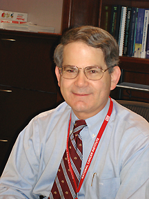
James
Doroshow
|
James Doroshow received his M.D. degree in 1973 from Harvard Medical School in Boston. He was chairman of the Department of Medical Oncology and Therapeutics Research and associate director for clinical research at the City of Hope Comprehensive Cancer Center in Los Angeles for 23 years before joining NIH in 2004. He is currently director of the Division of Cancer Treatment and Diagnosis and a senior investigator in the Laboratory of Molecular Pharmacology, NCI.
My laboratory will focus on the role of oxidant-mediated signal transduction in cell proliferation and cell death. I will pursue three lines of research within the theme of oxidant stress-mediated effects on cell growth: NADPH oxidase 1, the glutathione peroxidase gene family, and the mechanism of action of anticancer quinones.
First, we will be studying NADPH oxidase 1, a member of a recently discovered epithelial NADPH oxidase gene family that shares substantial homology with the NADPH oxidase of polymorphonuclear leukocytes. NADPH oxidase 1 catalyzes the NADPH-dependent reduction of oxygen to superoxide after binding of ligands for receptor tyrosine kinases (including EGF, PDGF, and VEGF).
Using stable shRNA constructs, we have found that NADPH-dependent reactive oxygen species play a critical role in growth factor–mediated signal transduction in tissues, such as the colon, that abundantly express NADPH oxidase isoforms.
Recent studies have demonstrated that a series of flavin dehydrogenase–binding iodonium analogs block constitutive oxidant production and cell growth in human colon cancer cells in the nanomolar concentration range; at the same time, they induce apoptosis, p27 accumulation, and blockade of the G1-S transition.
These effects are ameliorated by exogenous hydrogen peroxide and are also associated with alterations in signaling by the Met pathway.
These agents also have a unique pattern of growth inhibition in the NCI 60-cell line-screening assay. In vivo, the iodonium analogs have substantive therapeutic activity in two different human colon cancer xenograft models.
Future studies will involve the synthesis of novel members of the iodonium drug class as potential therapeutic agents. We will also evaluate the effects of those drugs on the expression of genes regulated by oxidant stress in human colon cancer cells.
In a second project, we are examining the role of the glutathione peroxidase gene family in the prevention of oxidant-mediated gastrointestinal carcinogenesis. Glutathione peroxidases are the major intracellular proteins that catalyze the detoxification of hydrogen peroxide and lipid hydroperoxides.
In collaboration with my former colleagues at the City of Hope, I identified a novel cytosolic glutathione peroxidase—named GPX-GI—encoded by the Gpx2 gene. GPx2 is expressed primarily in the distal small and large intestinal epithelium and is highly modulated by transretinoic acid, g-irradiation, and selenium.
Approximately 80 percent of the peroxidatic activity of the distal intestine in the mouse in provided by GPX-GI and 20 percent by the constitutive GPX-1 enzyme.
When the Gpx1 and Gpx2 genes are knocked out, mice develop severe inflammation of the small and large intestine that is indistinguishable from Crohn’s disease by light microscopy. In recent studies with this model, double- knockout mice at five months of age and older demonstrated a high (about 30-40 percent) incidence of adenocarcinomas in the distal intestine, at the site of the lowest glutathione peroxidase activity. Mice raised in a germ-free environment did not develop signs of inflammation after weaning and were cancer-free.
More than 90 percent of tumors in this model system demonstrate altered intracellular localization of b-catenin, suggesting that mutations in the Apc gene pathway may play a role in the intestinal carcinogenesis observed for Gpx1/Gpx2 double-knockout mice.
This model system is one of the first to demonstrate by genetic means that chronic oxidant stress, produced by inhibiting the expression of antioxidant genes, is associated with the development of intestinal neoplasms indistinguishable from human adenocarcinomas.
Future studies with this model will focus on the molecular evolution of oxidant-induced tumors and the development of novel molecules that could interfere with the inflammatory and carcinogenic processes.
Finally, we are evaluating the role of reactive oxygen radical formation in the mechanism of cell death produced by anticancer quinones, including the anthracycline antibiotics (such as doxorubicin) and other small molecules with clinically proven antineoplastic activity. An evaluation of the spectrum and mechanism of DNA base damage produced by anticancer quinone-induced free radical formation is ongoing for both in vivo and in vitro systems.
We have also explored the role of reactive oxygen production by the anthra-cycline antibiotics, arsenic trioxide, and bisphosphonate anticancer agents in modifying EGF-mediated signal transduction.
As a result of pronounced (more than 80 percent) protein tyrosine phosphatase inhibition by arsenic trioxide–, anthracycline-, or bisphosphonate-induced oxidant stress, the phosphorylation of EGFR after exposure to EGF is dramatically prolonged, leading to a continuous, and potentially lethal, upregulation of EGF-mediated signal transduction.
These observations suggest an important role for drug-enhanced oxidant stress in modulating cell proliferation signals by a variety of small molecules, and a novel strategy for therapeutic combinations based on the drug-related modulation of oxidant-mediated signal transduction.
 |
|
Peter
Kwong
|
Peter Kwong received his Ph.D. from Columbia University in New York in 1995 and continued his postdoctoral training there under the mentorship of Wayne Hendrickson. In 2000, he joined the Vaccine Research Center, NIAID, NIH, where he is currently chief of the Structural Biology Section.
My group is attempting to apply structural biology to the development of an effective HIV-1 vaccine. Despite the enormous potential of atomic-level design—successfully used, for example, in the development of the HIV-1 protease inhibitors—current vaccine development makes little use of atomic-level information. We are trying to change this.
One area in which we and others have already made an impact is in understanding how HIV-1 is able to evade the humoral immune system.
Determination of the structure of the HIV-1 gp120 envelope glycoprotein, the primary target of neutralizing antibodies against HIV-1, showed how N-linked carbohydrate can form both an immunologically silent face—with carbohydrate masquerading as "self"—and also can protect neighboring epitopes through an "evolving glycan shield."
We also showed how conformational flexibility of gp120 can combine with quaternary restrictions within the viral spike to prevent antibody neutralization.
These studies served to define the underlying mechanisms that protect HIV-1 from the humoral immune response.
But can one use structural biology in actual vaccine design? Currently, my group is following two lines of investigation.
One line involves the precise delineation of functional constraints to identify potential footholds of conservation and exposure.
In a collaborative study, we investigated antibodies that bound to the co-receptor–binding site on gp120 and found them capable of neutralizing not only HIV-1, but also the even more evolutionary divergent HIV-2. We found such CD4i antibodies to develop to high titers in most HIV-1–infected individuals.
Unfortunately, our analysis also found that the virus hides the site of co-receptor binding, so that before engagement of the primary HIV-1 receptor, CD4, the co-receptor site is not formed.
These studies demonstrate the strength of functional constraints in restricting epitope variation. But they also identify an important weakness: Functional conservation does not necessarily engender epitope exposure, which is required for antibody neutralization.
We are currently exploring how function might constrain the site of CD4 binding, which—unlike the co-receptor–binding site—must be available as an initial site of attachment.
A second line of investigation involves structural analysis of the broadly neutralizing antibodies thus far identified that have the ability to neutralize diverse isolates of primary HIV-1.
Only four antibodies of such ability have thus far been identified—2F5, 2G12, 4E10, and b12.
We have determined the structures of both 2F5 and b12, each with their HIV-1 envelope epitopes.
In a collaborative study, primarily with David Baker’s group at the University of Washington, Seattle, Joe Sodroski’s group at the Dana-Farber Cancer Institute in Boston, and also with Gary Nabel’s and Rich Wyatt’s groups at the Vaccine Research Center, we are currently generating epitope mimics unencumbered by known mechanisms of humoral evasion.
Tests of these epitope mimics in small animals should reveal their potential to elicit antibodies with abilities similar to the template broadly neutralizing ones.
Whether the confluence of structural information that we are generating is sufficient to elicit broadly neutralizing antibodies will depend in part on our ability to iteratively optimize immunogenicity and also on structural parameters of conformational mimicry, epitope accessibility, elicited potency, neutralization breadth, and target specificity.
Our investigations have already led to insight into the parameters governing antibody elicitation and neutralization. True success, however, will depend on whether or not we succeed in creating immunogens capable of substantially reducing the incidence of HIV-1 infection in humans.
 |
|
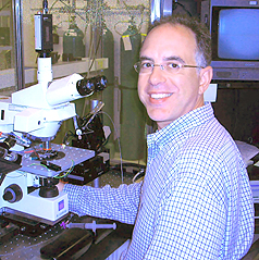
Carl
Lupica
|
Carl Lupica received his Ph.D. in psychobiology and neuroscience from Wayne State University, Detroit, Mich., in 1989. He did postdoctoral work at the University of Colorado Health Sciences Center in Denver and held faculty positions in the pharmacology department there and at the University of Arizona Medical School at Tucson before joining NIH in 2002 as an investigator in the Cellular Neurobiology Research Branch, NIDA. He is currently a senior investigator and chief of the Electrophysiology Research Unit, NIDA.
During my time at the University of Colorado, NIH funding launched my career as an independent researcher studying the physiological mechanisms through which opiate drugs alter synaptic transmission in brain areas involved in pain perception and cognition.
This work resulted in several novel discoveries, including the identification of a new signal-transduction mechanism coupled to opiate receptors and the observation that m and d opiate receptors are segregated to distinct classes of inhibitory neurons in the hippocampus.
After that, while at the University of Arizona Medical School, I began NIH-funded studies to determine the physiological mechanisms through which the newly discovered endogenous cannabinoid molecules altered brain function.
These studies, now continued at the NIDA intramural research program, have identified the substrates upon which cannabinoid CB1 receptors and endogenous cannabinoids act to regulate the function of brain reward circuits and memory processes in the hippocampus.
Among the most significant findings attributed to my work are (1) the identification of voltage-dependent calcium channels as the mechanism through which CB1 receptors transduce their signal in inhibitory neurons in the hippocampus and (2) the discovery that dopamine neurons located in the central reward circuit of the ventral tegmental area (VTA) synthesize and release endogenous cannabinoid molecules.
These latter studies also determined that under conditions of increased excitability and during sustained bursting activity, the endogenous cannabinoid molecules released from VTA dopamine neurons regulate the strength of synaptic input to this nucleus.
This action provides a likely explanation for the ability of cannabinoid CB1 receptor antagonists to diminish craving for several classes of abused drugs, such as opiates and nicotine, because all these drugs share the ability to increase VTA dopamine neuron activity and bursting.
Additional ongoing studies in my laboratory combine modern physiological techniques such as patch clamp electrophysiology and confocal microscopy to identify the molecular changes that occur in the brain as a result of the prolonged use of marijuana and exposure to its psychoactive ingredient, Æ9-tetrahydrocannabinol.
One of our goals is to identify the mechanism through which prolonged marijuana use in humans can cause persistent cognitive deficits, and to gain a further understanding of the role of endogenous cannabinoids in regulating the activity of these cognitive neural circuits.
 |
|
Alan
Michelson
|
Alan Michelson received his M.D. and Ph.D. degrees from Harvard Medical School in Boston in 1986. His graduate thesis work was undertaken under the supervision of Stuart Orkin at Children’s Hospital Boston. He subsequently completed an internship in internal medicine at Brigham and Women’s Hospital in Boston, followed by a postdoctoral fellowship with Tom Maniatis at Harvard University. In 1992, he joined the faculty at Harvard Medical School and Brigham and Women’s Hospital and was appointed an investigator of the Howard Hughes Medical Institute. Earlier this year, he moved to NHLBI as associate director for basic research; he is also a senior investigator in the Genetics and Developmental Biology Center, NHLBI.
My laboratory has had a longstanding interest in the mechanisms that govern the specification, differentiation, and assembly of diverse cell types into complex tissues and organs during embryogenesis. We have been addressing these issues using development of the heart and body wall muscles of the Drosophila embryo as a model system.
Our initial studies demonstrated that homeotic genes act autonomously in the early mesoderm to establish the unique identities of muscle founder cells, thereby contributing to the diversification of the mature muscle pattern.
Using unbiased genetic screens, we then uncovered separate roles for signaling by two receptor tyrosine kinases (RTKs)—epidermal and fibroblast growth factor receptors (EGFR and FGFR, respectively)—in mesoderm development: FGFR activity is required for the migration of mesodermal cells after gastrulation, whereas both EGFR and FGFR signaling contribute directly to the commitment of muscle and cardiac cell fates.
We further showed that the RTKs act together with three other signaling pathways—Wingless (Wg, a WNT family member), Decapentaplegic (Dpp, a bone morphogenetic protein superfamily member), and Notch—to promote the progressive determination of uncommitted mesodermal cells. Wg, Dpp, EGFR, and FGFR exert positive, cooperative effects on cell fate specification, whereas Notch functions in an inhibitory manner.
These findings prompted us to investigate how individual cells interpret multiple convergent signals in a tissue-specific manner. Results revealed that a considerable degree of signal integration occurs in the nucleus. Using a particular muscle and cardiac identity gene as a model, we found that a single transcriptional enhancer is directly regulated by the transcription factors acting downstream of all three positive signals that specify the fate of the cell in which this enhancer functions.
Moreover, tissue specificity of the generic signaling pathways is conferred not only by their combinatorial effects, but also by two mesoderm-restricted transcription factors that cooperate with the three signal-activated regulators bound to the same enhancer.
A key principle emerging from these studies is that subtypes of related cells are specified by unique combinations of a limited number of signals and tissue-restricted selectors. A major focus of our current work is dedicated to testing and refining this hypothesis using a more comprehensive systems-level approach. To this end, we have undertaken genome-wide expression-profiling studies of purified subpopulations of both wild-type and informative mutant primary embryonic cells.
A meta-analysis of the combined microarray datasets derived from these experiments led to the assignment of several hundred genes to either of two myoblast subclasses, predictions that we independently validated by whole-embryo in situ hybridization. We are now determining whether these large sets of co-expressed genes are also co-regulated by similar mechanisms.
In an ongoing collaboration with Martha Bulyk and her research group at Brigham and Women’s Hospital, we are applying computational algorithms that predict the locations of cis-regulatory modules on a genome-wide scale and that assess the fit of a particular transcriptional regulatory model to a set of co-expressed genes. These strategies have enabled us to identify a subset of transcription factors that comprise a core code for the regulation of a subset of myoblast genes, a model that we validated using transgenic reporter assays to test the functions of candidate enhancers.
The next question that we are addressing is how cell-type specificity is superimposed on the core transcriptional code. Central to these investigations is our prior discovery of previously unchar-acterized transcription factors that are candidate cell-specific selectors.
We are using protein-binding microarray technology to characterize the DNA binding specificities of these factors, with the resulting motifs incorporated into our computational framework to predict novel transcription factor combinations that are responsible for differential gene expression in muscle and cardiac cells.
Classical genetics and RNA interference directed against these factors are also being used to selectively perturb the associated transcriptional networks, thereby assessing the functional significance of newly identified regulators and their predicted combinatorial interactions.
Additional work is in progress in my laboratory to examine how co-expressed target genes function in the later differentiation and morphogenesis of the heart and body wall muscles.
 |
|
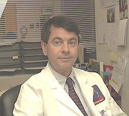
Forbes
Porter
|
Forbes D. Porter received his M.D. and Ph.D. from Washington University in St. Louis in 1989 and then trained in pediatrics and clinical genetics at St. Louis Children’s Hospital. After a postdoctoral fellowship in the lab of Heiner Westphal (LMGD, NICHD), he became a tenure-track investigator in the Heritable Disorders Branch, NICHD, in 1996. He is currently a senior investigator in that branch.
Any given birth defect or genetic syndrome is relatively rare, but viewed in the aggregate, genetic defects are a signficant component of child health care. My interest has been in understanding the biological processes that underlie birth defects and using these rare syndromes to understand more-common genetic problems.
My laboratory has been studying a group of human malformation and mental retardation syndromes caused by inborn errors of cholesterol synthesis. The most common of these disorders is the Smith-Lemli-Opitz syndrome, or SLOS, an extremely variable disorder with a broad clinical spectrum. Severely affected infants often die before or soon after birth, whereas the findings in children with milder cases can be limited to minor physical abnormalities associated with learning problems or autistic-like behavior.
Children with SLOS have low cholesterol levels and elevated levels of a cholesterol precursor molecule known as 7-dehydrocholesterol. Because sufficient cholesterol does not cross the placenta, normal development is impaired. My group studies SLOS both in clinical protocols and in the laboratory.
One of our first accomplishments was to clone the gene responsible for SLOS. To date more than 100 different mutations of this gene have been identified in SLOS patients. After cloning the human gene, we identified the mouse gene and produced mouse models of SLOS to study the biological changes associated with the syndrome and to test various therapeutic interventions.
In collaboration with Phil Beach’s laboratory at Johns Hopkins in Baltimore, we have shown that the hedgehog signaling pathway is impaired in SLOS cells. Proper hedgehog signaling is important for multiple developmental processes, and disruption of this signaling pathway can cause birth defects.
We have also developed mouse models of desmosterolosis and lathosterolosis—two rare SLOS-like human malformation syndromes—which have helped us separate the effects of low cholesterol from those due to precursor accumulation.
In addition to our basic science work, I currently follow more than 50 patients with SLOS in whom we are studying the beneficial effects of increasing dietary cholesterol intake. Cholesterol therapy enhances the overall health of SLOS patients, many of whom in the past died in the first few years of life. Growth and nutritional status are improved, and the children also appear to be less irritable. However, dietary cholesterol therapy does not improve learning because it does not get into the brain.
We are currently studying the safety and efficacy of simvastatin to alter brain cholesterol levels in these children. In blocking cholesterol synthesis, simvastatin also decreases the synthesis of abnormal sterol 7-dehydrocholesterol—and may also paradoxically increase cholesterol levels. We would like to determine if alleviating the biochemical disturbance in the brain exerts a positive effect on either learning or behavior.
 |
|
Peter
Schmidt
|
Peter Schmidt received his M.D. degree in 1982 from the Royal College of Surgeons and the National University of Ireland in Dublin. He completed a residency in psychiatry at the University of Toronto and joined the Biological Psychiatry Branch at NIMH in 1987. He is currently a senior investigator in the Section of Behavioral Endocrinology, NIMH.
The goals of my program are threefold:
![]() To identify the sources of vulnerability for the development of mood disorders
during periods of altered reproductive function
To identify the sources of vulnerability for the development of mood disorders
during periods of altered reproductive function
![]() To evaluate the potential role of hormonal therapies in reproductive endocrine-related
mood disorders
To evaluate the potential role of hormonal therapies in reproductive endocrine-related
mood disorders
![]() To determine the neural mechanisms underlying the mood-regulating effects of
gonadal steroids
To determine the neural mechanisms underlying the mood-regulating effects of
gonadal steroids
My early work, much of it in collaboration with David Rubinow at NIMH and Lynnette Nieman at NICHD, focused on women with severe premenstrual dysphoria (PMD).
We used the progesterone receptor antagonist RU486 and GnRH agonists to demonstrate that the symptoms of PMD could occur in the absence of normal luteal-phase progesterone levels.
Nonetheless, symptoms depended on exposure to physiologic levels of ovarian steroids, with behavioral sensitivity manifested in those women with PMD but not those without PMD.
These observations of differential behavioral sensitivity informed my subsequent studies of the relationship between changes in reproductive aging and affective adaptation.
My group has used several strategies to test the hypothesis that declining reproductive function plays no role in the onset of mood disorders that occur in women at midlife.
We initiated what is still an ongoing history study in which we prospectively evaluate asymptomatic premenopausal women to determine whether an onset of depression is coincident with a specific phase of the menopause transition.
We have also examined the effects on mood of reversing selected aspects of age-related hypogonadism by administering reproductive hormones, the secretion of which decline by or during midlife. Specifically, we demonstrated the short-term antidepressant effects of estradiol therapy in women who developed depression during the menopause transition.
In an ongoing study, we are examining the effects of the selective estrogen-receptor modulator raloxifene and the phytoestrogen rimostil in a separate group of depressed perimenopausal women.
Additionally, we have documented the antidepressant efficacy of the adrenal androgen DHEA in men and women with midlife-onset depression.
In future studies of the potential relationship between reproductive senescence and mood disorders, we will study selective estrogen-receptor antagonists as they become available for human use. We will examine the specific roles of estrogen receptors alpha and beta in the psychotropic actions of estradiol and in the effects of estrogen withdrawal on the onset of perimenopause-related depression.
Finally, my group is conducting studies of experimentally induced hypogonadism. In one study, we examined the effects on mood of estradiol withdrawal in asymptomatic women with a past history of perimenopausal depression.
In another study, we administered a GnRH agonist—a depot brand of leuprolide acetate—in normal volunteers to investigate the symptomatic and physiologic consequences of hypogonadism.
In this latter study, performed in collaboration with Karen Berman at NIMH, we demonstrated that hypogonadism eliminates—and physiologic ovarian steroid replacement restores—the cognition-activated regional cerebral blood flow in the dorsolateral prefrontal cortex, inferior parietal lobule, and posterior inferior temporal cortices of the human brain—regions implicated in the regulation of cognitive function and emotional state.
Considerable effort is now underway to reconcile these and related findings on the neuroregulatory effects of ovarian steroids with findings from the Women’s Health Initiative of an increased risk of dementia in women taking combined estrogen-progestin therapy
Reproductive endocrine-related mood disorders have a well-defined biological stimulus that we can demonstrate is critical in the precipitation of affective disturbances.
Thus, these conditions afford a unique opportunity to identify neural substrates involved in the regulation of affective state.
The availability for study of known triggers (such as gonadal steroids) and associated signaling pathways dramatically increases the likelihood of identifying mechanisms of both affective-state regulation and illness susceptibility.
The impact of these studies, therefore, extends beyond reproductive endocrine-related mood disorders to mood disorders in general, as well as to our understanding of the relevance of brain development and plasticity in affective disturbance.
 |
|
Jeffery
Taubenberger
|
Jeffery Taubenberger received an M.D. in 1986 and a Ph.D. in 1987 from the Medical College of Virginia in Richmond. He completed a residency in anatomic pathology at NCI and served as chairman of the Department of Molecular Pathology at the Armed Forces Institute of Pathology before joining NIAID in 2006. He is currently a senior investigator in the Respiratory Viruses Section, Laboratory of Infectious Diseases, NIAID.
My group is interested in studying the pathogenesis and evolution of influenza A viruses, with special emphasis on pandemic influenza. Influenza A viruses are found in a wide variety of hosts, including wild birds, domestic poultry, horse, pigs, humans, and other mammals.
Influenza causes yearly outbreaks and, occasionally, novel pandemic influenza strains emerge in humans ultimately derived from bird influenza viruses. The most devastating pandemic on record was the 1918 "Spanish" influenza, which may have killed as many as 50 million people globally in 1918–1919.
There is concern that a new influenza pandemic might emerge if the current H5N1 avian influenza virus ultimately adapts fully to humans.
Before coming to NIAID, my laboratory group was able to determine the complete genetic sequence of the 1918 virus using tiny fragments of viral RNA recovered from the lungs of victims of the 1918 pandemic. With colleagues in several institutions, we reconstructed the 1918 virus using reverse genetics, and the first experiments with the virus were conducted in high-containment labs at the CDC.
As do some of the virulent H5N1 viruses in animal models, the 1918 virus induces a strong acute inflammatory response, suggesting that the immune response itself may be a component of the pathogenesis of these viral infections.
At NIH, my laboratory is based in the new C.W. Bill Young Center for Biodefense and Emerging Infectious Diseases (Building 33). We will focus on different experimental models to map the genetic basis of the virulence of the 1918 influenza virus and compare it with recent highly pathogenic avian influenza viruses that have caused fatal infections in humans. We also hope to identify genetic changes that are associated with host switching in influenza viruses.
To do this, we will also need to expand the number of influenza genome sequences available, both from human and animal strains, and my laboratory will continue to collaborate with the NIAID Influenza Genome Sequencing Project.
Analyses of hundreds of human influenza virus genomes are demonstrating that the evolution of human influenza strains is more complex than previously thought. Influenza viruses in humans evolve rapidly, and frequent antigenic changes in the surface proteins require routine reformulation of the vaccine.
With our collaborators around the country we are also engaged in prospective avian influenza surveillance in wild birds. We are working to develop rapid molecular genetic diagnostic assays to improve influenza surveillance in humans and in animal populations.
We are also developing models to examine the significance of other mutations identified by genomic analyses of contemporary human influenza viruses to understand selection in the context of viral fitness.
We are continuing to derive sequence information from archival influenza cases, some as early as 1907. By placing the emergence of the 1918 pandemic virus in the context in which it emerged, we may be able to more fully understand how pandemic strains emerge in a form that allows them to spread efficiently to humans.
We have identified several influenza-positive pre-1918 cases and will be screening influenza cases from the 1920s as well to help identify unique genetic signatures of virulence of the pandemic virus. The goal of all of these projects is to better understand how pandemic strains emerge in humans, as well as the genetic basis of how influenza viruses cause disease in humans. These findings may ultimately lead to new avenues for treatment and may help mitigate the impact of a future pandemic.
 |
|
Yingzi
Yang
|
Yingzi Yang received a Ph.D. in molecular biology from the Weill Medical College of Cornell University in New York in 1996. After completing a postdoctoral fellowship in the laboratory of Andy McMahon at Harvard University in Cambridge, she joined the Genetic Disease Research Branch, NHGRI, as a tenure-track investigator in 2000. She is currently head of the Developmental Genetics Section and a senior investigator in the Genetic Disease Research Branch, NHGRI.
My laboratory studies vertebrate limb development and skeletal morphogenesis. Our goals are to understand the mechanisms by which molecular signals are transduced and integrated in a regulatory network to control cell proliferation, differentiation, and organization and to gain insight into key regulatory events during limb and skeletal morphogenesis.
To that end, we focus on the signaling mechanisms of Wnts and Hhs (hedgehogs) in the limb and skeletal system and how abnormalities in these two signaling pathways lead to birth defects, skeletal diseases, and tumor formation. Our multidisciplinary approach combines the strength of mouse molecular genetics with embryology, functional genomics, and the cell and organ culture systems in the developing limb.
My previous work elucidated the regulation of early limb-patterning events that provide temporal and spatial information for the initial formation of the skeletal elements. My current research addresses how signaling molecules regulate skeletal development in the limb, a later morphogenetic process.
I seek to understand several fundamental events in this process: the regulation of mesenchymal condensation, the differentiation of chondrocytes vs. osteoblasts from mesenchymal progenitor cells, the coordination of chondrocyte proliferation with maturation, and the induction and maintenance of synovial joint.
I have made several major discoveries in this current research effort:
![]() Different Wnts play distinct roles in regulating chondrocyte differentiation.
Different Wnts play distinct roles in regulating chondrocyte differentiation.
![]() The canonical Wnt pathway is both necessary and sufficient for synovial joint
induction and determines whether mesenchymal progenitors differentiate into
chondrocytes or osteoblasts.
The canonical Wnt pathway is both necessary and sufficient for synovial joint
induction and determines whether mesenchymal progenitors differentiate into
chondrocytes or osteoblasts.
![]() The noncanonical Wnt5a signaling promotes chondrocyte differentiation by inhibiting
the canonical Wnt signaling activity.
The noncanonical Wnt5a signaling promotes chondrocyte differentiation by inhibiting
the canonical Wnt signaling activity.
![]() The canonical Wnt pathway interacts with the Indian hedgehog (Ihh) signaling
pathway in distinct ways during different processes of skeletal morphogenesis,
and Wnt5a acts in parallel pathways with Ihh to coordinate chondrocyte maturation.
The canonical Wnt pathway interacts with the Indian hedgehog (Ihh) signaling
pathway in distinct ways during different processes of skeletal morphogenesis,
and Wnt5a acts in parallel pathways with Ihh to coordinate chondrocyte maturation.
My lab is continuing to study how Wnt and Hh signaling pathways interact with other pathways in skeletal development, and we are now exploring the role of Wnt and Ihh signaling in bone diseases, such as osteoarthritis and bone tumors. My lab is also actively investigating the molecular mechanism underlying the control of cell and tissue organization by the planar cell polarity pathway in both embryonic development and adult tissue homeostasis.
My hope is that our research
in basic skeletal developmental biology will lead to clinical studies that advance
disease prevention, diagnosis, and treatment. ![]()