| T H E N I H C A T A L Y S T | J U L Y – A U G U S T 2005 |
|
|
|
| P E O P L E |
RECENTLY TENURED
 |
|
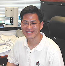
Kyung
Lee
|
Kyung Lee received his Ph.D. in 1994 from the Department of Biochemistry at the Johns Hopkins University in Baltimore. He then worked with Raymond Erikson at Harvard University in Cambridge, Mass., as a postdoctoral fellow and studied in the fields of cellular proliferation and mitotic controls. In 1998, he joined NIH as a tenure-track investigator in the Laboratory of Metabolism at NCI and is now a senior investigator and head of the Chemistry Section.
During the eukaryotic cell cycle, reversible protein phosphorylation by protein kinases is a fundamental regulatory mechanism. The G/M phases of the cell cycle comprise a series of regulatory biochemical steps and coordinated cellular events that ensure the faithful partitioning of genetic and cytoplasmic components.
It is now clear that, in addition to the much-studied cyclin-dependent protein kinases, the polo subfamily of serine/threonine protein kinases (collectively, polo-like kinases, or Plks) plays a pivotal role in regulating various mitotic events. Plks cooperate with a master cell-cycle kinase, Cdc2, to bring about essential mitotic events at mitotic entry; Plks also regulate pathways leading to the inactivation of Cdc2 at mitotic exit.
Loss or depletion of Plks activity in various organisms results in mitotic arrest followed by cell death, suggesting that the function of Plks is essential for mitotic progression in all eukaryotic organisms.
Using both budding yeast and cultured mammalian cells, my laboratory has investigated the mitotic functions of Plks. We have shown that the polo-box motif, which resides in the C-terminal noncata-lytic domain, is essential for the localization of both budding yeast polo kinase Cdc5 and mammalian Plk1 to specific subcellular structures.
In line with this notion, mutations of the polo-box of either Cdc5 or Plk1 in its native organism abolished the function of these enzymes and induced mitotic arrest, indicating that polo-box–dependent subcellular localization is critical for the mitotic functions of these enzymes.
One of the critical events that budding yeast polo kinase Cdc5 mediates is mitotic entry, a process that requires activation of Cdc28 (homolog of mammalian Cdc2). We set out to clarify the molecular mechanisms of Cdc5 regulation of this event.
Studies have shown that Swe1 (ortholog of mammalian Wee1) inhibits mitotic entry by negatively regulating Cdc28. We found that Cdc5 directly phosphorylates and downregulates Swe1, thus permitting the activation of Cdc28 and thereby mitotic entry. The conserved polo-box domain of Cdc5 appeared to play a crucial role for this biochemical step by targeting the catalytic activity of Cdc5 to its substrate Swe1.
Similar regulation occurs in human cells, where down-regulation of Wee1 by Plk1 is critical for entry into mitosis. These findings demonstrate the importance of the polo-box domain for specifically targeting the Plks to their substrates through protein-protein interactions.
Dynamic subcellular localization and the multitude of Plks functions predict that the polo-box interacts with multiple cellular proteins at specific stages of the cell cycle. To gain a deeper understanding of the function of the polo-box, we have been focusing on the identification of novel polo-box–binding proteins in cultured mammalian cells.
From a yeast two-hybrid screen using the polo-box domain of Plk1 as bait, we isolated a novel kinetochore protein that we termed PBIP1 (polo-box interacting protein 1). Consistent with the two-hybrid analyses, PBIP1 interacted with endogenous Plk1 in vivo under physiological conditions.
Preliminary characterization of PBIP1 suggested that it is a kinetochore-specific mitotic inhibitor that is phosphorylated and degraded by Plk1. Expression of the nondegradable PBIP1 lacking the Plk1-dependent phosphorylation sites induced a mitotic arrest, indicating that degradation of PBIP1 is required for proper mitotic progression.
Intriguingly, PBIP1 interacted with cellular proteins whose deregulation is implicated for tumorigenesis, suggesting that the Plk1-dependent downregulation of PBIP1 may contribute to the development of cancers in humans. In line with these observations, Plk1 was both upregulated and multiply mutated in about 80 percent of human cancers.
Our recent results raise the possibility that PBIP1 is a novel tumor suppressor. Precocious degradation of PBIP1 by deregulated Plk1 activity may lead to chromosome missegregation and genomic instability, thus promoting tumorigenesis. We expect that understanding Plk1-dependent PBIP1 regulation may provide new insights into how Plk1 deregulation promotes cancers in humans and how to approach the development of anti-Plk1 therapeutic agents.
 |
|
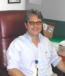
Francesco
Marincola
|
Francesco Marincola received his MD from the University of Milan (Italy) in 1978 and completed a general surgery residency at Stanford University (Stanford, Calif.) in 1990. He joined the Surgery Branch, NCI, in 1990 as an immunotherapy fellow and subsequently became a surgical oncology fellow. In 1993, he was appointed to the staff of the Surgery Branch; in 2001, he assumed two additional positions—director of the Immunogenetics Program in the Clinical Center Department of Transfusion Medicine and director of the HLA Laboratory.
Since my immunotherapy fellowship in 1990 with Steve Rosenberg in the NCI Surgery Branch, I have been intrigued by the rare yet overwhelming observation of dramatic tumor regressions mediated by immune manipulation with the systemic administration of interleukin-2 (IL-2)—with or without tumor-infiltrating lymphocytes. When the fellowship ended, I agreed to continue in the staff of the Surgery Branch to work on the identification of the algorithm regulating tumor rejection in humans exposed to this kind of therapy.
As the tumor antigens (TA) recognized by lymphocytes became molecularly characterized in the ’90s, TA-specific vaccines were developed that yielded the unprecedented opportunity to study tumor-host interactions specific to a unique TA and in some cases to a specific TA-human leukocyte antigen (HLA)–specific combination. This ability to minimize experimental variables in humans simplified the study of the effect of immunization in clinical trials.
We discovered that TA-specific immunization can reproducibly induce cytotoxic T cells (CTL) in patients that are capable of recognizing tumor cells. In addition, we observed that the CTL induced by vaccines localized at tumor sites, where they recognized tumor cells and produced cytokines such as IFN-g.
Such interactions, however, were not sufficient to induce tumor regression. In addition, we observed that the level of expression of TA or HLA molecules responsible for the presentation of the TA to CTL did not predict response, suggesting that other factors modulate immune responsiveness at the tumor site.
The development of high-resolution molecular testing of HLA molecules, using high-throughput sequencing technology, was critical in these early vaccine studies and was made possible by Harvey Klein in the CC Department of Transfusion Medicine (DTM), who offered me a joint appointment as director of the HLA Laboratory in addition to my original appointment in the NCI Surgery Branch.
We employed a novel strategy to analyze T cell localization at tumor sites during immunization and the interactions of T cells with their target (the HLA-TA combination): With multiple fine-needle-aspiration biopsies, we could monitor tumor-host interactions in the target tissue ex vivo while leaving the tumor in situ, allowing us to follow the natural history of the disease and serially evaluate response to therapy.
In 2000, Ena Wang in my laboratory developed and validated a technique for RNA amplification that allowed the use of very small amounts of starting RNA (obtained from fine-needle aspirates) in the study of tumor-host interactions in conjunction with high-throughput technologies such as microarrays. This research expanded our ability to monitor immune responses during immunization and other immune therapies.
These original studies suggested that the tumors most likely to respond to immunotherapy are characterized by an immunologically active (chronically inflamed) transcriptional profile. When successful, immune therapy—whether TA-specific immunization or the systemic administration of immune stimulants such as IL-2—acts by turning a chronic inflammatory process into an acute one quite similar to that observed by others in the context of acute transplant rejection.
Subsequently, Monica Panelli, also in my group, compared fine-needle aspirates obtained before and during IL-2 therapy and showed that this cytokine acts by inducing a general inflammatory process at the tumor site. This inflammatory process activates innate effector mechanisms in mononuclear phagocytes and natural killer cells and induces activation of antigen-presenting cells and production of chemoattractants capable of recruiting and activating T cells at the tumor site.
In 2001, Klein invited my group to develop the Immunogenetics Program in the DTM. We are now developing high-throughput, hypothesis-searching tools for the direct ex vivo analysis of tumor-host interactions during vaccine therapy. Our aim is to identify factors predictive of the immune responsiveness of patients and/or their tumors.
These tools include the combined analysis of DNA, RNA, and protein to identify genetic and epigenetic differences between patients who do and do not respond to therapy.
Using affordable, custom-made oligonucleotide-based chips, we are developing high-throughput screening tools to analyze cytokine polymorphisms and test whether genotype can be linked to phenotypic responses and to clinical parameters. We are also continuing to improve our proteomics and functional genomics tools for the global assessment of patients’ response to treatment during clinical trials.
 |
|
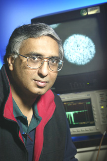
Mahendra
Rao
|
Mahendra Rao completed his medical training at Bombay University in India in 1983 and received his Ph.D. from the California Institute of Technology in Pasadena in 1991. After postdoctoral training with Story Landis, then at Case Western Reserve University in Cleveland, and David Anderson at the California Institute of Technology, he joined the Department of Neurobiology and Anatomy at the University of Utah School of Medicine in Salt Lake City in 1994. In 2001, he joined NIA as chief of the Stem Cell Biology Unit in the Laboratory of Neurosciences. He is currently also an adjunct associate professor at the National Center for the Biological Sciences in Bangalore, India, and holds a joint appointment as an associate professor at Johns Hopkins University, Baltimore.
At both the University of Utah and NIA, my laboratory has focused on understanding how embryonic and adult stem cells self-renew and differentiate. Our early work with fetal and adult neural stem cells revealed that they change over time with respect to their self-renewal capabilities, expression of markers, positional information, and differentiation ability.
We then showed that stem cells do not differentiate into fully mature cells directly but do so by first generating intermediate precursors, which we termed lineage-restricted precursors. We subsequently isolated specific lineage-restricted precursors and showed that they play an integral part in the normal process of development. Indeed, their action at specific stages can explain the effects of many genes.
Our ability to identify specific populations of cells allowed us to use the large-scale analytical procedures—such as massively parallel signature sequencing, microarray, and EST scan—developed at NIH and elsewhere to identify novel genes and new signaling pathways that regulate self-renewal. Using such methods, we were able to cross-correlate information to obtain a global view of cell function and start to identify context-dependent and cell type–specific signaling pathways. For example, we identified Lin41 as a microrna-regulated gene that is important in regulating embryonic stem-cell self-renewal.
In the past couple of years, we have extended this strategy to examine human embryonic stem-cell populations—with equally exciting results. Most of our findings have followed as a logical consequence of the key observation that all stem-cell populations generate differentiated progeny by first generating more restricted precursors.
For example, based on our initial finding, we were able to hypothesize and then confirm that repair in the adult nervous system likely involves a larger role for restricted progenitors than for true multipotent stem cells.
This result has important implications for both embryonic stem-cell and neural stem-cell biologists. It suggests both that stem-cell populations need to be differentiated prior to implantation and that for specific diseases, subsets of cell types may be more important—glial for glial disorders and neuronal cells for diseases that include neuronal loss.
We then proceeded with transplant experiments using rodent models of Parkinson’s disease and spinal cord injury to show that these restricted precursors have a therapeutic benefit. This opened up an entirely new field of endeavor, new collaborations, and interactions with new colleagues.
I have benefited enormously from the efforts and intellectual input of people in my own laboratory and from Mark Mattson and other colleagues in the Laboratory of Neurosciences—and from NIA and NIH resources and infrastructure that allow relatively small laboratories to undertake large-scale analyses.
In the coming years, I
hope to build on the wealth of information we have generated to begin to understand
how stem cells maintain their self-renewal capability and avoid senescence.
Further, comparisons across species will allow us to determine what common and/or
distinct mechanisms of self-renewal have evolved over time. ![]()