| T H E N I H C A T A L Y S T | J A N U A R Y – F E B R U A R Y 2005 |
|
|
|
| P E O P L E |
RECENTLY TENURED
 |
|
Nilanjan
Chatterjee
|
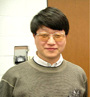
Nilanjan Chatterjee was trained in mathematical statistics at the Indian Statistical Institute, Calcutta, and received a Ph.D. in statistics from the University of Washington, Seattle, in 1999. In his thesis, he developed efficient methods for analyzing data from two-phase studies in which inexpensive exposure information is collected on a relatively large number of phase I subjects and more expensive exposure data are collected on a smaller subset of efficiently selected subjects. Chatterjee joined NIH as a research fellow in the Division of Cancer Epidemiology and Genetics (DCEG), NCI. Since 2001, he has been a principal investigator in the Biostatistics Branch.
Advances in molecular and cellular technologies have given epidemiologic researchers new opportunities to study the pathogenesis of complex diseases through population-based studies. The advent of these data in epidemiologic studies also has given rise to challenging theoretical and methodological problems in statistics. In recent years, I have initiated methodologic projects in three major areas of molecular epidemiologic studies, including gene-environment interactions, genetic association, and etiologic heterogeneity of diseases using molecular and pathologic markers.
It is now thought that the risks of many complex diseases are determined by the joint effect of genetic susceptibility and environmental exposures, and study of the interplay of these two factors can greatly enhance our understanding of the etiology of these diseases. Because studies of interactions, especially for rare exposures, typically require a large sample size, efficient designs and analytic principles for gene-environment interaction studies are an important area of epidemiological and statistical research.
In traditional logistic regression analysis of case-control studies, the information on the odds-ratio parameters is derived by studying the association between the disease status and the exposure variables of interest. In this approach, the associations between the exposure variables themselves are not utilized for estimation of the disease odds- ratio parameters. However, if we assume two exposures (for example gene and environment) are independent in the underlying population, then the presence of an association between these two exposure variables in the case-control sample, which is enriched by cases compared to the population, indicates the presence of an association of the disease with the exposures. I have developed methods that can utilize this additional information hidden in the distribution of the exposure variables in the case-control data, allowing for more precise estimation of various odds-ratio parameters than the standard logistic regression analysis.
One of my long-term research goals is to study efficient design and analytic methods for using single nucleotide polymorphisms (SNPs) to detect disease susceptibility genes. Once the SNPs to be studied have been selected and genotyping has been completed, an important issue is how to analyze data efficiently on multiple SNPs within a genomic region.
It has often been argued that haplotype-based association analysis can be powerful for this purpose, because haplotypes can capture interactions between SNPs as well as association due to linkage disequilibrium of the genotyped SNPs with untyped causal variant(s) in the same region. I have collaborated with Jinbo Chen in developing methods for haplotype-based genetic association studies in the context of cohort and nested case-control studies. Previous methods for haplotype analysis relied on binary logistic regression models. We have developed methodologies for testing and estimation in the Cox proportional hazard model that is more appropriate for analysis of prospective cohort-based studies.
It is becoming increasingly clear that many cancers (and other complex diseases) show phenotypic variation, yet the standard approaches for etiologic analysis do not readily allow for the evaluation of risk factors in relation to disease heterogeneity. Besides traditional pathologic data, such as stage, histology, and grade, various molecular technologies are now increasingly available for disease classification.
Analogous to multivariate statistical methods for the study of multiple interrelated risk factors, I have developed a method for risk factor evaluation in relation to multiple interrelated disease characteristics. Using this method, one can, for example, study whether the effects of exposure to particular breast cancer risk factors vary according to estrogen-receptor (ER) and progesterone-receptor (PR) status and whether or not any variation in the effect of an exposure among ER categories is modified by PR status and vice versa. The method has proven useful in a series of studies to identify heterogeneity in the effect of various genetic and environmental risk factors on the size, histologic characteristics, and multiplicity of adenomatous colorectal polyps.
All of my work has been directly or indirectly motivated by collaboration with my epidemiologist colleagues at the DCEG. My training in mathematical statistics and the scientific environment of DCEG have yielded a rich statistical research program for modern epidemiological studies.
 |
|
Heping
Cheng
|
Heping (Peace) Cheng received degrees in applied mathematics and mechanics, physiology, and biomedical engineering from Peking University, China, where he served as a faculty member in the Department of Electrical Engineering before earning his Ph.D. degree in Physiology in 1995 from the University of Maryland in College Park. He joined the NIH Intramural Research Program as a senior staff fellow in 1995 and was selected as a tenure-track investigator in 1998. He is now a senior investigator and the head of the Ca2+ Signaling Unit in the Laboratory of Cardiovascular Science, NIA.
In my early years at Peking University, recognizing the power of multidisciplinary integration, my mentors and I devised a unique career path beginning with rigorous training in physiology, mathematics, physics, and computer science. My dream was to pursue fundamental biomedical questions by seamless integration of the philosophy, theory, and craftsmanship of these different fields.
As a Ph.D. student at the University of Maryland, I was fascinated with the economy and simplicity of Ca2+ in biological systems. As a divalent cation, calcium undergoes neither catabolism or anabolism, yet it plays pivotal roles in nearly every aspect of biology. This paradox of simplicity and complexity became even more profound as I realized that the list for second messengers at work in any biological system is extremely short—cAMP, IP3, ROS, for example. What mechanisms bestow Ca2+, or any second messenger, with such amazing signaling specificity and versatility?
In my first English publication, my co-workers and I reported the discovery of "Ca2+ sparks" as the elementary events of intracellular Ca2+ signaling. Ca2+ sparks are brief openings of variable cohorts of from one to eight ryanodine receptor (RyR) Ca2+ release channels in the endoplasmic or sarcoplasmic reticulum (ER or SR). The summation of coordinated activation of Ca2+ sparks in space and time generates complex global Ca2+ signals.
Subsequent research in "sparkology" has unraveled exquisite hierarchal architecture of Ca2+ signaling. On the basis of these findings, we have proposed that Ca2+ signaling is, in essence, a discrete, stochastic, and digital system, rather than a continuous, deterministic, analog system, as previously thought. This concept not only sheds new light on calcium’s complex simplicity, but also allows for unprecedented precision in the detection and definition of disease-related aberrant Ca2+ signaling.
In collaboration with M.T. Nelson, we uncovered a novel Ca2+ signaling pathway in which sparks relax vascular smooth muscles. In this pathway, subsurface sparks activate large-conductance Ca2+-sensitive K+ channels, which shut off L-type Ca2+ influx through membrane hyperpolarization. This leads to reduction of intracellular Ca2+ and muscle relaxation. This finding vividly illustrates that a single simple messenger, Ca2+, can serve different and even opposing roles in the same cell.
In heart muscle cells, Ca2+ entering through L-type Ca2+ channels traverses a 12-nm junctional cleft to activate RyRs in the SR, liberating stored Ca2+. This process is known as Ca2+-induced Ca2+ release (CICR). For years, many physiologists dreamed of "seeing" nanoscale, intermolecular CICR. Our team has now painstakingly accomplished the optical recording of single L-type channel Ca2+ currents or "Ca2+ sparklets." We went on to demonstrate that a single sparklet can trigger a spark in an all-or-none fashion. These steps made it possible to define the stoichiometry, kinetics, and fidelity of intermolecular signaling in real time and in live, intact cells.
Most recently, we found that when a spark ignites, rapid and substantial decreases in Ca2+, called "Ca2+ blinks," develop within nanometer-sized stores—the junctional cisternae of the SR. The complementary spark-blink signal pairs in heart may be a prototype for similar reciprocal signals and suggest space-time organization of signaling from Ca2+ stores, including capacitive Ca2+ entry and ER/SR-dependent apoptotic signaling.
The aims of our current and future Ca2+ signaling research are to discover new phenomena, functions, and mechanisms—leading to new concepts and theories—as we develop novel methods, analytic tools, special reagents, and instruments for Ca2+ studies. We hope these "nuts and bolts" will broaden the frontier of technology for the field.
We will continue to focus on Ca2+ signaling in subcellular compartments and organelles (mitochondria, ER/SR, and nuclei) and in vivo imaging of biosensors at single-cell and single-molecule resolutions. But beyond this, we will consider the Ca2+ signalome as a whole, including synthesizing information gleaned from molecules, pathways, subcellular organelles, cells and organisms. This integration enlists the powerful addition of bioinformatics and system theory to our current research portfolio. In addition, through collaboration, we also hope to translate our findings to pertinent disease models, thereby advancing the understanding of the etiology and enlightening the treatment of human diseases.
 |
|
Karl
Csaky
|
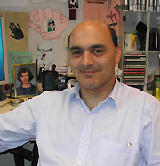
Karl Csaky received his M.D. degree in 1983 and his Ph.D. in pharmacology in 1987 from the University of Louisville in Kentucky. Following an internship in medicine at Duke University in Durham, N.C., he spent 15 months as a Fulbright Scholar in the University Eye Clinic in Essen, Germany, with Gerd Meyer-Schwickerath. He completed his ophthalmology residency at Washington University in St. Louis in 1990 and spent one year as a fellow in the Retinal Vascular Center at the Wilmer Eye Institute, Johns Hopkins Hospital, Baltimore. From 1991 through 1994, he completed a postdoctoral fellowship at the NCI laboratory of Stuart Aaronson. He joined the NEI in 1994 and currently heads the Section on Retinal Diseases and Therapeutics.
Age-related macular degeneration (AMD) is the leading cause of blindness in patients over the age of 60 years in the United States. It is estimated that 1/3 of all people over the age of 75 will develop AMD.
AMD has three major clinical presentations: the early dry form, which is characterized by extracellular deposits under the retina; the severe dry or atrophic form in which retinal cells undergo apoptosis; and the wet form, where neovascularization develops in the normally avascular subretinal space, so-called choroidal neovascularization (CNV).
My laboratory and clinical efforts are focused on elucidating the pathogenesis and developing new therapies for all three forms of AMD. As a clinician-scientist, much of the focus in my lab is on research that has direct disease relevance, drawing insights from clinical presentations or therapeutics that can be taken into clinical trials.
For example, in the atrophic form of AMD, it is known that cell death begins in one unique cell layer of the retina termed the retinal pigment epithelium (RPE). The RPE is a nonrenewable cell that maintains retinal function from birth through death. However, the natural history of RPE cell death in patients is one of an end-stage phenomenon that occurs late in the course of AMD. Therefore, we hypothesized that RPE cell apoptosis might be different from apoptosis in cells that undergo a more rapid time course of cell death.
Indeed, we discovered that unlike immune cells, RPE cells use primarily the release of apoptosis-inducing factor (AIF) and not activation of caspases to modulate cell death. In addition, the RPE cell has unique mechanisms for self-protection from various insults. After an oxidative injury, for example, a microarray profile of RNA expression in RPE cells demonstrated activation of both antioxidative defense mechanisms as well as direct downregulation of proapoptotic pathways. We are now studying how the loss of these survival mechanisms interplays with AIF and how this process may be involved in the development of the late atrophic form of AMD.
In examining the development of the neovascular form of AMD in various animal models, my laboratory discovered that bone marrow–derived endothelial cells (EPCs) are involved in this process. A key question about this and other forms of neovascularization is whether EPCs have some important role in this process. We hope to answer this question in part by examining EPCs from patients with neovascular AMD.
Unlike neovascularization in other diseases, such as cancer or cardiac ischemia, development of CNV can be precisely documented through multiple imaging techniques showing rates of growth, extent of growth, phenotypic characteristics, and various aspects of blood flow. In fact, when we analyze various characteristics of neovascularization in AMD patients, we find a large variety of clinical presentations. We hope to determine, from patients, specific functions of circulating EPC’s and correlate these to the various phenotypes of neovascularization. Our goal is to develop a predictive and therapeutic blood test that would identify patients most at risk for aggressive neovascularization.
Because only a small amount of tissue needs to be treated in patients with AMD, my laboratory is also involved in targeted sustained drug delivery through the use of drug implant delivery systems. We have developed a novel implant device that can deliver various antiangiogenic agents for up to three years. Clinical trials of these devices are now being planned.
In another study of neovascular AMD, in collaboration with Scott Cousins at the University of Miami, we identified the importance of innate immunity in the development of CNV in animals. This led us to develop a therapy by optimizing a steroid formulation that could be injected into the eye. This novel formulation has been produced and successfully injected into patients and is now in Phase I and II trials at NIH. A multicenter Phase III trial using this steroid formulation is now underway in 300 patients with neovascular AMD.
If the results of these trials are positive, they will be used for a new drug application through the FDA. Thus, beyond its direct translation from lab to clinic, our work is giving us useful insights into FDA regulatory issues and the economics of drug development.
 |
|
Peter
Inskip
|
Peter Inskip received his Sc.D. in epidemiology from the Harvard School of Public Health in Boston in 1989. He then joined NCI as a fellow in the Division of Cancer Epidemiology and Genetics (DCEG). In 1995, he left NCI to take a position as associate professor at the Texas A&M College of Veterinary Medicine in College Station. He returned to NCI in 1998 and is currently a senior investigator in the Radiation Epidemiology Branch of DCEG.
My research focuses primarily on quantifying radiation-related cancer risks and clarifying susceptibility factors that influence these risks. For example, I am conducting studies of therapy-related second primary cancers in a population of more than 14,000 five-year survivors of childhood cancer diagnosed between 1970 and 1986.
These studies include detailed information on radiotherapy and chemotherapy and anatomic site–specific radiation dosimetry. Our analyses have demonstrated very large relative risks for second neoplasms of the thyroid gland, brain, and breast due to radiotherapy. Interestingly, risk of thyroid cancer increased with dose at low to moderate doses but declined at very high doses, probably due to cell-killing effects of high-dose radiotherapy on the thyroid gland.
Although relative risks associated with radiotherapy for childhood cancer are very large, most patients who receive high partial-body doses of radiation do not develop a second cancer. It is unclear to what extent this is random or dependent on differences in host susceptibility. We are evaluating whether polymorphisms in genes involved in DNA protection, DNA repair, cell-cycle control, and apoptosis are predictive of second cancer risk.
In an earlier study of nearly 13,000 women treated with radiation for benign gynecologic disorders between 1925 and 1966, I found radiation-related excesses of acute and myelocytic leukemias but not chronic lymphocytic leukemia, Hodgkin or non-Hodgkin lymphoma, or myeloma.
Radiation dose-response relationships also were apparent for cancers of the bladder, colon, and ovary but not for cancers of the uterus or rectum, which appear to be relatively radioresistant organs.
I also am studying cancer risks associated with lower-dose protracted or fractionated radiation exposures. Taking advantage of unique features of the Swedish healthcare system, I evaluated the risk of thyroid cancer associated with lifetime history of diagnostic X-rays based on medical records; no association was found. In one of the first systematic studies of cancer among Chernobyl clean-up workers, we foundno excess of leukemia among 4,833 Estonian clean-up workers followed through 1993. We have expanded the study to include an additional 12,500 workers from Latvia and Lithuania and are extending the follow-up to further evaluate leukemia and solid cancers.
When the public became concerned that radiofrequency radiation emitted by cellular telephones might cause brain cancer, I initiated a case-control study to explore a wide range of hypotheses about the etiology of these poorly understood and often fatal tumors. Our article in the New England Journal of Medicine reported no evidence of increased risk of glioma, meningioma, or vestibular schwannoma associated with use of cellular phones. Unlike ionizing radiation, nonionizing radiation shows little evidence of causing cancer.
Cellular telephones might not cause brain tumors, but something does. The etiology of brain tumors has become my second major research focus.
In our study of brain tumors, we observed a significant inverse association between the risk of glioma and history of allergies—a finding that has been confirmed in subsequent studies.
A surprising observation was the lower risk of glioma among left-handed compared with right-handed individuals, which, if not due to chance, may relate to a longstanding hypothesis that prenatal exposure to high testosterone levels affects the developing brain and thymus, resulting in an "anomalous" pattern of hemispheric dominance associated with left-handedness, developmental language disorders, and immune dysfunction.
Glioma incidence was also positively associated with age at menarche and inversely associated with age at first live birth. To follow up on these observations, I am collaborating with other NCI investigators to explore associations of brain cancer risk with genetic polymorphisms involved in inflammation, immune response, and hormonal pathways. We also are studying brain tumor risk with respect to occupational exposures, including solvents, lead, and pesticides.
Because brain tumors are rare, and no single existing epidemiologic study of brain tumors has adequate statistical power to evaluate gene-environment or gene-gene interactions, it is important to combine results from multiple studies of brain tumors.
Towards that end, I organized a workshop that launched an international consortium of investigators conducting etiologic studies of brain tumors. Our aim is to establish a network that will enable investigators to collaboratively address etiologic hypotheses that otherwise could not be studied effectively.
 |
|
Caroline
Philpott
|
Caroline Philpott received her M.D. from Duke University, Durham, N.C., in 1987. Following an internal medicine residency at the Johns Hopkins Hospital in Baltimore, she joined NICHD in 1990 and became a clinical associate in the Genetics, Cell Biology, and Metabolism Branch. In 1998, she joined NIDDK, where she is currently a senior investigator in the Liver Diseases Branch.
I am interested in how cells take up and use iron, which is important for human health in a number of ways.
Worldwide, iron deficiency is the most common nutritional disorder, and yet genetic forms of iron overload are also very common. We are only beginning to understand the role that iron plays in the chronic neurodegenerative diseases, such as Friedreich ataxia and Parkinsonism. Pathogenic microorganisms must overcome the iron-withholding systems of the human host to establish infection. These systems are our first line of defense against microbial invaders, and microbial iron uptake mechanisms that overcome these systems are often important virulence factors.
Almost every organism on the planet requires iron because of its versatility in picking up and releasing electrons. This reliance on iron comes at a cost, however, because the same properties that make iron useful also make it very chemically reactive and potentially damaging for cells. Think of iron as cellular dynamite—useful, but dangerous.
Cells use iron to build heme and iron sulfur clusters, which are inserted into a variety of enzymes that carry out essential functions, such as respiration; sterol, lipid, and amino acid biosynthesis; and DNA repair. Most of the iron inside a cell is in the reduced form, which can catalyze the formation of damaging free radicals. Cells face a bioavailability problem with iron, as most iron in an oxygen-containing environment is in the oxidized form, which forms large, insoluble polymers with oxygen and water—rust. Thus, all organisms have evolved sophisticated mechanisms to control the uptake and use of iron.
We began our studies in the one-celled eukaryote Saccharomyces cerevisiae, an excellent organism in which to understand basic processes of both eukaryotic cells and pathogenic fungi. We used cDNA microarrays to ask the question "How do cells respond to iron deficiency?" Our studies of the transcriptional response allowed us to identify the set of genes involved in the primary response to iron deficiency and to understand the cell’s strategy for surviving iron deprivation. We found that the cellular response took three forms: 1) increasing the expression of iron uptake systems, 2) mobilizing stored iron, and 3) adjusting cellular metabolism to conserve iron.
We identified a new fungal system for iron transport that relied on the uptake of siderophores, which are small molecules that chelate iron. Most bacteria and fungi secrete these compounds in their iron-free form and take them back up once they contain bound iron. Budding yeast don’t make siderophores, but they readily take up those made by other microbes.
Siderophore uptake occurs through a family of transporters that exhibit a unique intracellular trafficking pattern that is controlled by the presence of the siderophore substrate. We found that the transporter itself contained a domain that acted as the receptor for siderophores. Binding of siderophores to this domain affected the cellular trafficking of the transporter, directing it to the cell surface only when the appropriate siderophore substrate was available. The regulated intracellular trafficking of transporters has emerged as an important mechanism for controlling transport of many molecules in various cells. We hope to uncover the processes that control the trafficking of transporters in yeast.
One surprising finding of our studies in yeast was how cells adjust to dwindling levels of iron. At first glance, one might suppose that cells simply continue to use iron in their iron-containing proteins until they run out. But we found that far more sophisticated adjustments take place.
As iron is depleted, yeast turn off transcription of certain nonessential pathways that rely on iron-sulfur cluster proteins and turn on transcription of parallel iron-independent systems. For example, yeast can either synthesize biotin from intermediates, or take it up from the medium. Synthesis requires iron, but uptake does not. Under iron deficiency, biotin synthesis is switched off and uptake is switched on.
Heme utilization is also adjusted in iron deficiency. As cells become iron depleted, they synthesize a heme-degrading enzyme, and this degradation of heme serves two purposes. First, iron is released and reused, and second, heme-dependent transcription is decreased. Heme is an important regulatory molecule, and the degradation of heme results in the downregulation of many proteins that contain iron-rich complexes.
We don’t yet know what types of adjustments are made in human cells in response to iron depletion. A new focus of our lab is to use yeast to identify new human proteins involved in iron homeostasis. Yeast and human cells have some clear differences in how they take up and use iron, and by selecting human genes that confer new iron-handling properties on yeast, we hope to identify novel human genes of iron metabolism.
 |
|
Brant
Weinstein
|
Brant Weinstein received his Ph.D. from the Massachusetts Institute of Technology, Cambridge, Mass., in 1992. After completing his postdoctoral work at the Massachusetts General Hospital, Boston, he joined the Laboratory of Molecular Genetics, NICHD, in 1997. He is currently a senior investigator in that lab and head of the Unit on Vertebrate Organogenesis.
My laboratory is trying to understand the cellular and molecular mechanisms responsible for the specification, patterning, and differentiation of blood vessels.
Blood vessels are ubiquitous and vital components of all vertebrates, forming an elaborate network that supplies all tissues and organs with oxygen, nutrients, and cellular and humoral factors. The mechanisms controlling blood vessel growth and regression are still poorly understood, but they have become a subject of intense clinical interest in recent years because of the great promise shown by antiangiogenic therapies for combating cancer. http://dir2.nichd.nih.gov/labs/lab.php3?15
Many of the critical insights into regulation of blood vessel formation have come from developmental studies, and the zebrafish has emerged as an important new vertebrate model organism for investigating early vascular development. This small tropical freshwater fish is genetically tractable and has a physically accessible, optically clear embryo. These features permit noninvasive high-resolution visualization of every vessel in the living animal and simple, rapid screening for even subtle vascular-specific mutants.
Several experimental tools that we developed allowed us to amplify the basic advantages of this system. A microangiographic method for imaging patent blood vessels allowed us to compile a comprehensive atlas of the vascular anatomy of the developing zebrafish. Our transgenic zebrafish lines expressing green fluorescent protein in vascular endothelial cells make it possible to visualize blood vessel formation in intact, living embryos. We devised methods for long-term, multiphoton time-lapse imaging of growing vessels in these transgenic zebrafish and used them to examine the morphogenesis of developing trunk and cranial vessels and adult fin blood vessels.
We used forward genetic mutational screening to identify and characterize zebrafish mutations in developing vasculature. We positionally cloned the defective genes from several vascular patterning mutants, including violet beauregarde (defective in Alk1/acvrl1), plcgly10 (defective in phospholipase C-gamma–1), and kurzschluss (defective in a novel chaperonin).
We are currently carrying out large-scale genetic screens for chemically induced mutants. These have yielded a large number of new mutations, with phenotypes including loss of all or subsets of vessels, increased sprouting or branching, and vessel mispatterning. Characterization and molecular cloning of these mutants is in progress.
Probably the most fundamental fate choice for endothelial cells is arterial vs. venous differentiation. We were the first to describe a molecular pathway regulating acquisition of arterial-venous identity: sonic hedgehog, vascular endothelial growth factor (VEGF), and Notch signaling, acting in series. Using genetic screening and positional and candidate cloning methods, we identified a mutation in phospholipase C-gamma–1 leading to defects in angiogenesis and arterial differentiation. We showed that this gene is a major downstream effector of VEGF signaling in vivo. We are currently using microarrays, mutant screens, and other methods to further explore the molecular mechanisms underlying this important cell fate decision.
We are also studying the mechanisms underlying vascular patterning and morphogenesis during development, in particular, how early vascular networks assemble with defined, stereotypic anatomical patterns. We used multiphoton time-lapse imaging of transgenic zebra-fish to obtain evidence supporting a novel model for genetically programmed vascular network formation in the embryonic trunk.
Subsequently, we showed that semaphorin signaling—an important mediator of axonal guidance in the nervous system—acts in an analogous fashion as a repulsive guidance factor for these developing trunk vessels via a novel endothelial-specific plexin receptor. Our results thus highlight similarities between the establishment of anatomical pattern in the developing vascular and nervous systems.