Patrice
Morin
| T H E N I H C A T A L Y S T | N O V E M B E R – D E C E M B E R 2004 |
|
|
|
| P E O P L E |
RECENTLY TENURED
|
|
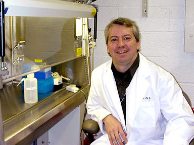
Patrice
Morin
|
Patrice J. Morin earned his Ph.D. degree at Boston University in 1995. He then did his postdoctoral research training at the Johns Hopkins Oncology Center in Baltimore. He joined the NIH as a tenure-track investigator in 1998. He is now a senior investigator at NIA in the Laboratory of Cellular and Molecular Biology and is head of the Cancer Genomics and Signaling Section.
Our research focuses on the molecular mechanisms of ovarian tumorigenesis. In particular, we are interested in identifying genes and pathways that may become novel targets for detection and therapy.
The American Cancer Society estimates that in the United States this year more than 23,000 women will be diagnosed with ovarian cancer, and approximately 14,000 will die of the disease. Yet very little is known about the biology of ovarian cancer.
Ovarian cancer affects older women disproportionately, with more than half the cases diagnosed in women older than 60. In spite of the recent introduction of aggressive treatments, five-year survival rates for patients with advanced ovarian cancer are low, ranging between 5 and 30 percent.
Five-year survival can reach 95 percent if the diagnosis is made at an early stage, but only about one-quarter of patients are diagnosed early, when there are no symptoms. These facts make ovarian cancer a disease for which early detection represents an intervention of choice in reducing morbidity.
It is likely that the discovery of highly sensitive and specific tumor markers will greatly affect ovarian cancer management and lead to a significant increase in overall survival.
In the past few years, we have used serial analysis of gene expression (SAGE) and microarrays to examine gene expression in ovarian cancer and normal ovarian tissues.
We have identified several thousand genes expressed in each tissue and found numerous genes differentially expressed between normal and malignant ovarian cells, including novel transcripts that we have named HOSTs (human ovarian cancer–specific transcripts). Genes whose expression is elevated in ovarian cancer, especially those that encode secreted and/or surface proteins, may become targets for early diagnosis and various therapeutic strategies, such as immunotherapy. We are evaluating promising candidates and generating antibodies to investigate their clinical potential.
The large number of genes abnormally expressed in ovarian cancer may also provide clues to which molecular pathways may be relevant to ovarian tumorigenesis. We are using a variety of molecular biological tools to validate our SAGE results and dissect the molecular pathways responsible for the aberrant gene expression.
Of particular interest is the pathway involving the claudin tight junction proteins. We have found that these proteins are consistently elevated in ovarian tumors. Since our initial findings, other research groups have found that claudins are dysregulated in many other cancers.
We are currently examining signal transduction upstream and downstream of the claudin proteins in order to clarify their functions in cancer. Our current data suggest that claudins may be important for cell motility and invasion. Overall, the identification and characterization of pathways crucial for ovarian tumorigenesis will provide a better understanding of this disease at the molecular level and may suggest novels targets for therapy.
Aside from the difficulties of ovarian cancer detection, ovarian cancer treatment is also plagued by drug resistance. Approximately half of ovarian tumors are intrinsically resistant to chemotherapy, and a significant fraction of women with tumors that initially respond to chemotherapy eventually relapse with drug-resistant disease.
We have created an in vitro model of drug resistance consisting of isogenic cell lines differing in their sensitivity to cisplatin. Using SAGE, we have identified genes whose expression is altered in cisplatin-resistant cells. These genes encode proteins of the extracellular matrix and cell adhesion, such as collagen VI, collagen X1, and decorin.
We are currently investigating the mechanisms of cell adhesion-mediated drug resistance in ovarian cancer. We are also studying the roles of the phosphatidylinositol 3 kinase/Akt pathway, as well as the X-linked inhibitor of apoptosis pathway in ovarian cancer drug resistance using, among other approaches, siRNAs and somatic gene-targeting strategies.
 |
|
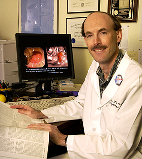
Ronald
Summers
|
Ronald Summers received his M.D. and Ph.D. degrees from the University of Pennsylvania School of Medicine in Philadelphia in 1988. After a medical internship, he completed a radiology residency at the University of Michigan Hospitals in Ann Arbor and a body magnetic resonance imaging fellowship at Duke University in Durham, N.C. He joined NIH in 1994 as a staff radiologist in the Clinical Center and is currently chief of the Virtual Endoscopy and Computer-Aided Diagnosis Laboratory in the Diagnostic Radiology Department of the CC Imaging Sciences Program.
While at Duke, I attended a lecture by David Vining, a radiologist who was touting a new way of detecting colorectal cancer using computed tomography (CT) scans.
He called the test "virtual colonoscopy" and highlighted his lecture with "Fantastic Voyage"–like movies of the inside of the large intestine.
Here was the potential to make new types of radiologic diagnoses noninvasively. Vining’s presentation sparked my imagination as I realized by performing research in virtual endoscopy I could combine my interests in medical imaging and computing to solve important clinical problems.
Upon arrival at NIH, I first explored virtual endoscopy’s potential for detecting disease of the human airways.
In collaboration with James Shelhamer of the Critical Care Medicine Department and Michael Sneller and Steven Holland of NIAID, I embarked on a protocol to use virtual bronchoscopy to study patients with cavitary lung disease and airway stenosis.
Later, with David Schrump and Steven Finkelstein of the NCI Surgical Oncology Branch, I used virtual bronchoscopy to study patients with lung cancer.
We wrote a series of articles presenting our methodology for producing the virtual bronchoscopy images. We also conducted clinical validation studies that showed that virtual bronchoscopy could reveal morphologic abnormalities such as airway occlusions and endobronchial masses.
Based on these results, we moved virtual bronchoscopy from the bench to the bedside—from an experimental project to a routine clinical service offered to patients at the CC.
About 1998, I became interested in computer-aided detection (CAD). Medical images were becoming more complex, and radiologic interpretation of these images became more labor-intensive. I saw potential clinical value in automating the process of lesion detection.
Our approach was to determine those structural features, such as shape, that the radiologist and endoscopist have traditionally identified. Then we identified methods such as differential geometry and other mathematical and image processing techniques to isolate and quantify these features and, ultimately, improve upon them.
The outcome of this work was the first computer-aided detection system for virtual bronchoscopy. This system enabled us to locate endobronchial polypoid lesions with automated software that had high sensitivity and a low false- positive rate.
Simultaneously with this work, interest in virtual endoscopy had accelerated in the medical community. CT scanners were acquiring images faster, and image quality was improving. These advances allowed more accurate three-dimensional reconstructions and better virtual depictions of internal anatomy.
For example, several clinical trials showed that virtual colonoscopy had potentially high sensitivity and specificity for detecting colorectal polyps, precancerous growths that are the precursor lesion to colorectal cancer.
While polypoid endobronchial lesions are rare, colonic polyps are not. In addition, polyp detection is important, and detection and removal of them is the centerpiece of colorectal cancer prevention and treatment. It soon became clear to me that we could apply similar CAD technologies to find polyps on virtual colonoscopy and that there was clinical value in doing so.
In 2000, in collaboration with colleagues at Stanford University (Stanford, Calif.), my lab published a feasibility study that showed CAD could locate simulated polyps in human virtual colonoscopy scans.
One year later, in collaboration with colleagues at the Mayo Clinic (Rochester, Minn.), we published results of a clinical trial that showed CAD could find real colonic polyps in patients. These papers, published in the widely read journal Radiology, initiated a ground-swell of interest in computer-aided colonic polyp detection.
Since then, researchers from institutions around the world have published on virtual colonoscopy CAD. Commercialization of computer-aided polyp detection is likely in the near future.
Currently my lab is working on improving the sensitivity and decreasing the false-positive rate of computer-aided polyp detection.
We are studying ways to help the colonoscopist precisely locate abnormal tissue found at virtual colonoscopy so that these abnormalities can be removed. We are verifying the performance of CAD in a screening population of patients and refining CAD to detect different histopathologic polyp types.
Other research areas we are working on include advanced image segmentation techniques to distinguish organs and lesions from normal background tissue and the application of CAD to detect other abnormalities on CT scans such as subcutaneous melanoma and bone metastases.
Our work would not be possible without a multidisciplinary approach that involves collaboration of computer scientists, engineers, statisticians, and clinicians.
Together we can bring the power of noninvasive imaging and computational methods to bear on health problems that affect many Americans. The NIH CC is an ideal place to do this kind of multidisciplinary work. n