Luca
Di Noto, NHLBI
| T H E N I H C A T A L Y S T | N O V E M B E R – D E C E M B E R 2004 |
|
|
|
Research Festival—Poster SessionPLUMBING
THE CELL
BIOLOGY OF LOU GERHIG'S
DISEASE,
|
by Aarthi Ashok |
|
|
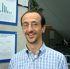
Luca
Di Noto, NHLBI
|
More than 300 posters, placed under 25 different scientific umbrellas, were presented during the Research Festival. Below are reports on three of the posters from the cell biology session.
Luca
Di Noto, Laboratory
of Biochemistry, NHLBI; Principal Investigator: Rodney
Levine
Susceptibility
of mutant superoxide dismutase to degradation by the proteasome and correlation
with amyotrophic lateral sclerosis
Lou Gehrig’s disease, or amyotrophic lateral sclerosis (ALS), results in loss of motility due to neuronal degeneration and is the most common motor neuron disease in adults.
The familial form of ALS (fALS), which constitutes about 10 percent of all ALS cases, has been linked to more than 100 mutations in the sod1 gene that encodes copper/zinc superoxide dismutase (SOD1).
Previous studies have shown that mutations of the ALS type decrease the half-life of the mutant protein in vivo. Di Noto and colleagues hypothesized that the cell’s multicatalytic protease, the 20S proteasome, may be responsible for the degradation of SOD1 with fALS-type mutations.
To test this hypothesis, they used purified 20S proteasomes from rat liver to examine the degradative susceptibility of the "apo" or metal-free forms of both normal and fALS mutation-bearing SOD1 proteins.
Quantification of degradation by reverse-phase chromatography and mass spectroscopy led them to conclude that some SOD1 mutant proteins were better substrates for the proteasome than the normal protein.
Further, because the cleavage sites were unaltered in these mutants, the differences between the normal and mutant forms were clearly quantitative.
When they looked at the temperatures at which each of the mutant proteins became completely unfolded (melting temperature), they noticed that some of the mutant SOD1 proteins had lower thermal stability than their normal counterpart, and this lower stability had a linear correlation with proteasomal degradation.
The unfolding of proteins at or near their melting temperature should lead to the exposure of the buried hydrophobic residues, and hydrophobic patches on proteins have been proposed as signals for proteasomal recognition and degradation. Using the fluorescent dye ANS, which binds hydrophobic patches, Di Noto noted that an increase in surface hydrophobicity correlated with increased degradation.
To drive home the point that thermal stability governs proteasomal susceptibility, Di Noto reduced an intramolecular disulfide bond in the normal protein that caused its melting temperature to decrease dramatically.
He then showed that this reduced form was a better substrate for proteasomal degradation than the normal unreduced form.
Taken together, the data point to the absence of metal ions, increased thermal instability, and surface hydrophobicity as the susceptibility factors for proteasomal degradation.
Di Noto suggests that the accumulation of toxic degradation products due to the increased turnover of the mutant SOD1 proteins may be toxic to neurons and thereby result in fALS.
Many degenerative diseases are hypothesized to occur as a result of the accumulation or aggregation of mutant protein.
Based on their studies, Di Noto and colleagues propose an alternative mechanism whereby rapid turnover of mutant proteins into toxic degradation products underlies the pathology of the disease.
 |
|
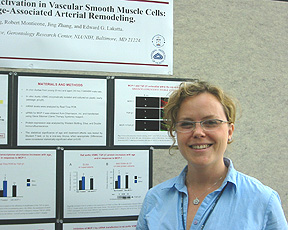
Gaia
Spinetti, NIA
|
Gaia
Spinetti, Laboratory
of Cardiovascular Science, NIA; Principal Investigator: Edward
Lakatta.
MCP-1 induces TGF-b activation in vascular smooth
muscle cells: implication for age-associated arterial remodeling
Remodeling of the arteries contributes to increased susceptibility to the diseases of aging, such as atherosclerosis. Fibrosis involves the accumulation of collagen, fibronectin, and other extracellular matrix components in the arterial wall and is a component of the vascular remodeling process.
Monocyte chemotactic protein 1 (MCP-1), metalloproteinase type II (MMP2), and transforming growth factor–b1 (TGF-b1) are all increased in expression in the aging arterial wall and are deemed to be molecular players in the fibrotic process. Previous work has shown that MCP-1 activates MMP2, and MMP2 can activate TGF-b1, in the rat aorta.
Spinetti and co-workers hypothesized that the increased levels of MCP-1 may be linked to the increase in TGF-b1 levels in vitro and in vivo.
In testing this hypothesis, they first showed that immunofluorescent detection of MCP-1 and TGF-b1 in young (8-month-old) and old (30-month-old) rat aortas led to a complete overlap in their signals, pointing to co-localization of these proteins in the vascular wall.
Using CD31 as a marker of endothelial cells and a-SMA as a marker of aortic vascular smooth muscle cells (VSMC), they further showed that the TGF-b1 signal was present within endothelial and VSMC regions of the arterial wall.
Real time PCR analysis using early passage VSMC showed that TGF-b1 transcript levels were increased in the old rat aortas and in young aortas exposed to MCP-1 for 24 hours, compared with the untreated young aortas. ELISA and western blot analysis showed that the TGF-b1 protein levels mirrored the age dependency and MCP-1 responsiveness of the TGF-b1 transcripts. Subcellular fractionation assays demonstrated the localization of TGF-b1 protein in these cells in the organelles, cytoplasm, and nuclei.
These data point to TGF-b1 production as a novel effect of MCP-1 signaling in VSMC and provide insight into the complex molecular regulation of arterial remodeling.
Spinetti and co-workers are currently analyzing the effect of MCP-1 siRNA transfection on TGF-b1 expression levels in VSMC to further define the relationship between these proteins.
 |
|
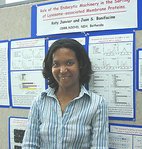
Katy
Janvier, NICHD
|
Katy
Janvier, Cell
Biology and Metabolism Branch, NICHD; Principal Investigator: Juan
Bonifacino
Role
of the endocytic machinery in the sorting of lysosome-associated membrane proteins
(LAMPs)
Lysosomal membranes are highly enriched in glycosylated transmembrane proteins that are appropriately termed lysosome-associated membrane proteins (LAMPs). Janvier and colleagues have set about to explore the cellular mechanisms behind the sorting of these proteins to the lysosomal membrane.
The most abundant members of the LAMP family are LAMP-1, LAMP-2, and CD63; they all carry tyrosine-based cytosolic sequences that are recognized by adaptor proteins (AP) involved in sorting proteins to endocytic and secretory compartments.
Previous work pointed to the existence of two possible pathways for the sorting of LAMPs to the lysosome: the direct and the indirect pathways. The former pathway involves the movement of LAMPs from the trans-Golgi network to endosomes and then to lysosomes, while the latter includes a movement of these proteins to the plasma membrane prior to their transport to endosomal compartments.
Janvier designed several experiments to dissect the contributions of these pathways to the trafficking of LAMPs in HeLa cells.
She first examined whether the expression of a dominant negative mutant form of dynamin 2, which blocks internalization from the plasma membrane, would affect the sorting of LAMPs.
FACS analysis revealed an increased cell surface expression of LAMPs in the presence of the dominant negative dynamin 2 compared with the levels present in cells expressing normal dynamin.
Next, using siRNA transfection, Janvier selectively depleted the m subunit of each of the AP complexes (AP1, 2, 3, and 4) and found that depletion of the AP2 m subunit (m2) resulted in a significant increase in the cell surface levels of all three LAMPs.
Because AP2 is the adaptor protein responsible for internalization of cargo in clathrin-coated vesicles from the plasma membrane, Janvier compared the internalization rate of the LAMPs with that of a marker for the AP2 pathway, transferrin, in both the presence and absence of the m2 siRNA.
The internalization rates of the LAMPs and transferrin were significantly diminished in the presence of siRNA, confirming the role of AP2 in the trafficking of LAMPs.
This finding was supported by experiments in which newly synthesized CD63, upon release from a brefeldin A block that held it in the ER, was sorted to the lysosomes in normal cells and was mostly present at the plasma membrane in cells transfected with the m2 siRNA.
Finally, Percoll gradient fractionation of HeLa cells followed by immunoprecipitation of LAMPs showed that 40–60 percent of the LAMPs were prevented from trafficking to lysosomes upon AP2 depletion.
Janvier therefore concludes that about half of the LAMP proteins in the cell are sorted to the lysosomes via the indirect pathway, in which AP2 and clathrin play a key role. n