| T
H E N I H C A T A L
Y S T |
J U L Y – A
U G U S T 2004 |
|
Workshop
Explores Adult Stem-Cell Research
NIH
CORE FACILITY SUPPORT
SOUGHT
FOR 'TRANSITIONAL' STEM-CELL
RESEARCH
|
by
Celia Hooper |
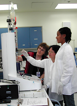 |
Cell
Isolation: E.
J. Read (left), chief of the CC Cell Processing Section, watches as
medical technologists Quyen
Chau and Tamara
Felton run the CliniMACS, a closed, sterile system that uses magnetically
labeled
antibody to separate enriched hematopoietic progenitor cells from a donor’s
mobilized peripheral white blood cells. A typical donor sample, containing
40 billion white blood cells, provides about 400 million progenitor cells
for one allogeneic transplant.
|
Radical
new therapies using today’s government-approved embryo
stem-cell lines may still be five or more years from trials in patients. In
the meantime, NIH scientists developing therapies based on adult cells are making
interesting strides in basic and clinical studies.
At an NIH workshop in
late May, these investigators said they hope their real-world experience turning
scientific concepts about stem cells into human cell-therapy trials will also
ultimately facilitate embryo stem-cell treatments.
Speaking at the May 24
workshop, "Clinical Applications of Stem Cells at the NIH," John
Barrett of NHLBI acknowledged that he had "a slightly crusading purpose"
behind his talk—not just to show promising clinical trial results, but
also to highlight the challenges of his 10-year effort to take a promising idea
from theory to clinical trial.
Effecting
Smooth Transitions
From this experience,
Barrett sees a need for a core facility to assist what he prefers to call "transitional"
research—the work needed to go from "proof of principle" experiments
with human cells to the manufacture of an FDA-approved clinical-grade cell product.
A transitional research
laboratory would facilitate interactions with industry partners to study and
perfect the systems for cell separation, selection, culture, and preservation
so that the techniques for making new cell products could be brought within
reach of cell-processing laboratories and blood banks nationwide.
The Cell
Processing Section in the CC’s Transfusion Medicine Department, which
operates under rigorous GMP (good manufacturing practice) conditions, would
be an essential partner in that effort, he emphasized.
Creating a transitional
research facility might be expensive, he said, but the ultimate savings from
transplants using sophisticated cell products that could emanate from such a
facility would be enormous.
Barrett cited comparative
costs within his own area of hematopoietic stem-cell transplantation for malignant
diseases: The creation of complication-free transplants—purified stem cells
free of cells that cause graft-vs-host (GVH) disease and enriched for immune
cells that specifically react against troublesome viruses and the patient’s
cancer—could cost as much as $20,000 per patient. But that expense pales
in comparison to the possible $250,000 price tag for the prolonged hospitalization
that often attends allogeneic stem cell transplantation and the cost of treating
recurring disease if the transplant fails.
Cancer
and
Graft-vs-Host Disease
Barrett, along with NCI’s
Michael Bishop,
described research that uses hematopoietic stem cells in treating cancer patients.
Barrett uses allogeneic
transplants of peripherally harvested bone marrow cells to treat leukemia. The
HLA-matched donor stem cells repopulate the leukemia patient’s blood-producing
system after their own cancerous blood cells are killed with anticancer chemotherapy.
But beyond that, Barrett’s
team is trying to improve their patients’ odds by delicate application
of one edge of a dangerous two-edged sword.
Investigators have seen
that immune reactions by grafted donor immune cells can have a powerful antileukemic
effect. But even in apparently perfectly matched donor-recipient pairs, some
immune cells attack the patient’s normal tissues, causing potentially fatal
GVH disease.
Because older leukemia
patients are especially vulnerable–about half may die of GVH disease—the
team targets this older cohort (whose average age is 64) for clinical trials
of measures aimed at minimizing GVH reactions, while maximizing the allogeneic
attack on leukemia cells that may have withstood chemotherapy.
In 14 patients thus far,
the investigators have seen 100 percent transplant engraftment, with significant
reduction in GVH disease and no deaths to date attributable to that cause.
The key to this GVH reduction
in Barrett’s work, as well as in Bishop’s, has been to temper immune
reactions by selectively eliminating the cells responsible for them—GVH-specific
T cells. Studies with mice showed that low doses of fludarabine and cyclophosphamide
effect a slow depletion of T cells in a transplant recipient, permitting engraftment
of even HLA-mismatched cells without having to completely destroy the recipient’s
immune system.
Working with patients
with metastatic breast cancer, Bishop is employing chemotherapy regimens that
gently, slowly deplete T cells as they kill breast cancer cells. Patients are
left with disease-attacking natural killer (NK) and other immune cells, but
free of the T cells’ destructive reactions.
Bishop says a key may
be promoting Type 2 cytokine interactions that suppress GVH reactions, while
squelching Type 1 reactions that promote them.
He emphasizes that hematopoietic
stem cell therapy (HSCT) goals differ among patients. In some patients, the
target of therapy may not be cancer but, rather, anemia and other nonmalignant
blood diseases in which there is no benefit in retaining potential for graft-vs-tumor
reactions.
Among cancer patients,
the blood cells themselves may be cancerous or blood may harbor cancer cells.
Similarly, T-cell numbers and reactivity may be naturally high or low.
"We don’t believe
one regimen fits all," Bishop said He envisions blood stem-cell therapy
will be pretty much "designer therapy."
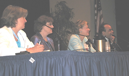 |
Wrap-Up:
(left
to right) Cynthia
Dunbar, senior clinical investigator, NHLBI; E.
J. Read, chief of the Cell Processing Section, Department of Transfusion
Medicine, CC; Betsy
Nabel, director of clinical research programs, NHLBI; and Ron
McKay, senior investigator, Laboratory of Molecular Biology, NINDS,
lead a general discussion at the end of the daylong workshop on "Clinical
Applications of Stem Cells at the NIH"
|
Array
of Applications
Roland
Martin of NINDS is examining the use of HSCT for a completely different
purpose—stopping the decline of patients with multiple sclerosis (MS).
Martin’s group shows that HSCT halts the progress of the autoimmune disease
by eliminating MS patients’ immune cells that are programmed to attack
their own myelin, causing inflammation and subsequent brain-cell loss. The patients’
immune systems are rescued with naive cells from transplanted hematopoietic
stem cells.
In the seven patients
treated thus far, this approach has reset the immunological clock, leaving patients
free of the inflammatory component of MS.
Stopping the clock, however,
doesn’t reverse neurodegeneration that has already occurred. For this,
Martin says, one has to look at a second transplant of some other stem cells—oligodendrocyte
precursor cells alone or with local delivery of growth factors. Transplantation
of these precursors in mouse models of MS has restored myelinated cells and
appropriate brain architecture, left no scarring, and, as a result, improved
motor function.
Other research presentations
in the center ring of the workshop featured a diverse array of results, ranging
from NIDCR’s Pam
Robey’s elaboration of stem cells in the dental pulp of children’s
baby teeth to NIAID’s Harry
Malech’s description of progress toward effective gene therapy for
chronic granulomatous disease.
For example, Rocky
Tuan of NIAMS described continuing refinements in a bag of innovative tricks
designed to charm mesenchymal stem cells into making replacements for tissues
damaged by arthritis and other ravages of age and degenerative joint disease.
Tuan has been inching
ever closer to producing lab-grown tissues that closely mimic human bone and
cartilage in texture, strength, ability to support appropriate growth and differentiation
of cells, and even transmission of critical signals for cell-cell interactions.
Tuan noted that understanding
these signals—and learning how to control them—may someday obviate
the need for transplants of stem cells or lab-grown tissues.
The ideal course would
be to reprogram cells or call in residual mesenchymal stem cells already within
the body to repair damaged joints. Look at the salamander’s ability to
regrow complete limbs, Tuan urges. "It does all these things without any
government intervention or support!"
Do
Reparative Cells Come
from Near or Far?
One key question for scientists
working with some adult stem cells is where the cells actually come from. Do
repairing cells already reside in tissues and multiply when needed to repair
adjacent damage? Or are the cells summoned from a stem-cell source elsewhere
in the body?
NIDDK’S David
Harlan and Nadya
Lumelsky say that their studies aimed at diabetes suggest that insulin-producing
b-cells may not arise, at least not to date with
clinically relevant efficiency, from circulating stem cells or other pancreatic
cells. Harlan presented work that did not support the existence of bone-marrow
resident stem cells capable of differentiating into insulin-producing cells.
In other work with human
pancreatic cells, Lumelsky had variable success in cultivating new insulin-producing
cells in comparatively short-term cultures. The success of the culture varied
greatly depending on the specific human islet isolation.
That said, her laboratory
takes cellular clusters from the disaggregated human pancreas and grows cells
from the mixture on fibronectin in defined culture medium (with a cocktail of
growth factors), promoting cellular re-aggregation.
With this system, Lumelsky
finds that a minority of such preparations make more C-peptide (a marker for
insulin production) than what she started with, while most make less.
Evidence of remote sources
of stem cells came from NEI’s Karl
Csaky, who presented data on bone marrow as one key source of cells involved
in neovascularization in the eye.
Csaky’s mouse studies
on ocular overexpression of VEG-F or laser-induced ocular damage showed that
70 percent of macrophages, 58 percent of endothelial cells, and 60 percent of
smooth muscle cells involved in neovascularization and repair came from the
bone marrow.
In contrast, none of the
retinal cells in the repaired tissue came from the bone marrow. Csaky is interested
in identifying factors responsible for summoning and triggering appropriate
differentiation of stem cells at sites of damage in the eye.
The
Heart of the Matter
Focusing on heart repair,
Richard Cannon
of NHLBI recalled studies from 1997 that detected endothelial progenitor cells
in peripheral blood and evidence from animal models of hind-limb ischemia that
injection of these cells can stimulate growth of new blood vessels and improve
blood flow to the affected extremity.
Attempts to use such cells
to revascularize human hearts suggest benefit to myocardial blood flow and function,
but the nonrandomized clinical trials reported to date have involved small numbers
of patients who often underwent coronary revascularization at the time of treatment.
Studies performed in the
NHLBI Cardiovascular Branch
suggest that there may be variation in the ability of people’s stem cells
to accomplish the feat of repairing heart damage.
The researchers isolated
mononuclear cells from blood samples of middle-aged men with varying levels
of risk for heart disease and examined the ability of progenitor cells to produce
endothelial-like cells needed to form new vessels in damaged hearts.
Cells from subjects with
the fewest risk factors produced the most colonies of reparative cells, and
those with the greatest number of risk factors had the lowest numbers of functional
colonies.
Further, endothelial function,
as tested by nitric oxide–mediated vasodilation, correlated strongly with
subjects’ endothelial colony-forming capacity. The researchers speculate
that loss of the cells’ ability to repair endothelial damage in the heart
may be yet another independent contributing factor in cardiovascular disease.
Cannon and his colleagues
studied whether just mobilizing stem cells from bone marrow with the cytokine
granulocyte–colony-stimulating factor (G-CSF) might be sufficient to help
repair hearts damaged by severe cardiovascular disease. Although they were successful
in augmenting endothelial progenitor cells in the circulation after G-CSF administration,
the numbers of cells were small, the duration of mobilization short, and the
benefits to patient health undetectable on cardiac MRI and treadmill stress
tests.
Cannon will soon be trying
direct myocardial administration of stem cells to patients’ damaged hearts.
In a collaborative study
with Suburban Hospital’s (Bethesda, Md.) Cardiopulmonary Rehabilitation
Unit, Cannon is exploring interrelationships of exercise, endothelial function,
endothelial progenitor cell mobilization, and nitric oxide bioactivity in hopes
of giving heart patients a vascular repair profile more like that of healthy
individuals.
More
Tools for the Trade
Still other NIH investigators
are developing tools and facilities to aid stem-cell research. New tools include
in vivo imaging and clinical-grade reagents that permit investigators to "see"
stem cells at work inside the human body.
Robert
Lederman of NHLBI described sophisticated MRI techniques that could be used
to watch precise delivery of bone marrow stromal cells to the margins of an
infarct, for example, or to calculate peripheral vascular function by measuring
reperfusion of blood into tissue when a restraint on circulation is abruptly
removed.
"These images are
incredibly sensitive tools," Lederman finds. But frustrations in working
with industry partners to develop the techniques have led Lederman to agree
with Barrett that NIH needs a "transitional" core. If there were one,
"I’d be discussing results rather than experimental design,"
Lederman said.
Lederman says one key
to his work is a technique for magnetically labeling cells developed by the
CC’s Laboratory of Diagnostic Radiology
Research. Joe
Frank, who leads LDRR, described ongoing development of the superparamagnetic
ironoxides that are taken up by cultured stem cells, for example.
As few as 50,000 labeled
cells can be injected into a living animal and then observed via 1.5 Tesla MRI—a
field strength that could also be used in human studies.
Frank says nondividing
cells, such as T cells, retain label for 40 to 60 days, and there is no change
in apoptosis or reactive oxygen species in labeled cells. As paperwork wends
its way through the FDA to get the magnetic label approved for clinical use,
Frank has been collaborating on studies that put the vital stain to work to
study angiogenesis, stroke, MS, and cardiovascular disease.
The
Challenges of
Product Development
If NIH wanted to take
up Barrett’s call for a transitional research core, perhaps the best advice
on such a facility would come from E.
J. Read, chief of the Cell
Processing Section of the CC Department of Transfusion Medicine.
"Product development,"
Read says categorically, "is different from basic science." Product
development studies aim to design cellular products that meet the needs of the
clinical trial as well as FDA’s regulatory requirements. The new products
must ultimately be prepared in a GMP environment to ensure that they are safe
and effective for patients who receive them.
Translation of cell therapies
requires working collaboratively with investigators, starting in the preclinical
phase, to define and characterize specific details of each product.
Challenges abound. The
most striking, Read says, is the variability of cells–starting material
from different patients. This variability means contingency planning is critical
because things will go wrong. Scale-up and movement from lab to clinic may not
be straightforward. Closed, automated systems are more desirable, she said,
because they reduce human error and the risk of contamination from microbes
in the environment when cells are processed.
Collaboration with industry
brings in lots of additional challenges. Read says her best allies in coping
with FDA oversight have been the facility’s masterfile of operational and
quality procedures, written in responses to FDA’s formal queries and published
guidelines, and copious documentation during the preparation of each clinical
product. "This has really helped a lot," Read says.
NINDS’ Ron
McKay gave the workshop a view on another core facility with an update on
the NIH Human Embryonic Stem Cell Unit (see The
NIH Catalyst,
March-April 2004).
That facility is growing
and characterizing human embryo stem cells, "currently the most useful"
stem cell of all, by virtue of its capacity "for infinite expansion to
lots of cell types," McKay said. "Somatic stem cells are not designed
for infinite expansion."
McKay says studies of
embryo stem cells in rats show that they can be differentiated into dopamine-secreting
neural precursor cells and implanted into the ventral midbrain, where they engraft
beautifully and are electrophysiologically and behaviorally functional.
"In five years,"
McKay predicted, "human embryo stem cells will routinely be put to many
clinically relevant uses."
Regulatory
Hurdles
Whether the prediction
is borne out depends in part on reasonably swift approval of stem-cell procedures
by the FDA. FDA’s Steve
Bauer acknowledged that stem-cell–based therapy is a relatively new
regulatory area for the agency, but he noted that development of specifications
for stem-cell–based products is evolving rapidly as scientific information
increases.
The agency will be looking
not only at the end products but also at initial starting materials, culture,
and other processing of cells. Bauer said key issues might include:
 Potency of cells
Freedom of starting materials from infectious disease
Derivation of the cells
Stability of the cells
Propagation conditions
Characterization of the cells
Where implanted cells go and how long they live
Tumorigenicity of implanted cells
Ectopic development of tissue from implanted cells
In a panel discussion
concluding the workshop, Barrett said the NIH intramural program has a unique
opportunity in stem-cell research. "We have all the pieces in one place
and lots of expertise to see the research through to clinical trials."
NHLBI Clinical Director Betsy
Nabel concurred, citing the workshop’s display of "incredible
breadth and depth, from basic to applied research."
Return to Table of Contents