| T H E N I H C A T A L Y S T | M A Y – J U N E 2004 |
|
|
|
| P E O P L E |
RECENTLY TENURED
 |
|
Jeffrey
Baron
|
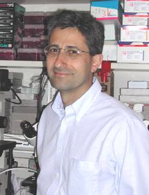
Jeffrey Baron received his M.D. from Southwestern Medical School, University of Texas Health Science Center at Dallas, in 1983. He completed a residency in pediatrics at Yale—New Haven Hospital in New Haven, Conn., in 1986. In 1989, he completed a fellowship in pediatric endocrinology in the Developmental Endocrinology Branch, NICHD, where he currently heads the Unit on Growth and Development.
The primary research interest of the Unit on Growth and Development is the cellular and molecular mechanisms governing skeletal growth and development. We have focused particularly on the process of longitudinal bone growth, which occurs at the growth plate—a thin layer of cartilage found near the ends of long bones and vertebrae.
The growth plate consists of three principal layers: the resting zone, the proliferative zone, and the hypertrophic zone. In the proliferative and hypertrophic zones, clones of chondrocytes are arranged in columns parallel to the long axis of the bone. Within these columns, the cells undergo clonal expansion followed by cellular hypertrophy. The hypertrophic cartilage is then remodeled into bone tissue. The net effect is that new bone is progressively created at the bottom of the growth plate, lengthening the bone.
The function of the resting zone is not well understood. We recently demonstrated that the resting zone can regenerate the proliferative and hypertrophic zones, suggesting that the resting zone contains chondrocytic stem-like cells that are capable of generating new clones of proliferative chondrocytes. We have also shown that ectopic resting-zone cartilage can induce a shift in the spatial orientation of nearby proliferative-zone chondrocytes, suggesting that the normal resting zone directs the spatial orientation of the proliferative clones, causing them to form columns parallel to the long axis of the bone.
The overall body proportions of vertebrates are determined by the size of the skeleton, which in turn is determined by the rate and duration of longitudinal bone growth. This rate falls progressively with age. In humans, fetal growth exceeds 100 cm/year. By birth, the growth rate has decreased to 50 cm/year, and by mid-childhood, 5 cm/year. A similar progressive decline in bone growth occurs in other mammals.
This decline in growth rate with increasing age is due primarily to a decrease in the rate of growth-plate chondrocyte proliferation.
In addition to functional changes, the growth plate also undergoes structural changes with age. We have termed these structural and functional changes "growth-plate senescence."
Our in vivo studies suggest that senescence occurs because the growth-plate stem-like cells have a finite proliferative capacity that is gradually exhausted. We are currently investigating the cellular and molecular mechanisms that limit proliferation of growth-plate chondrocytes.
Following a period of growth inhibition, the rate of longitudinal bone growth often does not just return to normal but actually exceeds normal. This phenomenon, known as catch-up growth, has been observed in humans and other mammals, following a wide variety of growth-inhibiting conditions.
It has long been speculated that catch-up growth is due to a central nervous system mechanism. However, we have shown that catch-up growth is due, at least in part, to a mechanism intrinsic to the growth plate, with evidence in particular that catch-up growth is caused by a delay in growth-plate senescence.
Growth-inhibiting conditions slow the proliferation of growth-plate chondrocytes, thus conserving the proliferative capacity of these cells and slowing growth plate senescence. If the growth-inhibiting condition resolves, the chondrocytes will have retained greater proliferative capacity than normal, will be less senescent than normal, and therefore will proliferate more rapidly and for a longer period of time than normal, resulting in catch-up growth.
Eventually, growth ceases and the growth plate is replaced by bone, a process known as epiphyseal fusion.
Our findings suggest that epiphyseal fusion is triggered when the proliferative capacity of the growth-plate chondrocytes is finally exhausted. We have found evidence that estrogen accelerates the proliferative exhaustion of these cells. As a result, estrogen leads to early termination of linear growth and early epiphyseal fusion. We recently found clinical evidence that estrogen accelerates growth plate senescence in girls exposed to estrogen because of precocious puberty.
One goal of this work is to improve medical treatment of growth disorders and childhood metabolic bone diseases. In addition, we seek to uncover general principles of developmental biology since the cellular processes underlying bone growth—such as cell proliferation, terminal differentiation, angiogenesis, and cell migration—are also essential for development in other tissues.
 |
|
Thomas
Bugge
|
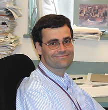
Thomas Bugge received his Ph.D. from the European Molecular Biology Laboratory/University of Copenhagen in 1993. He performed his postdoctoral studies jointly at the University of Copenhagen and the University of Cincinnati from 1993 to 1995. He held faculty appointments as research leader at the University of Copenhagen and associate professor of pediatrics at the University of Cincinnati from 1995 to 1999 and was recruited to NIDCR in 1999. He is currently chief of the Proteases and Tissue Remodeling Unit, NIDCR.
Extracellular proteolysis is essential to human development, homeostasis, tissue remodeling, tissue repair, learning, immunity, and fertility; excessive or impaired extracellular proteolysis is the cause of many human ailments. For example, abnormal extracellular proteolysis is the hallmark of cancer and enables tumor cell growth, survival, motility, invasion, and angiogenesis. Furthermore, inappropriate extracellular proteolysis by itself leads to genetic instability and can directly drive the malignant transformation of cells.
This "pruning" of the extracellular environment in health and during disease is performed by an array of proteases, several hundred in number, whose activities are tightly regulated by a series of inhibitors, receptors, associated proteins, and small molecules.
Research into how extracellular proteases modify their environment faces unique technical limitations, such as the lack of appropriate tissue-culture models and general methods to identify downstream protease targets. Moreover, a large number of extracellular proteases were discovered only very recently through the completion of the human, mouse, and rat genome sequences. Therefore, despite its imminent importance to human health and disease, the field of extracellular proteolysis is characterized by vast "frontiers of unknowns," which make the research particularly exciting and challenging.
Our laboratory has long been interested in understanding the functions of proteases that are bound directly to the surface of cells through membrane attachment or via the binding to specific cell-surface receptors. We use a collaborative, multidisciplinary approach that combines bioinformatics with targeted gene inactivation and overexpression in mice, advanced histology, proteomics, and degradomics to unravel the exceptionally diverse spectrum of functions of cell surface proteolysis in life.
We previously performed an analysis of the overall biological function of the plasminogen activation system, a sophisticated cell-surface proteolytic cascade. We found that the system had a universal role in postnatal tissue homeostasis, remodeling, and repair, and that in most, but not all, cases the critical substrate was the provisional matrix protein fibrin.
More recently, we identified the molecular function of a new receptor for the urokinase plasminogen activator called uPARAP. Surprisingly, we found that the novel receptor has a dual role as a protease receptor and as a receptor for the connective tissue protein collagen that shuttles collagen from the outside of the cell to the phagolysosomal compartment of cells for proteolytic degradation. Moreover, we have shown that tumor cells trick normal stromal cells located around a tumor into expressing uPARAP, thereby aiding the invasion and destruction of healthy tissue by the tumor.
We have performed fundamental gene discovery and functional studies of a curious new family of transmembrane proteases that display an unusual orientation in the cell membrane, with the NH2-terminus located inside the cell and the COOH-terminus containing the catalytic domain located outside the cell. This family of proteases, the type II transmembrane serine proteases, emerged from obscurity just a few years ago to represent one of the largest protease families known today.
By gene targeting in mice, we have shown that the absence of one member of this new protease family, matriptase/MT-SP1, impairs the maturation of epidermal surfaces and prevents the processing of the large epidermis-specific polyprotein profilaggrin, leading to perinatal death. Conversely, overexpression of matriptase/MT-SP1 in mice causes malignant transformation and the formation of invasive and metastatic carcinoma, which may explain the almost ubiquitous overexpression of this novel protease in human epithelial tumors.
We are also pursuing a quite different line of research in a close collaboration with anthrax researcher Stephen Leppla of NIAID. We exploit the fact that tumor cells vastly overexpress certain cell-surface proteases to engineer "made-to-order" bacterial toxins that are selectively activated on the surface of tumor cells.
We have found that the engineered toxins are much less toxic to to normal cells, but are endowed with potent tumor cell toxicity and can eradicate established tumors in animals. Our laboratories are working together toward the goal of introducing the modified bacterial cytotoxins into the growing arsenal of agents used for the treatment of cancer.
 |
|
Fabio
Candotti
|
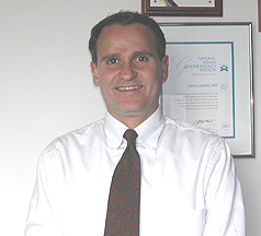
Fabio Candotti earned his M.D. degree at the University of Brescia, Italy, in 1987. He did his postgraduate clinical training in pediatrics and pediatric allergy and immunology in Italy and postdoctoral research training at NCI and NHGRI. He was an assistant professor of pediatrics at the University of Brescia from 1996 to 1998, when he returned to NIH. He is now a senior investigator in NHGRI’s Genetics and Molecular Biology Branch and is head of the Disorders of Immunity Section.
My laboratory studies the molecular basis of inherited disorders of the immune system and works to develop gene replacement strategies for this heterogeneous group of diseases.
Severe inherited immunodeficiencies can be cured with hematopoietic stem cell transplantation, which is most successful if performed using a perfectly matched sibling as a donor. However, most children with these disorders do not have matched siblings, which makes transplantation generally less successful and more risky.
As a pediatrician, I have long had an interest in finding alternatives to hematopoietic stem cell transplantation for the treatment of immunodeficient children.
Currently, we are focusing our research on adenosine deaminase (ADA) deficiency, Wiskott-Aldrich syndrome (WAS), and IL-12 receptor b1 deficiency.
Patients with ADA deficiency are unable to produce significant numbers of mature T or B lymphocytes and thus have a severe combined immune deficiency and no protection against viruses and bacteria. WAS is an X-linked recessive disorder characterized by a less profound form of immunodeficiency, along with eczema and thrombocytopenia. IL-12 receptor b1–deficient patients are extremely susceptible to salmonella and atypical mycobacterial infections.
For the past few years, we have been following a group of patients affected with ADA deficiency and WAS to learn about the natural history of these rare disorders and to evaluate whether genetic correction is a viable therapeutic option.
In addition, we are using in vitro and in vivo models to study the efficacy of corrective gene transfer into lymphocytes and hematopoietic stem cells, using viral vectors based on murine oncoretroviruses and lentiviruses.
At the bedside, we are evaluating novel retroviral vectors as gene-transfer tools for the genetic correction of ADA deficiency. A clinical gene-transfer trial is ongoing to test the hypothesis that these vectors will provide better reconstitution of the immune system than has been observed in previous trials.
In addition, this trial directly compares two viral promoters in the same patients to determine which confers higher expression in humans. The results of this clinical trial will provide important additional safety and biological information to the field of corrective gene transfer into human hematopoietic progenitors.
For WAS and IL-12 receptor b1 deficiency, we are performing preclinical gene-correction studies that have shown that retroviral-mediated gene transfer can correct the biological defects observed in lymphocytes and cell lines obtained from affected patients. We are evaluating similar strategies using in vivo models in knockout animals to test safety and efficacy of the gene transfer procedure in preparation for clinical applications.
Studies of primary immunodeficiency disease have been instrumental in defining multiple players and pathways critical for the correct development and function of the immune system. These unique experiments of nature have provided fertile ground for collaboration among clinical researchers and basic scientists not only in the field of immunology but also genetics and molecular and cellular biology.
Our ongoing explorations include
![]() The role of WASP, the protein mutated in WAS patients, in B lymphocyte development
and autoimmunity onset
The role of WASP, the protein mutated in WAS patients, in B lymphocyte development
and autoimmunity onset
![]() The significance of the IL-12 receptor b-1 chain
in the development and peripheral homeostasis of T cells
The significance of the IL-12 receptor b-1 chain
in the development and peripheral homeostasis of T cells
These subjects cut across
the expertise of several research groups at NIH, which should afford the opportunity
for idea exchange and collaboration. ![]()