
Monica
Ter-Minassian
| T H E N I H C A T A L Y S T | N O V E M B E R – D E C E M B E R 2003 |
|
|
|
|
A RANDOM SAMPLE OF FESTIVAL FARE |
text
and photos
|
|
|
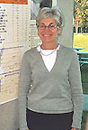
Monica
Ter-Minassian
|
Sun, Genes, and Melanoma
The sun-drenched Italian climate is good for growing both olives and skin cancer. Melanomas have been linked to mutations in two genes involved in cell cycle regulation, CDKN2A (protein p16) and CDK4.
These genes, however, are not mutated in the population of Northeastern Italian melanoma patients studied by NCI’s Monica Ter-Minassian.
Ter-Minassian and her NCI colleagues in the Genetic Epidemiology Branch, DCEG, have now begun whole-genome scans of pedigrees to find linked loci, and eventually genes, in hopes of harvesting the genetic source of melanomas in this region.
 |
|
Ruth
Kleinerman
|
Retinoblastoma Mutants
Retinoblastoma is caused by mutations in the RB-1 tumor suppressor gene.
In the familial form of the disease, which is characterized by disease in both eyes, children inherit one germline mutation from a parent and acquire a second somatic mutation. Both mutations are acquired somatically in the sporadic form of the disease, which affects only one eye. Sporadic and hereditary RB have been effectively treated by high-dose radiation, a treatment that can induce additional cancers.
Ruth Kleinerman and colleagues at NCI's Division of Cancer Epidemiology and Genetics wanted to determine whether patients with familial forms of RB were more susceptible to additional cancers than those with the sporadic version. A retrospective study of 1601 patients—some diagnosed as early as 1914—revealed that inherited mutations in RB-1 do confer increased sensitivity to radiation-induced cancers, especially in the head and neck.
Kleinerman also observed an elevated risk of other cancers, unrelated to radiation exposure, in children inheriting a mutation in RB-1.
 |
|
Fei
Xu
|
Presenting Cancer E-mice And More…
A favorite model for studying disease, mutant mice have now been created and examined by the hundreds of thousands. NCI’s Mouse Models of Human Cancer Consortium created a new website to help scientists navigate the sea of mouse models for cancers. In his poster, NCI’s Fei Xu, of NCI’s Center for Bioinformatics, pointed out some of the site features.
Scientists can search through a cancer model database containing information on all cancer mice by gene, lab, diagnosis, and tumor location. Links interconnect genes with publications, therapeutic strategies, images, and microarray data. Sister sites provide clinical trial information, point mutation databases (human and mouse), SAGE profiles for cancerous and normal tissues, and clickable pathway diagrams. Many of these data are interlinked, allowing scientists to identify new research targets, plan experiments—and predict results.
 |
|
Maria
Campos
|
Autofluoresence—Turning Lemons Into Lemonade
Fixing tissue samples in formaldehyde or glutaraldehyde has long been the best way to preserve cellular structure. Unfortunately, prolonged exposure to these fixatives causes samples to fluoresce, and this complicates analysis using newer imaging techniques such as confocal microscopy. But Maria Campos and her colleagues at NEI have turned this disadvantage into a strength.
Campos uses differential interference contrast (DIC) to identify blood vessels of interest in preserved eye tissue from people with diabetes and experimental rat models. She then switches to confocal microscopy, using specific wavelengths to exploit the autofluorescence characteristics of aldehydes.
Campos' technique opens up repositories of eye and other tissues—collected for decades—to study via contemporary imaging techniques.
 |
|
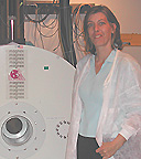
Stasia
Anderson
|
Tracking MS with MRI
Being able to track immune cells in vivo would improve understanding of autoimmune disease. Early-stage multiple sclerosis (MS) is thought to involve a reaction to myelin.
Stasia Anderson, an NINDS staff scientist working in the CC's Experimental Neuroimaging Section, is developing a method to visualize anti-mylelin T cells via MRI. She used ferumoxides-poly-L-lysine iron nanoparticles to label T-cell endosomes (iron cannot be applied to the cell membrane because it might interfere with antigen binding).
Anderson and her colleagues demonstrated that labeled T cells are functionally identical to their unlabeled counterparts. Using the experimental allergic encephalomyelitis mouse model of MS, she visualized labeled T cells in spinal columns of live mice at a resolution of 78–80 mM; higher resolutions can be achieved on sacrificed animals.
Due to the limitations of in vivo MRI, individual cells could not be visualized. However, Anderson did observe the temporospatial migration of groups of T cells that initiate myelin damage in the spinal cord in the live mice—suggesting that this technique may allow monitoring of disease course, as well as therapy used to change the immune response.
 |
|
Amy
Miyoshi
|
Glucosamine and Chondrocytes
Many people take glucosamine as a nutritional supplement to ward off, or at least lessen, the effects of arthritis. Amy Miyoshi, working in Rocky Tuan's NIAMS laboratory, wanted to examine glucosamine’s effect on human cells.
Collagen and proteoglycan deposition, measures of chrondrocyte differentiation, declined when glucosamine was added to mesenchymal stem cells. Mesenchymal progenitor cells have the potential to differentiate into chondrocytes.
Miyoshi and her colleagues added glucosamine to high-density mesenchymal stem cell cultures undergoing chondrogenesis and saw an inhibition of collagen and proteoglycan expression. In contrast, collagen and proteoglycan levels rose in primary chondrocytes, which were prepared from normal and osteoarthritic cartilage taken from human knee replacement surgeries.
The next step will be to test mesenchymal stem cells and chondrocytes growing on a synthetic matrix in order to mimic an in vivo environment. It may be, she speculated, that glucosamine works to maintain a correct balance between mesenchymal stem cells and existing chondrocytes.
 |
|
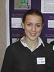
Beatrice
Del Riccio
|
Sniper Attacks and Stress
Completing everyday errands, such as buying gasoline, in the greater Washington, D.C., area last fall was complicated by random sniper attacks. But did the shootings increase depression and anxiety?
Summer intern Beatrice Del Riccio, a senior at Georgetown Day High School in Washington, and her colleagues at NIMH tried to answer that question in a study involving women with depression who were already participants in the POWER (premenopausal, osteoporosis, women, alendronate, depression) study and a control group of women.
Depression and anxiety
levels, as well as biological measures of stress, including plasma cortisol
and ACTH, were assessed before and during the sniper attacks. In neither group
did the biological and psychological parameters of stress change during the
sniper attacks, Del Riccio reported. ![]()