Andrew
Blauvelt
| T H E N I H C A T A L Y S T | M A Y – J U N E 2003 |
|
|
|
| P E O P L E |
RECENTLY TENURED
|
|
Andrew
Blauvelt
|
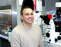
Andrew Blauvelt received his medical degree from Michigan State University, East Lansing, in 1988 and completed his dermatology residency training at the University of Miami, Coral Gables, Fla., in 1992. He then began his research career as an NIH fellow, working first with Stephen Katz at NCI on Langerhans cell–HIV studies and later with Kuan-Teh Jeang at NIAID performing Kaposi’s sarcoma–associated herpesvirus research. He is currently a senior investigator in the Dermatology Branch, NCI.
Scientific work in my laboratory has focused on defining interactions between HIV, herpesviruses, and skin.
Specifically, my laboratory has contributed to two major research areas: 1) examining the role of Langerhans cells and other types of dendritic cells in the pathogenesis of HIV and 2) studying the role of Kaposi’s sarcoma–associated herpesvirus (KSHV) in the pathogenesis of Kaposi’s sarcoma.
In the first area, my lab has been involved in the delineation of the cellular and molecular events that occur when Langerhans cells encounter HIV. Langerhans cells are specialized types of dendritic cells (professional antigen-presenting cells) that are located within the skin and genital mucosal epithelial surfaces, where they serve as sentinels for the immune system.
After encountering complex antigens, Langerhans cells emigrate from epithelial tissues to draining lymph nodes, where they present processed antigenic peptides to T cells.
Most of my HIV research has been driven by the hypothesis that Langerhans cells also serve as initial "targets" for HIV after sexual exposure to virus.
Specifically, we have described HIV co-receptor expression, function, and regulation on Langerhans cells; assessed, in detail, HIV infection versus virion "capture" pathways in Langerhans cells and other dendritic cells; and identified genotypes that predispose to (or protect from) Langerhans cell infection.
An interesting new direction is my recent involvement in translating this basic knowledge into the development of microbicides, which are topical agents designed to block sexual transmission of HIV.
The most promising drug we have studied in this regard is a chemical analog of the chemokine RANTES, a drug that binds to the HIV co-receptor CCR5 on the surface of Langerhans cells and blocks subsequent infection of HIV.
I hope that my research findings on the biology of sexual transmission of HIV will some day prove to be important in decreasing the number of HIV transmissions that occur daily throughout the world.
In a second area of research interest, my lab has contributed to the understanding of how KSHV leads to the development of Kaposi’s sarcoma, the most common cancer found in AIDS patients. KSHV was discovered in 1994 as the long-sought etiologic agent of Kaposi’s sarcoma.
Our early work in this area involved the identification of clinically relevant drugs and biologic conditions that either blocked or induced KSHV reactivation within latently infected cells.
For example, we showed that KSHV was sensitive to cidofovir and ganci-clovir, yet relatively insensitive to acyclovir and its derivatives. Our prediction, although not yet realized, was that this information could ultimately be translated into clinically meaningful treatment advances for patients with Kaposi’s sarcoma.
With this same goal in mind, we have recently turned our attention to analyzing specific KSHV viral proteins that we believe are critically involved in the maintenance of Kaposi’s sarcoma tumors. We have successfully created novel transgenic mice that express two of these viral proteins—LANA and k-cyclin—in vivo.
My hope is that we can improve these mouse models of Kaposi’s sarcoma even further and that they will allow us to preclinically test agents that interfere with LANA and/or k-cyclin expression and function.
Identifying LANA and k-cyclin as novel molecular targets for Kaposi’s sarcoma may eventually help patients with this disease and may also serve as a paradigm for targeting key viral proteins that are involved in the formation of other virus-induced cancers.
 |
|
Allen
Braun
|
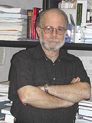
Allen R. Braun received his M.D. in 1980 from Rush Medical College in Chicago, where he also completed a residency in neurology. His postdoctoral training was at the NIH Clinical Center in the Experimental Therapeutics Branch of NINDS and in the Department of Nuclear Medicine, where he completed an additional residency with an emphasis on PET imaging. He joined NIDCD in 1991 and is currently acting chief of the Language Section, Voice, Speech, and Language Branch.
Our lab uses a variety of neuroimag-ing methods—functional and structural MRI, PET, electro- and magnetoencephalography—to study auditory processing, voice, speech, and language in the human brain. Each of these methods provides qualitatively different information. Our multimodal approach thus yields complementary and converging evidence that we use to pinpoint normal brain-language relationships and the ways in which these relationships are altered in neurological disorders that affect the ability to communicate.
We investigate the use of complex natural language, that is, language as it is used in everyday communication, rather than performance on highly structured artificial tasks commonly utilized in neuroimaging research. Thus, we study both comprehension and production—in the real world these are essentially inseparable. We look at language at multiple levels: We study receptive language from basic auditory and visual perception up to the level of discourse comprehension; we study production from the level of language formulation to overt articulation.
By studying language in this more natural context, we have been able to show that there are emergent features—activation of a host of regions outside what is traditionally considered "language" cortex—that are seen during the processing of narrative discourse. This activity is not apparent during simple processing of isolated sentences or words, typically investigated in neuro-imaging studies of language. Our approach also permitted us to demonstrate dynamic fluctuations in brain activity during discourse processing and to locate brain systems that are responsible for making inferences when subjects read a complex narrative text.
Beyond this, we’ve gone on to com pare the production of narrative in English and American Sign Language in bilingual subjects and have found that there is a core, unitary network for the production of language that is independent of the modality in which the language is expressed. Studies of overt speech production in monolinguals have capitalized on the complementary features of fMRI and EEG: fMRI shows us where in the brain lexical access or semantic decisions occur, and EEG shows us how information flows between these regions over time.
We are evaluating the use of gesture in conversational discourse and the ways in which the brain processes music—how we perceive and produce both melody and rhythm. We recently identified unique, lateralized differences in brain activity during singing (vs. speaking), an issue that may seem arcane but actually may be clinically important: Such mechanisms may enable fluent speech production in developmental stuttering and in certain types of aphasia.
Indeed, many of our approaches have been developed in control subjects with the idea of translating these into clinical studies of neurological illnesses affecting speech, voice, and language—in order to characterize the pathophysiology of these disorrders and to monitor the effects of treatment.
Examples of our translational work include investigation of ways in which dysfunction in the basal ganglia—likely mediated by dopaminergic mechanisms—affects speech and language. We have demonstrated abnormal patterns of activity in developmental stuttering that implicate the basal ganglia and their projections. We also characterized cerebral responses to dopamine agonists and deep brain stimulation during speech and language production in Parkinson’s disease. We described functional abnormalities in basal ganglia circuitry in Tourette’s syndrome and demonstrated clinically significant responses to botulinum toxin treatment in spasmodic dysphonia. We are currently using neurochemical PET methods to characterize pre- and postsynaptic dopamine mechanisms, which we hypothesize may play a central role in the pathophysiology of stuttering.
Functional neuroimaging can also be a powerful counterpart in the genetic investigation of communication disorders: In collaboration with NIDCD’s Dennis Drayna, our lab is investigating a heritable disorder of musical pitch perception. For those studies we are using electrophysiological and MRI methods to provide phenotypic markers for linkage studies. We hope ultimately to clarify the role a defective gene product may play within the CNS.
Our lab is increasingly interested in neuroplasticity—the capacity of the brain to adapt and reorganize in response to experience, trauma, or stress. One way we are pursuing this is through longitudinal PET studies of central auditory processing by deaf people who have received cochlear implants. Initially, following activation of the implant, most recipients cannot understand the auditory input they receive. Over months to years, however, their auditory understanding improves to a remarkable degree, although signals from the implant do not change. These individuals may thus provide a unique chance to study neural reorganization in response to sensory input.
Over the next several years, we also plan to use neuroimaging and neurobehavioral and pharmacological methods to study the natural history of language recovery in stroke patients with aphasia. In this work, we will follow patients with different degrees of language loss and recovery and monitor their progress over time. We will look for correspondences between linguistic performance and the neural processes that are visible with our imaging techniques—such as cortical map expansion or changes in neurochemical or electrophysiological function. Such correspondences could point to ways in which recovery of function may be facilitated by behavioral and pharmacological intervention.
The concept of use-dependent plasticity—recovery of function after training and repeated experience—is now well established. Recent studies have shown that some degree of motor recovery—likely mediated by synaptic plasticity or structural reorganization–can occur in stroke patients, even years after brain damage has occurred. We hypothesize that there will be similar, significant responses in aphasics who receive intensive training aimed at correcting specific psycholinguistic deficits.
If we can demonstrate this, we intend to conduct clinical trials of drugs that, by modulating signal transduction mechanisms, may facilitate use-dependent neuroplastic effects—by amplifying late-phase long-term-potential–like responses, stimulating neurotrophin transcription and synthesis, or enhancing the reorganization of axonal or dendritic architecture. In each case, we will use our armamentarium of imaging tools to evaluate outcome.
 |
|
Carter
Van Waes
|
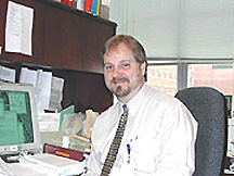
Carter Van Waes received his Ph.D. in tumor immunology in 1985 and his M.D. degree in 1987 under the NIH Medical Scientist Training Program at the University of Chicago. He completed a cancer research fellowship and residency in otolaryngology–head and neck surgery at the University of Michigan, Ann Arbor, between 1988 and 1993. He developed the Tumor Biology Section in the Head and Neck Surgery Branch, NIDCD, where he is now a senior investigator and acting clinical director.
In the process of trying to understand the molecular basis for immune recognition, inflammation, and angiogenesis in human and murine squamous cell carcinomas (SCC), my colleagues and I detected a diverse repertoire of cell recognition and cytokine molecules that are usually expressed in response to injury. I noted that the promoters of the genes encoding many of these molecules contain sites for activation by an injury-response transcription factor, nuclear factor–kappa B (NF-kB), originally identified in the laboratory of David Baltimore. I hypothesized that NF-kB may contribute to regulation of gene programs that contribute to the malignant phenotype, including proliferation, cell survival, tumorigenesis, angiogenesis, and inflammation.
My lab demonstrated that NF-kB is constitutively activated and that multiple genes related to the NF-kB pathway are expressed at increased levels with metastatic tumor progression (Cancer Res., 59:3495–3504, 1999; Mol. Carcinog., 26:119–129, 1999; Cancer Res., 61:4797–4808, 2001).
Inactivation by dominant negative mutants of inhibitor-kB or pharmacologic inhibitors of proteasome-mediated activation blocked proliferation and cell survival in vitro, and angiogenesis and tumorigenesis in syngeneic murine and human xenograft models (Cancer Res., 59:3468–3474, 1999; Clin. Cancer Res., 7:1419–1428, 2001)
Recently, my lab has used microarray profiling to confirm an important role of NF-kB in the cumulative changes in gene expression with progression of SCC, and after switching off NF-kB by an inhibitor-kB mutant.
Remarkably, inhibition of NF-kB restored the expression of more than 60 percent of the 308 genes differentially expressed in metastatic murine SCC cells compared with expression in normal keratinocytes.
Most of the NF-kB–upregulated genes contained NF-kB promoter sequences, and most of the downregulated genes contained homologous 7-nucleotide motifs involved in the NF-kB–dependent downregulation of mRNA. Inhibition of NF-kB corrected expression of representative mRNAs and proteins, and inhibited malignant phenotypic features, including cell survival, proliferation, migration, and angiogenesis. These results indicate that NF-kB is an important molecular switch for the gene expression program and malignant phenotype in SCC.
Inhibition of NF-kB also sensitized squamous carcinomas to radiation, one of the important therapies for patients with head and neck cancers.
In collaboration with colleagues in NCI medical and radiation oncology, I am engaged in a phase I clinical trial of concurrent therapy with a proteasome inhibitor and radiation in patients with inoperable SCC of the head and neck.
My lab is examining the ability of the drug to inhibit proteasome and NF-kB activation in tumor, and NF-kB–regulated cytokine levels in serum as a marker of response and recurrence.
There are several important questions raised by these observations:
![]() What molecular events affecting known or unknown oncogenes and tumor suppressor
genes can result in activation of NF-kB as an important
common pathway to the malignant phenotype?
What molecular events affecting known or unknown oncogenes and tumor suppressor
genes can result in activation of NF-kB as an important
common pathway to the malignant phenotype?
![]() Are most of the genes apparently regulated by NF-kB
regulated directly—or indirectly via activation of other transcriptional
programs?
Are most of the genes apparently regulated by NF-kB
regulated directly—or indirectly via activation of other transcriptional
programs?
![]() Which target genes are responsible for the key changes in proliferation, survival,
angiogenesis, and inflammation?
Which target genes are responsible for the key changes in proliferation, survival,
angiogenesis, and inflammation?
![]() Is NF-kB one of several pathways co-activated in
SCC that cooperate in activation of different repertoires of genes that together
determine the differences in malignant behavior and resistance to therapy?
Is NF-kB one of several pathways co-activated in
SCC that cooperate in activation of different repertoires of genes that together
determine the differences in malignant behavior and resistance to therapy?
I hope that studies directed
at these questions will help us develop improved methods for molecular diagnosis,
treatment selection, and molecularly targeted therapy of SCC and other cancers.
![]()