Sergey
Bezrukov
| T H E N I H C A T A L Y S T | N O V E M B E R – D E C E M B E R 2002 |
|
|
|
| P E O P L E |
RECENTLY TENURED
|
|
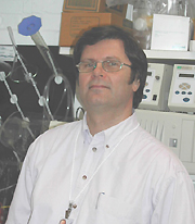
Sergey
Bezrukov
|
Sergey Bezrukov received his Ph.D. in physics and mathematics from Moscow State University in 1981 and did postdoctoral work at the St. Petersburg Institute of Nuclear Physics, Russian Academy of Sciences. In 1990 he joined the Laboratory of Biochemistry and Metabolism of NIDDK as a special volunteer and in 1992 as a Visiting Scientist. He is now a senior investigator in the Laboratory of Physical and Structural Biology, NICHD.
A physicist by education, I believe that interactions between physics and biology are mutually rewarding because observations on biological materials and in biology in general lead to discoveries of new physical principles.
Our Section on Molecular Transport approaches ion channels from two unique directions: (i) ion channels as molecular Coulter counters to probe metabolite and macromolecule transport; and (ii) ion channels as stochastic resonators to transduce weak sensory signals.
Molecular Coulter Counting. Bacterial porins, mitochondrial channels, gap junctions, some toxins, the nuclear pore complex, and protein-conducting channels of the endoplasmic reticulum are "mesoscopic" channels designed to transfer solutes larger than water and small ions. In order to observe the dynamics of this large-solute transport, we invented molecular Coulter counting.
The underlying idea for molecular Coulter counting is very similar to the resistive pulse principle used since 1953 in Coulter counters. The basic difference is that the pulses in current come from nanometer-sized molecules such as ATP or sugars rather than micrometer cells, as in the standard Coulter counter. Using mesoscopic channels, we are watching the passage of molecules with gyration radii as small as 5–15 Å. The dynamics and selectivity of transport demonstrate that many of these large channels, rather than being merely general-diffusion pores, are highly specialized to conduct metabolites across membranes.
We were recently able to use this approach to observe the details of a single drug molecule’s translocation through a target membrane. It is well known that membrane permeability barriers are among the main contributors to bacterial antibiotic resistance. Using the bacterial porin OmpF reconstituted into planar lipid bilayer membranes, we saw how addition of the betalactam antibiotic ampicillin created transient interruptions in small-ion current through the channel. The kinetics of these transients gave us reaction parameters needed to link antibiotic penetration to the molecular features of the bacterial channel and the drug. We found that the charge distribution of an efficient antibiotic complements the charge distribution at the narrowest part of the pore. Consequently, we have recognized molecular interactions that can reduce the membrane permeability barrier and increase antibiotic efficacy.
Stochastic Resonance. This term refers to the counterintuitive phenomenon in which random fluctuations added to weak signals actually bring out otherwise undetectable features by improving a transducer’s performance. Studying the influence of ambient noise on signal transfer through the ion channels of alamethicin, we found that external voltage fluctuations could play a constructive role via stochastic resonance.
With increasing intensities of white noise, the signal-to-noise ratio actually peaks at a particular added-noise level. Thus, we had demonstrated that this surprising phenomenon actually operates at the molecular level, not just in the instruments of physicists.
Our next major contribution was to show the generality of stochastic resonance. By reducing this phenomenon to its mathematical essence, we demonstrated that noise-facilitated signal transduction is a far more general statistical property of nonlinear systems than was previously believed. In this way we liberated the concept and highlighted its ubiquitous possibilities.
Keeping in mind the enormous structural and dynamic complexity of neural organization, our next logical step is to investigate whether and how noise-facilitated signal transduction reaches the molecular scale of intra- and intercell communication. We have begun to examine the electrosensitive organs of elasmobranchs. We hope that the study of these organs will elucidate the functioning of the central nervous system as profoundly as the study of the squid giant axon has served to reveal the process of action-potential conduction.
 |
|
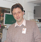
Sergey
Leikin
|
Sergey Leikin received his Ph.D. from the Moscow State University in 1987. He did his postdoctoral work at the Frumkin Institute of Electrochemistry in Moscow, Russia, and at DCRT (CIT), NIH. He joined NICHD in 1997 and is now a senior investigator and chief of the Section on Physical Biochemistry, NICHD.
My interest lies in understanding the physics of biomolecular recognition and assembly reactions and in development of treatments for diseases related to these reactions.
I focus on collagen fiber formation and function, particularly in brittle bone disease, or osteogenesis imperfecta (OI). Although many factors in bone disease—such as collagen mutations—are known, the exact molecular mechanisms of bone pathology are poorly understood. Treatments are scarce and rarely effective.
In my early work at NIH, my colleagues and I were the first to report direct measurement of collagen-collagen forces in fibers. We described the physical nature of these forces and demonstrated that the helical domain of collagen is responsible for the recognition that governs fibrillogenesis, whereas nonhelical terminal peptides play only a catalytic role in fiber formation.
Probably the most surprising discovery stemming from this work was that collagen fibrillogenesis is initiated by melting (micro-unfolding) of the most thermally labile regions of the protein triple helix. We further showed that triple-helical collagen monomers are unstable at body temperature. For instance, human lung collagen fully denatures within 2–3 days at 37 oC rather than being stable up to 41–42 oC as previously thought.
It now appears that procollagen is forcibly folded by specialized chaperonins (for instance, Hsp47) inside cells. Once it loses chaperonins and is secreted, it becomes unstable and begins to unfold. Initial melting triggers assembly of fibers, which in turn protects molecules from further unfolding. Proteins and processes susceptible to OI mutations include:
![]() Chaperonin-assisted procollagen folding
Chaperonin-assisted procollagen folding
![]() Extracellular enzymatic processing
Extracellular enzymatic processing
![]() Micro-unfolding and complete unfolding
Micro-unfolding and complete unfolding
![]() Fibrillogenesis
Fibrillogenesis
![]() Collagen-collagen interactions in fibers
Collagen-collagen interactions in fibers
![]() Collagen interactions with matrix proteoglycans
Collagen interactions with matrix proteoglycans
We continue investing a substantial fraction of our efforts in studies of complex interrelationships between these processes. As we gain more basic knowledge, we gradually increase our involvement in studies of specific bone disorders in collaboration with NICHD clinical researchers.
Careful thinking about interactions between collagen helices required a rigorous theory. We found a solution to this rather difficult problem and demonstrated that helical symmetry of molecular structure not only creates characteristic X-ray diffraction patterns, but also determines features of molecular interactions. This theory explained the measured forces not only between collagen helices, but also between DNA helices.
The theory also gave a rationale for DNA overwinding from 10.5 bp/turn in solution to 10 bp/turn in aggregates, and for hexagonal/cholesteric transition in hydrated DNA aggregates. It clarified electrostatic interactions responsible for changes in packing of DNA aggregates, and it suggested that DNA duplexes may recognize sequence homology at a distance via electrostatic interactions modulated by sequence-dependent variation in the twist between adjacent base pairs.
We often rely on analogy with DNA and other biological helices and on theory coupled with experiments as tools for understanding underlying common physical principles. We hope that such knowledge will be used someday to design new treatments for disorders associated with pathology of molecular recognition.
In the meantime, we are trying to bring this research directly to the bedside in diagnosing and monitoring patients. We assisted NICHD clinical researchers in determining the origin of unusually severe hyperextensibility and joint laxity in several OI patients at NIH. Although we do not have a treatment, we at least now know what is wrong and what should be fixed. As I recently learned at a meeting, even such basic understanding of the origins of their disease provides hope and tremendous psychological help for OI patients.
 |
|
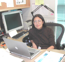
Cecilia
Lo
|
Cecilia Lo received her Ph.D. from Rockefeller University in New York City in 1979 and did postdoctoral work at Harvard Medical School in 1979–1980. In 1980, she was appointed to the faculty of the University of Pennsylvania, Philadelphia, where she rose through the ranks to full professor in the Biology Department in the School of Arts and Sciences before joining NHLBI in 2001 as chief of the new Laboratory of Developmental Biology.
A long-standing research interest in my laboratory is the role of gap junctions in mammalian development. Gap junctions are cell junctions containing membrane channels encoded by the connexin multigene family. They allow direct cell-to-cell movement of ions, metabolites, and cell-signaling molecules.
The gap junction gene connexin43 plays an essential role in heart development. Our studies indicate connexin43 modulates two extracardiac cell populations—the cardiac neural crest and proepicardially derived cells. These two cell populations play a critical role in development of the heart outflow tract and patterning of the coronary arteries.
Using transgenic and knockout mouse models, we determined that connexin43 regulates the deployment of these two cell populations to the heart. This involves modulation of cell motility by a novel signaling function of connexin43, one of which is separable from the channel-forming capacity of the connexin43 protein. Studies are currently underway using time- lapse videomicroscopy and laser capture microscopy to elucidate the cellular and molecular mechanisms regulating neural crest and proepicardial cell migration. We are also pursuing other genomic and proteomic approaches to further define cell signaling by connexin43.
Another line of investigation in my laboratory entails a discovery-based approach using chemical mutagenesis to identify genes and genetic pathways essential for mammalian cardiovascular development. For these studies, N-ethyl N-nitrosourea (ENU)–mutagenized mice are analyzed using dual genotype- and phenotype-based screens.
The genotype-based screen focuses on the identification of new mutations in the connexin multigene family and is carried out using a novel custom DNA resequencing microarray. The resequencing array provides DNA sequence information for all 20 mouse connexin genes and thus is an efficient method for identifying mutants with novel connexin mutations. These mouse mutants will be used to elucidate the cell-signaling function of connexins and their potential role in cardiovascular development and function.
For the phenotype-based screen, we use noninvasive in utero fetal Doppler echocardiography to identify mutants with structural and functional cardiovascular anomalies. This screen has identified mice with various forms of arrhythmias, outflow tract anomalies, congestive heart failure, and other cardiovascular defects. Efforts are underway to map and identify the genetic alterations underlying these cardiovascular phenotypes.
In summary, my lab’s research program focuses on genetic, cell, and molecular biology of mammalian cardiovascular development. We are integrating whole animal imaging, microscopy, and genomic and proteomic approaches to elucidate the role of connexins and other genes and genetic pathways essential for normal mammalian cardiovascular development. We plan to extend these studies to human patient populations and determine whether connexin mutations and mutations in other genes identified in our mutagenesis screen play a role in human congenital heart disease.
 |
|
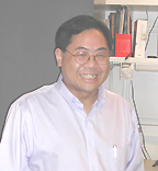
Michael
Quon
|
Michael Quon received his Ph.D. in biomedical engineering (1987) and M.D. (1988) from Northwestern University in Evanston, Ill., and Chicago, respectively. After completing an internship and residency in internal medicine at the University of Chicago, he entered the Interinstitute Endocrine Training Program at NIH in 1990 and did additional postdoctoral work in the Diabetes Branch, NIDDK. He then joined the Hypertension-Endocrine Branch, NHLBI, as a tenure-track Investigator in 1995 and was recently appointed chief of the Diabetes Unit, Laboratory of Clinical Investigation, NCCAM.
I have a long-standing interest in understanding the molecular mechanisms of insulin action and insulin resistance as they relate to the pathophysiology of diabetes, obesity, and cardiovascular diseases. My graduate school studies focused on developing detailed mathematical models of insulin signaling pathways regulating metabolic actions of insulin.
When I arrived at NIH as an Endocrine fellow in 1990, I complemented my mathematical biology background by acquiring experimental expertise in the molecular and cellular biology of insulin signal transduction in the laboratory of Simeon Taylor, Diabetes Branch, NIDDK.
In collaboration with Sam Cushman’s lab, I developed a novel electroporation method to transfect rat adipose cells in primary culture that enabled us to overexpress and inhibit various insulin signaling molecules in a bona fide insulin target cell. Using this technique, we identified the insulin receptor tyrosine kinase, members of the insulin receptor substrate (IRS) family, and phosphatidylinositol 3-kinase (PI 3-kinase) as essential signaling components. These molecules regulate insulin-stimulated translocation of the insulin-responsive glucose transporter GLUT4 from an intracellular compartment to the cell surface. We ruled out a role for Ras in this process.
After I established my own independent laboratory in the Hypertension-Endocrine Branch, NHLBI, in 1995, we continued studies along these lines. This led us to identify the protein tyrosine phosphatases PTP1B and PTP-a as negative regulators of metabolic actions of insulin that act by dephosphorylating the insulin receptor and IRS proteins. Downstream from PI 3-kinase, we demonstrated that PDK-1, Akt, and PKC-z also participate in the regulation of insulin-stimulated translocation of GLUT4. Moreover, we uncovered additional complexities in metabolic insulin signaling pathways by showing that IRS-1 is a novel substrate for PKC-z (an example of a negative feedback pathway) and by identifying PTP1B as a novel substrate for Akt (an example of a positive feedback pathway).
In NHLBI, we also initiated a new project to investigate vascular actions of insulin. Using a nitric-oxide selective electrode, we were the first to demonstrate directly that insulin can stimulate the production of the potent vasodilator nitric oxide (NO) from human vascular endothelial cells in primary culture. More recently, we developed a fluorescent dye–based method for directly visualizing NO production in single living cells. Using these novel techniques, we elucidated a complete biochemical pathway in endothelial cells involving the insulin receptor phosphorylating IRS-1. In this pathway, the insulin receptor phosphorylates IRS-1, leading to binding and activation of PI 3-kinase, subsequent activation of PDK-1, phosphorylation and activation of Akt, which then directly phosphorylates and activates endothelial nitric oxide synthase (eNOS). Moreover, we have shown that this phosphorylation-dependent mechanism for activation of eNOS is completely independent and separable from the classical calcium-dependent activation of eNOS used by most G-protein coupled receptors.
The striking parallels we discovered between signaling pathways related to metabolic actions of insulin and signaling pathways regulating vasoactive actions of insulin support our hypothesis that regulation of metabolic homeostasis and hemodynamic homeostasis are coupled. Thus, factors causing metabolic insulin resistance may also predispose to impaired vasodilator actions of insulin and provide a molecular explanation for the frequent association observed between diabetes and hypertension.
Our lab also conducts patient-oriented clinical investigation of metabolic and vascular physiology in diabetes, obesity, and hypertension. The gold-standard method for measuring insulin sensitivity in humans is the hyperinsulin-emic euglycemic glucose clamp. We use this method in conjunction with forearm blood flow studies to investigate the relationship between vascular and metabolic actions of insulin in vivo.
These labor-intensive procedures are not easily applied in large studies. Therefore, we recently devised and validated a novel index of insulin sensitivity based on a mathematical transformation of fasting glucose and insulin levels. This index, which we named the Quantitative Insulin-sensitivity Check Index (QUICKI), is much simpler to use than the glucose clamp and is useful for large epidemiological studies as well as for following changes in insulin sensitivity in individual patients after therapeutic interventions. QUICKI has now been used successfully by others in more than 40 published clinical studies.
Future studies in NCCAM will aim to identify and elucidate the molecular basis of promising therapies for diabetes, obesity, and hypertension within the realm of complementary and alternative medicine.
 |
|
Karen
Usdin
|
Karen Usdin received her Ph.D. in microbiology from the University of Cape Town (UCT), South Africa, in 1986 and did postdoctoral work at UCT and in the Laboratory of Biochemical Pharmacology, NIDDK. She is now a senior investigator in the Laboratory of Molecular and Cellular Biology,
I am interested in the repeat expansion diseases—genetic disorders caused by an increase in the size of a specific tandem repeat sequence. Expansion beyond a threshold size has pathological consequences that depend on the location of the repeat in the affected gene. When the repeat is in the coding sequence, the connection between expansion and disease symptoms is relatively straightforward: Expansion results in the production of an aberrant protein that is toxic. It is less straightforward when the repeat is outside the open reading frame.
I am interested in both the mechanism and the consequences of expansion in this latter group of disorders, which includes progressive myoclonus epilepsy type 1 (EPM1), fragile X mental retardation syndrome (FXS), and Friedreich’s ataxia (FRDA).
EPM1, which is characterized by tonic-clonic seizures and progressive stimulus-sensitive myoclonus, results from expansion of a C4GC4GCG•CGCG4CG repeat tract in the promoter of the cystatin B gene.
FXS, the most common heritable cause of mental retardation, is caused by expansion of a CGG•CCG-repeat in the 5' UTR of the FMR1 gene. Carriers of FXS "premutation" alleles—intermediate-sized alleles at risk of expansion to the full mutation on maternal transfer—have a much higher incidence of premature ovarian failure and cerebellar dysfunction than individuals with the "full mutation." Expansion has two apparently paradoxical effects on transcription in FXS: At "premutation" lengths, it leads to an increase in FMR1 transcription. In the "full mutation," it leads to hypermethylation of the promoter and a dramatic decrease in transcription. Expansion also results in a transcript that is translated less efficiently.
FRDA, a neurodegenerative disease associated with cardiomyopathy and diabetes, is caused by GAA•TTC-repeat expansion in the first intron of the frataxin gene.
My group initially set out to investigate the biochemical properties of the disease-causing repeats. We showed that in vitro DNA containing such repeats forms unusual secondary structures such as tetraplexes or triplexes, and that the ability to form such structures is a common property of hypervariable sequences in general. In addition, we showed that in vitro transcripts containing the FXS repeats form stable RNA hairpins containing a mixture of C•G and G•G base pairs.
These structures may have biological consequences. For example, we have shown that triplex formation occurs during transcription of the FRDA repeats. This traps RNA polymerase on the template, leading to a deficit of full-length transcript, which mirrors that seen in FRDA patients.
We also showed that structures formed by the FXS repeat block DNA synthesis. These blocks may force premature condensation of chromatin at mitosis, resulting in the fragile site seen at the FMR1 locus in FXS patients. These blocks may also cause expansion by promoting strand slippage or by recombino-genic repair of the stalled replication fork.
We showed a correlation between secondary structure formation and instability of the repeat tract in bacteria. In collaboration with Robert Nussbaum (NHGRI), we generated transgenic mice carrying part of an FXS allele with a 100 percent probability of expansion in humans. These mice partially recapitulate what occurs in FXS pedigrees, producing large deletions but no expansions. This suggests that expansion and deletion occur by different mechanisms and that specific cis- and/or trans-acting factors may be necessary for expansion.
However, crossing these mice with mice deficient in a number of important DNA repair and replication pathways also did not produce any large expansions. We are now generating mice containing a targeted insertion of a long repeat tract in the murine Fmr1 gene in hopes of creating a better model of expansion. These mice would be the murine equivalent of FXS "premutation" carriers and thus may also be useful for understanding disease pathology in these individuals.
RNA hairpins, such as those formed by the FXS repeat, may be responsible for the symptoms seen in both "premutation" and "full mutation" carriers. For example, certain double-stranded RNAs activate enzymes like the protein kinase PKR that can lead to apoptosis. The FXS-RNA hairpins are thus a potential source of RNA toxicity that could lead to the ovarian and cerebellar dysfunction seen in FXS "premutation" carriers. Our preliminary data showing the ability of CGG-RNA to activate PKR in mammalian cells support this hypothesis.
In addition, double-stranded RNA may be a trigger for epigenetic modifications of chromatin that lead to gene silencing. Thus the FXS RNA hairpins may also cause the transcription deficit in "full mutation" carriers. We now have the cell lines that will allow us to test this hypothesis. Finally, the RNA hairpins may also block scanning of the 5' untranslated region by the 40S ribosomal subunit, thus causing the translation deficit seen in alleles with large repeat tracts.
Since the coding sequence is intact in most FXS patients, it is possible that reversing or counteracting the epigenetic events that cause promoter silencing may have therapeutic value. My group has identified the transcription factors necessary for FMR1 gene expression and shown that the binding of one of them, NRF-1, is abolished by cytosine methylation. This suggests that DNA demethylation will be necessary to fully reactivate the gene. We are currently exploring different strategies for restoring FMR1 gene function.
 |
|
Thomas
Wynn
|
Thomas Wynn received his Ph.D. in Medical Microbiology and Immunology from the University of Wisconsin–Madison in 1991. He worked as an IRTA fellow in the Immunobiology Section of the NIAID Laboratory of Parasitic Diseases and as an NIH senior staff fellow before being appointed to NIAID’s tenure-track program. He is now a senior investigator in the Laboratory of Parasitic Diseases, NIAID, where he heads the Immunopathogenesis Section.
My research is focused on understanding the molecular and immunological mechanisms of pathogenesis in schistosomiasis and other parasitic diseases. Schistosomiasis is an important human disease, with about 200 million adults and children in 74 countries infected with the waterborne parasite. Approximately 20 million people annually develop serious complications because of chronic infection. More than 600 million people are at risk of infection, and recent estimates suggest that about 280,000 people die each year in sub-Saharan Africa alone. Schistosomiasis ranks second, just behind malaria, as a cause of morbidity and mortality from a parasitic disease. There is also emerging evidence that infection with helminthes, such as schistosomiasis, is a major aggravating factor in both AIDS and tuberculosis in the developing world. Thus, the disease imposes a high socioeconomic burden on many of the affected developing countries.
Although there are drugs for the treatment of schistosomiasis, people living in endemic areas are at a constant risk of reinfection. Indeed, a substantial portion of the population is reinfected within one to two years after treatment. Therefore, control of morbidity and mortality via drug therapy requires continuous surveillance of the population and intermittent chemotherapy. Control of schistosomiasis using conventional means is, therefore, life-long and expensive.
A relatively inexpensive alternative would be a vaccine that either prevents or reduces infection or significantly reduces the occurrence of severe disease. My work is focused on better understanding the pathogenesis of schistosomiasis, so that a more defined and rational approach to vaccination might be developed.
In murine schistosomiasis, the pathology resulting from infection with Schistosoma mansoni is predominantly caused by the host reaction to parasite eggs that are laid in the portal venous system and subsequently trapped in the liver and intestine. Egg-induced liver fibrosis can lead to portal hypertension, which causes much of the morbidity and mortality associated with the disease.
A major emphasis of my research program has been to dissect the contributions of type-1 (IFN-g) and type-2–associated cytokines (IL-4, IL-10, and IL-13) to the pathogenesis of schistosomiasis. The mechanisms controlling granulomatous inflammation and development of hepatic fibrosis have been the primary focus. Such studies are needed because it is the host response to the eggs, rather than the parasite itself, that is the major driving force in the development of hepatosplenic disease. An important finding in our studies was the discovery that the Th2-type cytokine IL-13 is the critical profibrogenic mediator in murine schistosomiasis.
Although there is a great deal of mechanistic information regarding the process of tissue remodeling and fibrosis, there are still large gaps in our understanding of the role of inflammatory cells and cytokines in the initiation and maintenance of the fibrotic process. In experiments conducted with a soluble IL-13 antagonist, we showed that IL-13 plays an indispensable role in the fibrotic process. In contrast, IFN-g, IL-12, and IL-10 all appear to antagonize the profibrotic effects of IL-13. Indeed, mice that exhibit strong IL-13 and weak IFN-g/IL-10 responses consistently display the most severe pathology from S. mansoni infection.
We have also identified macrophages and fibroblasts as major targets of IL-13 activity. Macrophages activated with IL-13 express high arginase-1 activity, which is critical for proline production, the basic building block of collagen. IL-13 receptors are also expressed on fibroblasts, and we showed that IL-13 could directly activate collagen deposition by these cells.
Looking ahead, a major objective of my research will be to determine whether IL-13 antagonism can be developed as a strategy to treat chronic fibrotic disease and to characterize the molecular pathway of IL-13–mediated fibrogenesis. Little information exists regarding the regulation of fibrogenesis by type-2 cytokines; therefore, a major effort of our research program in the coming years will be to further characterize this novel aspect of the Th1/Th2 paradigm.
We hope that better understanding
these basic mechanisms and disease processes in general will ultimately translate
into an effective disease intervention strategy for schistosomiasis. Beyond
that, fibrosis is also a major complication in asthma, chronic graft rejection,
and several autoimmune diseases. We therefore hope that the information we glean
for schistosomiasis may also provide new directions for the treatment of fibrotic
disease in general. ![]()