| T H E N I H C A T A L Y S T | S E P T E M B E R – O C T O B E R 2002 |
|
|
|
| P E O P L E |
RECENTLY TENURED
 |
|
Thomas
Dever
|
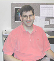
Thomas Dever received his Ph.D. from Case Western Reserve University in Cleveland in 1990 and did postdoctoral work in the Laboratory of Molecular Genetics, NICHD, before joining NICHD’s Laboratory of Eukaryotic Gene Regulation in 1994. He is now a senior investigator in the Laboratory of Gene Regulation and Development, NICHD.
My research interest is the mechanism and regulation of cellular protein synthesis—what I like to call the ultimate step of gene expression. As a graduate student, I used biochemical techniques to characterize translation initiation factors—proteins that facilitate binding of the ribosome to an mRNA and selection of the AUG start codon for protein synthesis.
As a postdoc with Alan Hinnebusch, I continued my studies on translation initiation, investigating the roles of the factor eIF2 in control of GCN4 expression in the yeast Saccharomyces cerevisiae.
We discovered that the yeast protein kinase GCN2 phosphorylates the a subunit of eIF2 to downregulate general translation while paradoxically stimulating GCN4 mRNA translation. We went on to show that the human dsRNA-activated, antiviral kinase PKR could substitute for GCN2 in yeast, phosphorylate eIF2a, and stimulate GCN4 expression.
In my lab, we continued to study the phosphorylation of eIF2a, focusing on the mechanism of kinase-substrate recognition. To overcome the inhibitory effects of PKR, most viruses express inhibitors of the kinase. We found that high-level expression of PKR in yeast is toxic and that co-expression of the vaccinia virus K3L protein, a pseudosubstrate inhibitor of PKR, suppresses this toxicity.
Mutational analyses demonstrated that a sequence motif shared between the K3L protein and eIF2a, and located around 30 residues from the Ser-51 phosphorylation site in eIF2a, is critical for eIF2a phosphorylation and for K3L protein inhibition of PKR. These results reveal an unexpected contribution of remote sequences to kinase-substrate recognition and indicate that phosphorylation site consensus sequences are not sufficient to characterize all kinase-substrate interactions.
In related studies, we used our yeast system to identify a novel eIF2a kinase inhibitor from baculovirus. This inhibitor resembles a truncated protein kinase. We are currently extending these studies using molecular genetic, biochemical, and structural techniques to identify the residues in PKR and GCN2 that contribute to the recognition of eIF2a and selection of Ser-51 as the phosphorylation site.
A second research focus in my lab is the GTP-binding protein IF2/eIF5B. In bacteria, the factor IF2 binds the initiator methionine tRNA to the ribosome, a function carried out by eIF2 in eukaryotic cells. We discovered an IF2 ortholog in yeast and humans and demonstrated that this protein, now called eIF5B, is a translation initiation factor.
Together with Tatyana Pestova at the State University of New York, Brooklyn, we showed that eIF5B is a ribosome-dependent GTPase that promotes subunit joining in the final step of translation initiation.
In collaboration with Stephen Burley at The Rockefeller University in New York, we determined the X-ray structure of eIF5B, revealing a chalice-shaped protein that undergoes lever-type domain movements upon GTP binding.
Our recent genetic and biochemical analyses revealed that GTP-binding regulates the ribosome affinity of eIF5B, and we are currently conducting structure-based mutational analyses to further our understanding of eIF5B function and the mechanism of GTP hydrolysis by eIF5B.
Protein synthesis is a somewhat underappreciated step in gene expression; however, recent studies have revealed the importance of translational regulation in cell growth, development, and human diseases.
By combining yeast genetics, molecular biology, biochemistry, and structural studies, we hope to provide new and critical insights into this final step of gene expression.
 |
|
Eric
Freed
|
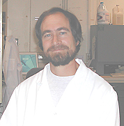
Eric Freed received his Ph.D. from the University of Wisconsin–Madison in 1990. He did postdoctoral work at UW–Madison before joining the Laboratory of Molecular Microbiology (LMM), NIAID, in 1992. He is now a senior investigator in the LMM.
For a number of years my work has focused on understanding the molecular biology of HIV replication. When I began at NIH, the functional properties of most HIV proteins were poorly understood.
Our initial efforts were therefore focused on characterizing viral protein function. We were particularly interested in the Gag proteins, which direct virus assembly and release, and the envelope glycoproteins, which bind receptor and co-receptor and catalyze the membrane fusion reaction as the virus enters a host cell. We identified key regions of the Gag and envelope glycoproteins involved in the targeting and binding of Gag to the plasma membrane, Gag multimerization, membrane fusion, envelope glycoprotein incorporation into virions, and particle budding from the cell.
As we learned more about the viral determinants of particle assembly and release, our work shifted to a greater emphasis on the complex interplay between viral and host factors. We are currently focused largely in two major areas: 1) elucidating the role that lipid rafts and other plasma membrane components play in HIV replication, and 2) defining the host cell machinery involved in virus particle budding and release from infected cells.
The plasma membrane, rather than being a uniform sea of lipid, contains a variety of microdomains with specific lipid and protein compositions. This concept sparked excitement in my lab. We have appreciated for many years that HIV virions contain high levels of cholesterol and glycosphingolipids relative to the host cell plasma membrane. Since cholesterol and sphingolipids are components of the so-called "lipid rafts," we asked whether HIV-1 Gag associates with these microdomains during virus assembly.
We found that a large portion of membrane-bound Gag is recovered in rafts and that cholesterol depletion, which disrupts raft structure, markedly and specifically inhibits virus release. These results identify the association of Gag with lipid rafts as an important step in HIV-1 particle production and suggest a link between rafts and virus assembly. Ongoing studies will define the role of rafts throughout the virus replication cycle and explore the possibility that the Gag-raft association may be a target for the development of novel antivirals.
Early work from my lab indicated that deletion of the p6 domain of Gag markedly inhibits virus particle production from Gag-expressing HeLa cells. Examination by electron microscopy reveals that p6 mutation blocks a very late step in virus release, such that p6-mutant particles fail to pinch off and instead remain tethered to the plasma membrane.
It was recently demonstrated that the host protein TSG101 binds HIV-1 Gag in a p6-dependent fashion. We examined the impact of overexpressing a transdominant negative form of TSG101 on HIV particle assembly and release. Intriguingly, we observed that this truncated protein potently and specifically inhibits virus production by blocking budding. These results indicate that TSG101, which functions in the endosomal sorting pathway, plays a central role in HIV-1 budding. The results also demonstrate that TSG101 derivatives can act as potent and specific inhibitors of HIV replication.
A high priority for my lab’s future research will be to understand fully the role that TSG101 and the endosomal sorting pathway play in HIV-1 release and to apply this knowledge to develop antivirals that block virus budding. We will also study in detail the role that host endosomal sorting pathways play in the budding of other enveloped viruses. Understanding the mechanism by which enveloped viruses have co-opted cellular machinery to promote budding from the plasma membrane will likely contribute important information to the fields of cell biology and virology.
 |
|
Okihide
Hikosaka
|
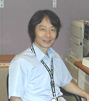
Okihide Hikosaka received his M.D. and Ph.D. from the University of Tokyo in 1973 and 1978 and did postdoctoral work at the NEI Laboratory of Sensorimotor Research (LSR) from 1979 to 1982. He held faculty positions at Toho University School of Medicine in Tokyo, the National Institute of Physiological Sciences in Okazaki, and Juntendo University School of Medicine in Tokyo before returning to the LSR in 2002 as a senior investigator.
My original interest in neuroscience was to discover the neural mechanisms for "voluntary movement." This is still my major interest and goal. However, it became clear to me that voluntary movement includes or is related to many brain functions. Voluntary movement is a final outcome of brain functions, including attention, intention, learning, memory, and motivation, in addition to reflexes and fixed motor patterns. Accordingly, I have studied neuronal mechanisms underlying the following functions: 1) brainstem and basal ganglia networks, 2) procedural and motor skill learning, 3) attention and response selection, and 4) motivational control of behavior. My early research with Hiroshi Shimazu at the University of Tokyo involved brainstem mechanisms for saccadic eye movement (or simply saccade). The eye typically makes saccades two to four times per second when a person is awake—for example, as you are reading this article. We discovered and characterized a group of saccadic burst neurons that are inhibitory burst neurons. These neurons are essential for reading this article, for example.
I then studied the neuronal mechanisms of the basal ganglia with Bob Wurtz at the LSR. We demonstrated that the basal ganglia use inhibitory mechanisms to control movements: Neurons in the substantia nigra pars reticulata (output of the basal ganglia) normally exert tonic inhibitory effects on the superior colliculus (a command area for saccadic eye movement) and remove the inhibition when a saccade is necessary.
Wurtz and I found that the basal ganglia mechanism is especially critical when a saccade is guided by memory, not by vision. The behavioral paradigm to induce memory-guided saccades, which we devised, is now widely used. We also devised a now-widely-used method for reversible blockade of local brain function by muscimol injection. In Japan, working with Satoru Miyauchi and Shin Shimojo, I devised a new psychophysical method to measure the spatial distribution of attention, which is called "line motion effect." Using this method, we characterized the time course and spatial extent of stimulus-induced (bottom-up) and voluntary (top-down) attention.
I then turned my interest to more complex motor functions. Almost any kind of movement or procedure is at first difficult to perform and requires volitional control. But after repeated practice, it becomes easy and eventually nearly automatic. This is usually called procedural or motor skill learning. With many people in Japan, I studied the neural mechanisms of procedural learning and memory involved in learning a new task. Based on behavioral and physiological experiments, we proposed a parallel neural network model in which a sequential procedure is acquired and stored independently by two loop networks, including the cerebral cortex, basal ganglia, and cerebellum.
My current research focuses on the neuronal mechanisms of decision-making. Probably the most fundamental reason for making a decision is based on biological needs or expectation of reward, as in the pursuit of food and sex. This kind of decision-making may be described as "motivational," while decision-making based on rules may be described as "cognitive."
Using a new saccade task, we found that neurons in the basal ganglia are specialized for, if not exclusively involved in, motivational decision-making. We now plan to investigate more detailed mechanisms underlying motivational decision-making by combining pharmacological and neurochemical methods. We also plan to investigate how the two different kinds of decision-making mechanisms conflict, interact, or negotiate with one another in the brain.
 |
|
Stephanie
London
|
Stephanie London received her M.D. degree from Harvard Medical School in Boston in 1983. After completing an internal medicine residency at Massachusetts General Hospital in Boston, she earned a D.P.H. degree in epidemiology from Harvard School of Public Health in 1989. She was an assistant professor in the Department of Preventive Medicine at the University of Southern California School of Medicine, Los Angeles, before joining NIEHS in 1995. She is currently an investigator in the Laboratory of Pulmonary Pathobiology, NIEHS.
My work deals with how genetic and environmental factors interact in the etiology of diseases of the lung. I began with the study of lung cancer over 10 years ago. During my six years at NIEHS, I have extended this effort to asthma—the major nonmalignant respiratory illness. I have developed a research program that is international in scope, taking advantage of variation in disease rates, allele frequencies, environmental exposures, and potential protective factors.
Although smoking is the major cause of lung cancer, diet and genetics may influence how an individual responds to tobacco carcinogens. When I began my research in this area, there was little work on genetic susceptibility to lung cancer. In 1990, I initiated a case-control study of genetic susceptibility to lung cancer among African-Americans and Caucasians in Los Angeles. I have published on several genetic polymorphisms in pathways relevant to lung cancer with early findings on myeloperoxidase, which may influence oxidative stress in the lung, and XRCC1, a gene involved in DNA repair.
I was also interested in dietary and hormonal factors that can best be tested in a prospective study where samples and other data are collected prior to development of cancer. To test these hypotheses I developed a relationship with the Shanghai Cohort, a study of 18,244 followed since the late 1980s. We found a novel gene-diet interaction. Subjects with higher levels of a urinary biomarker of isothiocyanate from dietary intake of cruciferous vegetables were at reduced risk of lung cancer, but this preventive effect was limited to subjects null for GSTM1, who would be predicted to excrete these beneficial compounds more slowly (Lancet 356:724–729, 2000). In these Shanghai samples I have also found that subjects with higher levels of insulin-like growth factor binding protein-3 are at reduced risk of lung cancer (J Natl Cancer Inst 94:749–754, 2002).
In the past few years, I have concentrated most of my efforts on developing a coherent program of research into environmental and genetic factors in asthma, an area central to the NIEHS mission. At the University of Southern California, I was part of a small group of investigators who established a cohort study of respiratory effects of air pollution in Southern California children. After coming to NIEHS, I established a genetic component to this study. This subsequently has become a large extramurally funded project.
Childhood asthma is notable for wide geographic variation in rates—with lower rates in developing countries. This variation in rates does not appear to be due to differences in diagnostic access. Taking advantage of this variability, I have also developed studies of genetic and environmental factors in childhood respiratory illness among school-aged children in Wuhan, China, and Mexico City, Mexico. I am also planning a birth cohort study in Mexico City to examine the role of early life factors in the etiology of asthma. In addition, I have recently begun a collaboration to study genetic, dietary, and environmental factors in adult asthma and chronic bronchitis in an NCI-funded cohort study of Chinese Singaporeans.
 |
|
Constantine
Stratakis
|
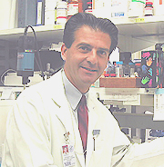
Constantine Stratakis received his M.D. and Doctor of Medical Sciences (Ph.D.) degrees from the National & Capodistrian University of Athens, Greece, in 1989 and 1994, respectively; he did predoctoral work there in the Unit of Endocrinology, Department of Experimental Pharmacology, before joining the Developmental Endocrinology Branch, NICHD, first as a student and then as a postdoctoral fellow in 1988. In 1990, he continued his postgraduate medical education at Georgetown University Medical School, Washington, D.C., where he finished a residency in pediatrics and two fellowships, in pediatric endocrinology (as part of the NICHD/GU training program) and in medical genetics and clinical dysmor-phology. In 1996, he returned to the Developmental Endocrinology Branch, where he is now a senior investigator and chief of the Section on Genetics and Endocrinology.
My interests are in the area of endocrine tumor cell development and function. This follows a long-time interest in how hormones and cancer relate.
During my research as a predoctoral student in Greece, I became interested in the regulation of growth hormone secretion and its function as a "growth factor." This work became the subject of my doctoral thesis.
During a subsequent medical clerkship in the Hospital Cochin, in Paris, France, I was introduced to adrenal gland genetics and physiology through Professor Jean-Pierre Luton, internationally known for his clinical studies on adrenal tumors.
I strengthened my ties to adrenocortical research in the late 1980s in the Developmental Endocrinology Branch, where pioneering research in adrenal development, oncology, and pharmacology was underway. As a postdoc, I was part of the team that identified the first mutations in the human glucocorticoid receptor in familial glucocorticoid resistance. I also was involved in some of the clinical studies on Cushing syndrome.
When I began my own laboratory in the branch, it was natural for me to pursue the work on adrenal tumor genetics. At this fortuitous time, the Human Genome Project (HGP) had just started providing geneticists with the tools to investigate questions that could not even be asked 20–30 years ago. These tools complemented NIH’s unique resources and access to patient populations.
One of the first patient groups that attracted my attention had a relatively unknown, newly described syndrome, called Carney complex (CNC). These patients had inherited adrenal tumors, but they also had a variety of other manifestations, including pigmented spots on the skin and the mucosae; skin, breast and heart tumors, known as "myxomas"; and other endocrine and nonendocrine neoplasms. The disease was inherited as an autosomal dominant.
There were only a few known kindreds with this disorder in the entire world, but I had already been to two of the three places where most of these families had been seen: the NIH Clinical Center and Hospital Cochin. The third place, and the one with the largest collection of patients, was Mayo Clinic in Rochester, Minn., where J. Aidan Carney worked.
Collaboration with Carney allowed us to apply the tools of the HGP to DNA of more than two dozen kindreds and led to the identification of the genetic loci of this disease in my lab. More recently, we cloned PRKAR1A as the gene that is mutated in approximately half of the patients with CNC (Nat Genet 26:89–92, 2000).
CNC’s complex clinical presentation and the involvement of many different organs suggested that the genetic defects responsible for CNC are important for basic functions of the human cells that are common among several tissues and organs. Indeed, the PRKAR1A gene that is mutated in CNC patients is present in almost all mammalian cells. It participates in the structure of an enzyme, protein kinase A (PKA), that regulates one of the most important cellular signaling pathways—that of cAMP. The defective gene encodes the most common regulatory subunit regulating PKA.
In the normal physiologic state, this regulatory subunit appears to act as a tumor suppressor (albeit not a classic one). When its action is abolished (as in patients with CNC who have inactivating mutations of that gene [Hum Mol Genet 9:3037–3046, 2000]), tumors of various organs ensue.
This knowledge added significantly to what was known about PKA and its possible involvement in tumorigenesis, but it also raised important questions. Currently, two animal models constructed by our laboratory are addressing the participation of PRKAR1A in endocrine cell growth and neoplastic development. We are also trying to find what other genes are mutated in CNC patients. Preliminary evidence suggests that two (or more) genes interact in the process of tumorigenesis in CNC tissues.
In addition, we are screening several sporadic tumors to identify mutations or functional dysregulation of the PRKAR1A gene and/or the PKA system. We recently found downregulation of this gene in thyroid carcinomas from patients who had no genetic syndromes (Genes, Chromosomes & Cancer, in press). We are also using microarray technology to identify genes that interact with PRKAR1A in mouse and human cell lines, as well as to investigate the genetics of noninherited adrenocortical tumors. We are studying forms of bilateral adrenal hyperplasia (both cortisol- and aldosterone- producing), adrenocortical cancer, and single adrenocortical adenomas.
We have identified a locus for idiopathic hyperaldosteronism on chromosome 7 (J Med Genet 37:831–835, 2000). Recently, in collaboration with a group in Brazil, we identified a novel TP53 genetic defect that predisposes individuals to the development of adrenal tumors (Proc Natl Acad Sci USA 98:9330–9335, 2001) in a unique way: Functional effects of this mutation depend on the pH of the cellular microenvironment.
Our work on adrenal cortex would not be complete without looking at patients with the opposite defects, namely, those with adrenocortical hypoplasia. The identification of genes with a role in fetal adrenal development is an important addition to the understanding of the neoplastic processes affecting this gland. Our group has identified mutations in both isolated glucocorticoid deficiency and triple A syndrome (J Clin Endocrinol Metab 86:5433–5437, 2001).
We hope that this work
will lead to better molecular elucidation of endocrine cell development and
neoplastic evolution and, ultimately, to better treatments for patients, especially
those with adrenal cancer, currently a disorder with dismal prognosis.
![]()