| T H E N I H C A T A L Y S T | J U L Y – A U G U S T 2002 |
|
|
|
| P E O P L E |
RECENTLY TENURED
 |
|
Julie
Donaldson
|
Julie Donaldson received her Ph.D. from the University of Maryland, Baltimore, in 1988 and did postdoctoral work at NICHD before joining the Laboratory of Cell Biology of NHLBI in 1995. She is now a senior investigator in the Laboratory of Cell Biology, NHLBI.
My research interests are in the organization of membrane compartments in cells. Membrane-bound organelles carry out distinct biochemical functions in eukaryotic cells. Understanding how these organelles maintain their properties and communicate with and transfer materials to other organelles is a fascinating area in cell biology, referred to as membrane traffic.
Over the past six years, my laboratory has focused on membrane traffic during endocytosis, in which plasma membrane proteins, receptors, and extracellular materials are brought into the cell as vesicles. The most studied and best understood type of endocytosis is clathrin-mediated endocytosis in the selective uptake of specific receptors and ligands into cells.
By contrast, very little is known about the various clathrin-independent forms of endocytosis. These alternative endocytic mechanisms are important as modes of entry for certain toxins, bacteria, and viruses and for the plasma membrane reorganization that accompanies phagocytosis, tissue differentiation, and cell migration.
We began to characterize a novel clathrin-independent endocytosis pathway through our studies on Arf6, a low- molecular-weight GTP-binding protein (GTPase). GTPases function as molecular switches, in the active form when GTP is bound and in the inactive form when GDP is bound after GTP hydrolysis. The Arfs comprise a family of proteins related to the Ras superfamily of GTPase regulators.
While I was a postdoctoral fellow in NICHD, I studied the role of Arf1 in regulating membrane traffic and organelle structure at the Golgi complex, a processing and sorting station for secretory proteins after they have left the endoplasmic reticulum. Then, as an independent investigator, my colleagues and I began studying Arf6 and found that this Arf localizes to the plasma membrane and to endosomal membranes that are distinct in protein and lipid composition from those endosomes derived from the clathrin-mediated endocytic pathway.
Among the proteins that travel along this endocytic pathway are important immunologic proteins such as the MHC Class I proteins and proteins associated with cell adhesion such as the integrins. We showed that Arf6, through its cycle of activation and inactivation, regulates the movement of membrane through this endosomal system. After internalization, a fraction of this membrane is recycled back to the plasma membrane in an Arf6-dependent manner, and another fraction of this membrane fuses with membrane internalized via the clathrin pathway. Thus, this Arf6-associated endosomal membrane system parallels and intersects with the clathrin pathway.
An interesting feature of Arf6 is that in addition to regulating membrane trafficking, it also regulates the actin cytoskeleton underlying the plasma membrane. We discovered several years ago that acute activation of Arf6 results in the formation of cell surface protrusions that are enriched in actin filaments. Furthermore, we found that the Rac GTPase implicated in plasma membrane ruffling and cell migration associates with the Arf6 endosome and that Rac’s ability to form membrane ruffles is dependent upon Arf6 function.
The finding that an Arf family member could regulate the actin cytoskeleton surprised researchers in the actin cytoskeletal field. This result was later corroborated by my group and others in demonstrating an Arf6 requirement for other actin-dependent processes such as cell spreading, Fc-mediated phagocytosis, and cell migration.
A key question for my lab is how Arf6 functions to affect both membrane traffic and the actin cytoskeleton. One important feature of Arfs is their ability to alter membrane lipid composition. We found that Arf6, through its regulation of a phosphatidylinositol kinase, modulates phosphatidylinositol 4,5-bisphosphate levels at the plasma membrane and on the Arf6-associated endosomes influencing membrane traffic and actin structures at the surface.
The roles played by membrane lipids in membrane trafficking pathways is now an area of intense research in my lab and in many others. We are also interested in identifying regulators of Arf6, identifying other proteins that travel through the Arf6 pathway, and understanding the molecular details of how membrane traffic is regulated in endocytic membrane systems.
 |
|
Gerhard
Hummer
|
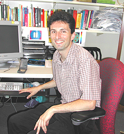
Gerhard Hummer did his graduate studies at the Max-Planck-Institute for Biophysical Chemistry in Goettingen, Germany, and at the University of Vienna, Austria, where he received his Ph.D. in 1992. He then moved to the Los Alamos National Laboratory as a postdoctoral fellow and became a principal investigator in 1996. He joined NIH in 1999 and is now a senior investigator in the Laboratory of Chemical Physics, NIDDK.
My interests are in the area of biomolecular structure, dynamics, and function, with a focus on solvent effects studied by theory and computer simulations.
Water penetration into proteins and transient fluctuations in hydration in the interior of proteins are important to their stability and function. Water-mediated proton transfer, in particular, is fundamental to bioenergetics and enzyme catalysis. An important goal driving my research at NIDDK is to understand how nonpolar channels function in protein-mediated proton transfer.
From computational studies of simple model channels, we found that single-file chains of water molecules can fill narrow molecular pores even if the hydrophobicity of the pore walls causes substantial losses of hydrogen-bonding energy. Water filling and emptying of hydrophobic channels can be regulated by small changes in channel polarity. In proteins such as cytochrome C oxidase, bacteriorhodopsin, and cytochrome C P450, the observed sensitivity to polarity can provide a tight coupling of water-mediated proton delivery to electron transfer and active-site chemistry.
This polarity-controlled filling and emptying of channels can help in particular to prevent proton leakage and unwanted side reactions by "dissolving" the proton wire after a successful proton transfer.
My lab’s studies also shed new light on the mechanism of rapid water conduction through biological water pores. In our simulations of nonpolar channels, we observed pulselike transport of water with an average flow rate comparable to that measured for a biological water channel, aquaporin-1.
Indeed, structures of aquaporin-1 show that relatively nonpolar residues line much of the pore. For optimal water transport, the polarity of the pore needs to be sufficiently high to ensure water filling, but low enough not to hinder water motion by strong directional interactions. Increased polarity would only slightly enhance the water occupancy but would greatly reduce the water mobility.
In cytochrome C oxidase, a proton pump in the respiratory chain, my collaborators and I have studied the side-chain and hydration dynamics of the active-site region. Solvent sites, identified through computation, establish a continuous proton path from the membrane inside to a critical glutamic acid residue. Our calculations further suggest that the glutamic acid shuttles a proton into the hydrophobic active-site cavity, to be picked up by transient water molecules. A combination of experiment, theory, and simulation allowed us to put important constraints on the proton pumping mechanism of cytochrome C oxidase.
My group is also working on fundamental aspects of the hydrophobic effect, in particular its role in ligand binding and protein folding. Other work with colleagues is leading to theories for single-molecule experiments. These are applied to nonequilibrium single-molecule pulling by atomic-force microscopes and laser tweezers, for example, to extract equilibrium thermodynamic properties, such as binding constants.
In future work, we will
build on this understanding of nonequilibrium processes and proton delivery
mechanisms to understand how cytochrome C oxidase converts energy and acts as
a "Maxwell demon" by pumping protons across membranes against an electrostatic
potential and chemical gradient. ![]()