| T H E N I H C A T A L Y S T | M A R C H – A P R I L 2002 |
|
|
|
| P E O P L E |
RECENTLY
TENURED
 |
|
Lawrence
Brody
|
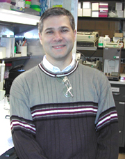
Lawrence Brody received his Ph.D. in Human Genetics from the Johns Hopkins University, Baltimore, in 1991. He was a Howard Hughes postdoctoral fellow at the University of Michigan before coming to NHGRI in 1994. He is currently a senior investigator in the Genome Technology Branch and head of the Molecular Pathogenesis Section, NHGRI.
Identification and evaluation of human gene variants important to health have not kept pace with the rush of gene discovery from the Human Genome Project, and our ability to deduce the function of novel genes remains limited. Research in my section is focused on defining genetic variations that underlie disease and understanding the mechanism by which these variant alleles produce disease.
To accomplish this, we use research tools traditionally associated with diverse disciplines such as genetic epidemiology, evolutionary biology, cell biology, biochemistry, and physiology. Our integrated approach, combining investigation into genetic variation and biological function, allows us to take maximum advantage of the wealth of data produced by the Human Genome Project.
One of our research interests is the study of the genes BRCA1 and BRCA2 and their role in inherited breast and ovarian cancer susceptibility. Although the link between these genes and cancer risk is well established, it is based on the averaging of data from the more than 600 different mutations.
We have very limited knowledge of the risk associated in carrying a specific BRCA1 mutation. In collaboration with investigators at NCI, our laboratory discovered that a small set of specific BRCA1 mutations are present at a high frequency in the Ashkenazi Jewish population.
This finding allowed us to measure the risk associated with these mutations in an unselected population. We are continuing to study this population in order to better understand the risk of cancer associated with these mutations and to identify additional genes that may modify this risk.
The biological function of the BRCA1 and BRCA2 genes remains poorly understood. Work from numerous labs suggests that BRCA1 controls some aspect of DNA repair. Our own investigations into the function of the BRCA1 gene revealed that BRCA1 interacts with the proteins of the chromatin-remodeling complex.
We recently found that BRCA1 plays an important role in the regulation of the cell cycle. By restoring BRCA1 to BRCA1-minus cells, we were able to demonstrate that BRCA1 is essential for maintaining the DNA damage checkpoint preceding the G2-to-M transition. BRCA1 appears to control this activity by directly interacting with the cell cycle regulator.
Our second area of interest is the identification of genes associated with the risk of neural tube defects (NTDs). These defects are a major public health concern, yet the pathogenesis of NTDs is poorly understood. In collaboration with investigators at the NICHD and Trinity College, Dublin, we are searching for genes controlling disease risk in a large series of NTD families.
In contrast to breast cancer, there are epidemiological clues to the identity of the genes responsible for the inherited component of NTD risk. Past research established that perturbations of the metabolic pathways involving folate, vitamin B12, and homocysteine can account for a large fraction of NTD cases. The genes making up these metabolic pathways are logical candidates for putative "NTD genes." We identified human genetic variants in the majority of the genes comprising these pathways. We are currently measuring the frequency of these variants in NTD families and control subjects. In addition to measuring the connection between specific variants and the risk of having an NTD, we plan to measure the biochemical and functional consequences of these variants in experimental models and in the patients who carry them.
 |
|
Steven
Kleeberger
|
Steven Kleeberger received his Ph.D. in ecology from Kent (Ohio) State University in 1982 and did his postdoctoral research at Johns Hopkins University, Baltimore, where he became a full professor. He was recruited to NIEHS as chief of the Laboratory of Pulmonary Pathobiology in 2001. He also directs a research group in environmental genetics.
Epidemiological studies have shown that exposures to outdoor and indoor pollutants are associated with increased morbidity and mortality in cities throughout the United States and other industrialized countries. In particular, ozone—a highly reactive and toxic oxidizing pollutant—and particulates have become prevalent and have received considerable attention.
Because of the potential effect that pollutants may have on public health, identification of the intrinsic and extrinsic factors that may influence the pulmonary response(s) to exposures is an important issue. Over the past few years, my laboratory has focused considerable effort on understanding the role of genetic background as a susceptibility factor in the toxic effects of common air pollutants. Our lab combines state-of-the-art methods in inhalation toxicology, pulmonary physiology, and molecular genetics to address this goal.
Our initial analyses of susceptibility to inflammation and injury induced by ozone indicated that susceptibility was controlled by a locus we termed Inf-2. We subsequently identified a quantitative trait locus (QTL) for ozone susceptibility on chromosome 17. Candidate genes for the locus include the pro-inflammatory cytokine tumor necrosis factor–a (Tnf). Functional analyses of this locus confirmed Tnf as a candidate gene.
More recently, we have identified another ozone susceptibility QTL on chromosome 4 that contains a candidate gene, toll-like receptor 4 (Tlr4). Tlr4 has recently been implicated in innate immunity and endotoxin susceptibility. Functional analyses confirmed an important role for Tlr4 in oxidant lung injury.
Results of our linkage analyses therefore suggest a genetic commonality between signal transduction pathways involved in determining ozone and endotoxin responsivity. We believe this work will have important implications for understanding mechanisms of oxidant lung injury as well as exacerbation of preexisting diseases, such as asthma.
We have also recently identified susceptibility loci for alveolar macrophage immune dysfunction induced by inhalation of sulfate-associated carbon particles in differentially susceptible inbred mice. A genome-wide linkage analysis identified significant and suggestive QTLs on chromosomes 17 and 11, respectively. Candidate susceptibility genes were identified for mouse and human by comparative mapping. Importantly, both QTLs overlap previously identified QTLs for susceptibility to another common pollutant, ozone. This is the first demonstration that genetic background is an important determinant of responsiveness to particle-induced immune dysfunction—which has important implications for understanding the epidemiological associations between particulates and morbidity and mortality.
Our research focus has expanded to include genetic pathogenesis of hyperoxia-induced acute lung injury. Hyperoxia exposure induces lung responses that resemble subphenotypes of acute respiratory distress syndrome (ARDS), a major lung disease characterized by noncardiogenic edema and inflammation. We have developed a mouse model of susceptibility to hyperoxia exposure and identified significant and suggestive susceptibility QTLs on chromosomes 2 and 3, respectively. We have further identified—within the chromosome 2 QTL—a candidate susceptibility gene, NF-E2 related factor 2 (Nrf2), an essential nuclear transcription factor involved in antioxidant gene expression and regulation. We used sequence anal-ysis to identify the polymorphism in the Nrf2 promoter that confers differential susceptibility to hyperoxic injury in this model. Functional analyses with a Nrf2 knockout mouse confirmed this observation. We believe this model has important implications for developing treatments for oxidant lung injury and ARDS.
Another aspect of my research is focused on gene-environment interaction and the pathogenesis of disease in human populations. In this work, we are participating in genetic analysis of asthma pathogenesis in a large population-based case-control study in Finland. We are also collaborating with Francine Kaufmann and Rachel Nadif of INSERM to identify the genetic basis of susceptibility to pneumoconiosis in a cohort of coal miners in France. These studies look at the interaction of polymorphisms in innate immunity and pro-inflammatory genes with occupational exposures to coal dust. We are also examining the role of innate immunity genes in determining susceptibility to HIV infection and AIDS progression in the NIH-sponsored Multicenter AIDS Cohort Study. The integration of predictive animal genetic models with population-based epidemiological studies positions my laboratory to take advantage of the exciting progress in genome sequencing and proteomics, the better to tackle the etiology of environmental lung diseases, identify genetically susceptible individuals, and design interventions.
 |
|
Ward
Odenwald
|
Ward Odenwald received his Ph.D. from the combined Johns Hopkins University–NIH FAES program in 1987 after having joined the NINDS Medical Neurology Branch in 1978. He is now a senior investigator for the Neurogenetics Unit, LNC, NINDS.
The long-term goal of my research is understanding the molecular events that guide cell fate decisions in the developing nervous system. Over the last four years, my unit’s studies on Drosophila CNS cell-fate patterning reveal that a transcription factor regulatory network acts temporally during stem cell lineage development in all CNS ganglia.
We have discovered that during neuroblast (NB) lineage formation many NBs undergo sequential transitions in the expression of the transcription factors: hunchback (hb), the POU domain 1 and 2 genes (pdm1/2), castor (cas), and grainy head (grh). As a result of the hb->pdm1/2->cas->grh (H-P-C-G) NB expression, sequentially formed basal (inner and first born)-to-apical (outer and last born), multilayered neuronal subpopulations arise that share expression of one of these factors.
Our loss-of-function analysis of four of the five H-P-C-G network components (hb, pdm1/2, and cas) indicate that this temporal regulatory cascade is required for the proper development of many, if not all, neuronal subtypes.
Given the global nature of the H-P-C-G cascade, we hypothesize that these transcription factor expression domains represent fundamental branch points in a genetic circuit controlling temporally sensitive functions common to the development of all CNS ganglia. Our analysis of POU gene regulation by Cas demonstrates that Cas performs at least two essential roles.
First, Cas insulates the developmental program(s) of the sublineages that express it by repressing neural identity regulators (such as pdm1/2) that participate in the cell fate decisions of neuronal subtypes born earlier. Second, Cas promotes neural identity decisions within its sublineages by directing and promoting expression of other neural identity POU genes.
Our in vitro analysis of the regulatory mechanisms controlling the orchestrated expression of the H-P-C-G network factors indicates that once NBs initiate lineage formation, no additional extrinsic cues from either the neuroectoderm or underlying mesoderm are required. Given the remarkable conservation observed in metazoan transcription factor networks and the identification of putative H-P-C-G cognate genes in mammals, we believe that all or part of the temporal regulatory network may function during vertebrate CNS development. In order to fully understand the biological significance of the H-P-C-G cascade and how its transcription factors integrate with upstream cell fate decisions, we need to identify the downstream targets of these regulators and to understand the time-dependent signals that regulate it.
To identify new components of the network, we have recently carried out differential gene expression, enhancer-trap, and mutant screens for novel genes that are either dynamically expressed during NB lineage development or required for proper expression of the H-P-C-G network. Our developmental expression screen of cDNAs prepared from staged embryonic fruit fly heads has thus far identified 57 new genes that are dynamically expressed during NB lineage development. Our experimental approach is now aimed at understanding their roles in NB lineage development. These genes—plus 1,837 other genes identified in the screen—are described at our website.
 |
|
Ignacio
Rodriguez
|
Ignacio Rodriguez received his Ph.D. in 1986 from the University of Miami in Coral Gables, Fla. He did his initial postdoctoral training at the Bascom Palmer Eye Institute in Miami, Fla. He joined the National Eye Institute in 1987 and continued his postdoctoral training in several laboratories. He is now a senior investigator at the Laboratory of Retinal Cell and Molecular Biology, NEI.
My main interests are in understanding the molecular mechanisms of age-related diseases. I am particularly interested in those affecting the eye, more specifically, age-related macular degeneration (AMD). AMD is the leading cause of blindness in the United States, responsible for severely reducing the quality of life of millions of our senior citizens.
This disease is complex and scientifically very challenging because it involves aging, genetics, and environmental factors. As biological systems, humans are not designed to live much beyond middle age. Thus, there are fundamental design flaws in our biology that make certain organ systems susceptible to age-related malfunctions. The eye is a particularly interesting system to study because AMD and cataracts are often found in individuals who have survived into their late 60s and 70s in relatively good health.
I originally trained as a carbohydrate biochemist and started working in vision research during my first postdoctoral fellowship at the Bascom Palmer Eye Institute. After joining NEI in late 1987, I trained as a molecular biologist, initially in the Ophthalmic Genetics and Visual Function Branch and later in the Laboratory of Mechanisms of Ocular Diseases (LMOD). While at the LMOD, I identified a splice site deletion in the guinea pig z-crystallin gene responsible for a hereditary cataract. I joined the Laboratory of Retinal Cell and Molecular Biology in 1990 and began cloning retina-related genes and genes differentially expressed between the macular and peripheral regions of the retina. This research led to the cloning and characterization of 10 novel human genes and several orthologs in other species.
My research program is now focused on determining the pathogenesis mechanism(s) of AMD. We have been searching for common age-related factors that may be adversely affecting the health of the retinal pigment epithelium (RPE) and the choriocapillaris. The observed accumulation of lipids and lipoproteins in the back of the eye as a function of aging is particularly interesting because of the known cytotoxic effects of oxidized LDL and oxysterols. The RPE expresses a variety of receptors capable of internalizing LDL and is believed to shuttle cholesterol to the photoreceptors. We have found that oxidized LDL and certain oxysterols are highly cytotoxic to cultured human RPE cells. We hypothesize that this lipid accumulation in Bruch’s membrane leads to its hyperoxidation, which can adversely affect the RPE and choriocapillaris.
The immune system may also play a role in the pathogenesis of AMD given that scavenging macrophages that encounter the oxidized LDL in Bruch’s membrane experience cytotoxicity. This could lead to an inflammatory response that may contribute to the drusen deposits observed during the early stages of the disease.
Oxysterols have potent pharmacological properties and are likely the most cytotoxic substances associated with LDL and other lipoproteins. Thus, we have been cloning and characterizing oxysterol-binding proteins (OSBPs). We recently characterized a novel OSBP, OSBP2, which is almost exclusively expressed in retina and has a high affinity for one of the most toxic oxysterols, 7-ketocholesterol. We have also identified and cloned 10 additional and previously uncharacterized OSBP genes.
The OSBPs, although highly similar in structure, have different expression patterns. In addition, 11 of the 12 known OSBPs contain Pleckstrin homology (PH) domains. These PH domains are known to bind phosphinositols and are likely serving to target the OSBPs to different cellular organelles. Future work will focus on elucidating the function(s) of the OSBPs and determining whether they mediate the pharmacological effects of oxysterols.
 |
|
William
Simonds
|
William Simonds received his M.D. degree from the University of Pittsburgh in 1981 and did postdoctoral work in the Laboratory of General and Comparative Biochemistry and Laboratory of Molecular Biology in NIMH before joining the Molecular Pathophysiology Branch of NIDDK in 1987. He is now a senior clinical investigator in the Metabolic Diseases Branch, NIDDK.
My basic science interests have centered on biochemical and signal-transducing properties of the G protein—bg complex and related heterodimers. The tightly associated bg complex of heterotrimeric G proteins is required for activation of G-protein pathways by seven transmembrane-spanning receptors in response to such diverse extracellular stimuli as neurotransmitters, hormones, pheromones, light, tastants, and odorants.
Activation of G proteins by receptors results in dissociation of the a subunit (in GTP-bound form) from the bg complex. The Ga-GTP and/or the free Gbg complex can regulate a variety of downstream effector molecules depending on the cellular context. The bg complex regulates the function of ion channels, isoforms of adenylyl cyclase (AC), isoforms of phospholipase C-b (PLC-b), and MAP kinase (MAPK) pathways and thus mediates critical cellular functions such as chemotaxis, response to growth factors, and regulation of neurotransmitter release.
Despite the multiplicity of potential Gbg dimers implied by the existence of multiple genes encoding Gb and Gg isoforms, Gbg heterodimers had been considered functionally interchangeable. This view prevailed until my colleagues and I provided the first clear demonstration of functional specialization among Gb subunits in an analysis of the effector properties of brain-specific Gb5. Gb5-Gg2 complexes stimulated PLC-b like Gb1-Gg2, but unlike the latter complex, Gb5-Gg2 failed to activate MAPK, Akt/PKB, and AC type II.
We hypothesized that the unique functional properties of Gb5 might indicate its specialization to interact with novel effectors. This was borne out when we purified native Gb5 complexes from mouse brain and discovered tightly bound regulator of G-protein signaling (RGS) proteins 6 and 7, not Ga or Gg subunits. This unexpected finding complemented work from other laboratories describing Gb5-RGS protein complexes in the retina and identifying a subset of RGS proteins containing a "Gg-like" domain that could mediate binding to Gb5 in a Gg-like fashion.
Using neuron-like PC12 cells, we discovered that Gb5 and RGS7 proteins are expressed in neuronal cell nuclei, a novel subcellular localization for G proteins that we confirmed in mouse brain.
These findings raise the possibility of information transfer by Gb5-RGS protein complexes between the plasma membrane and nuclear compartments in neurons. We are now looking for the signals that may regulate the function and/or subcellular localization of Gb5-RGS protein complexes in brain and trying to learn whether these complexes might regulate transcriptional events in a signal-dependent fashion.
My clinical interest involves familial isolated hyperparathyroidism (FIH) and related disorders. We recently reported a series of 36 FIH kindreds seen in the Clinical Center over the past 30 years. In these 36 families, genomic DNA mutational and biochemical testing largely excluded multiple endocrine neoplasia type 1 as an etiology. Among the total set of families, we identified three kindreds with the hyperparathyroidism—jaw tumor syndrome (HPT-JT), five kindreds with mutations in the gene for the calcium-sensing receptor, and 28 kindreds for whom no syndromic diagnosis could be made.
Future testing for HPT-JT mutations should clarify whether any of the 28 nonsyndromic families have currently unrecognized causes of FIH. We are working with colleagues at NHGRI and others in an international consortium to identify the gene on chromosome 1 responsible for the HPT-JT familial cancer syndrome.
 |
|
Stanislav
Tomarev
|
Stanislav Tomarev received his Ph.D. from the Russian Academy of Sciences in Moscow in 1977 and worked at the N.K. Koltzov Institute of Developmental Biology of the Russian Academy of Sciences as a group leader and leading investigator before joining the NEI Laboratory of Molecular and Developmental Biology in 1989. He is now a senior investigator in the same laboratory.
My scientific interests are in the area of molecular mechanisms underlying glaucoma. Glaucoma is the second leading cause of blindness in the United States and affects between one and two percent of people over the age of 40. Glaucoma is a group of neurodegenerative disorders characterized by the death of retinal ganglion cells and by a specific deformation of the optic nerve head, known as glaucomatous cupping. This disease is also often associated with elevated intraocular pressure (IOP). The molecular mechanisms underlying neuronal damage associated with elevated IOP are not understood.
My group is developing mouse and rat models of glaucoma using genetic approaches. In 1997, my colleagues and I isolated and characterized the mouse myocilin gene. Mutations in this gene lead to autosomal dominant juvenile open-angle glaucoma with elevated IOP in humans and may account for 2.6–4.3 percent of adult forms of open-angle glaucoma.
Using myocilin knockout mice, we demonstrated that the absence of myocilin does not lead to glaucoma and therefore glaucoma-causing mutations most probably act by a gain-of-function mechanism. Overproduction of myocilin in the ocular tissues, using myocilin-containing BAC clones, also fails to produce glaucomatous changes. We demonstrated that mutations in the mouse myocilin gene, corresponding to glaucoma-causing mutations in the human myocilin gene, reduce secretion of the protein. We are currently generating transgenic mice expressing mutated forms of myocilin in the ocular tissues and we believe that this approach will produce a useful new animal model of glaucoma.
To understand mechanisms of myocilin action, we are looking for proteins that interact with myocilin. We identified a new secreted protein, named optimedin, that interacts with myocilin. Both optimedin and myocilin contain the olfactomedin domain that was previously identified in several biologically active proteins, and we demonstrated that myocilin and optimedin interact through this domain. We showed that the presence of mutant myocilin interferes with secretion of optimedin in transfected cells. We are testing the hypothesis that changes in the expression level and distribution of optimedin in eye tissues in myocilin-related glaucoma contribute to the pathology of myocilin-related glaucoma.
My colleagues and I are using rat models of pressure-induced optic nerve damage to identify changes in the retina at different stages of retinal damage. We demonstrated that molecular pathways activated by elevated IOP overlap those induced by optic nerve transection. We showed that elevated IOP may stimulate a group of genes normally associated with inflammation and the immune response. These findings suggest new possibilities for treating glaucoma.
We are also looking for glaucoma-associated genes in the cDNAs from the human trabecular meshwork—a key component of the aqueous humor outflow pathway in the eye that is essential for maintaining normal IOP. This approach has allowed us to identify several new candidate genes that we are probing by examining families of patients with glaucoma.
We hope that by elucidating
the molecular changes in the eye associated with glaucoma, we may improve diagnostic
tools and treatments for this blinding disease. ![]()