| T H E N I H C A T A L Y S T | N O V E M B E R – D E C E M B E R 2001 |
|
|
|
| P E O P L E |
RECENTLY TENURED
 |
|
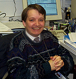
Leslie
Biesecker
|
Leslie Biesecker received his M.D. from the University of Illinois in 1983 and did postgraduate work at the University of Wisconsin, Madison (pediatrics), and the University of Michigan, Ann Arbor (medical and molecular genetics), before joining the Genetic Diseases Research Branch of NHGRI in 1993. He is now a senior investigator in that branch.
My research focuses on the clinical and molecular delineation of human malformation syndromes. Currently we are working on two classes of disorders, those that involve classic multiple congenital anomaly syndromes and disorders with progressive postnatal overgrowth.
The multiple congenital anomaly syndromes include Pallister-Hall syndrome, Greig cephalopolysyndactyly syndrome, McKusick-Kaufman syndrome, Bardet-Biedl syndrome, and the Lenz microphthalmia syndrome. These disorders include varying combinations of polydactyly and central nervous system and visceral malformations, and some also have functional complications such as mental retardation, seizures, and visual loss.
Our research is both clinical and molecular. The clinical component includes phenotypic characterization and natural history studies to delineate the range and consequences of the disorders. For many of these disorders, the range of severity and the long-term prognosis are unknown.
Several of the disorders under study are frequent in closed Anabaptist sects such as the Old Order Amish and Mennonites of Lancaster County, Pa., and certain regions of Ohio and Kentucky. These disorders are approached via field studies to evaluate patients, computerized genealogical analysis, and clinical testing and treatment.
In the laboratory, we perform classical positional cloning studies to find the genes that cause these syndromes, perform genotype-phenotype correlations, and delineate the pathogenesis of these disorders using animal models and cell biological approaches.
The first disorder we studied was the Pallister-Hall syndrome, which we showed is caused by mutations in the GLI3 zinc finger transcription factor. This discovery demonstrated that Pallister-Hall syndrome was allelic to Greig cephalopolysyndactyly syndrome, a distinct malformation syndrome. We have developed a model to explain the genesis of two distinct syndromes from mutations in a single gene and are carrying out ongoing studies to support that model.
The other disorders we study were selected because they have physical features that overlap with the Pallister-Hall and Greig cephalopolysyndactyly syndromes. We hypothesize that disorders that share manifestations will also share pathogenic mechanistic features in development.
We are also studying a disorder of postnatal overgrowth, the Proteus syndrome. This disorder includes severe disproportionate overgrowth of many tissues, occasional malformations of the limbs and central nervous system, cutaneous nevi, and other manifestations. It appears to be associated with two serious complications—massive pulmonary embolism and tumor predisposition. The disorder is hypothesized to be caused by somatic mosaicism for a mutation that is lethal in the nonmosaic state. This model explains the sporadic nature of the disorder, the patchy distribution of overgrowth, and its discordance in monozygotic twins.
We are testing this model by screening for alterations in gene structure or expression and comparing affected and unaffected tissues of patients with this condition.
This project also has significant clinical and laboratory components. Clinically, we are conducting a longitudinal natural history study to determine the range of manifestations, severity, and natural history of the condition. In the laboratory, we are using modern molecular techniques—including microarray expression analysis, representational difference analysis, and two-dimensional Southern analysis—to characterize gene alterations.
Understanding the cause of Proteus syndrome will be important both for developing specific therapies and for understanding the mechanisms of control of human postnatal growth.
 |
|
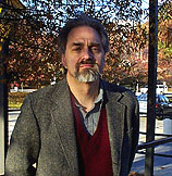
Kevin
Brown
|
Kevin Brown received his medical degrees from Cambridge University in England in 1982. He did internships in internal medicine and infectious diseases in London before specializing in virology. He joined the Clinical Hematology Branch in NHLBI as a visiting associate in 1992 and became an investigator in the Hematology Branch in 1996. He is now a senior investigator in the Hematology Branch, NHLBI.
The main focus of my research is the study of the interaction of viruses with hematopoiesis. My work can be broadly divided into two main areas: studying the interaction of known viruses—such as the small DNA viruses—with blood cells and their precursors; and looking for novel viruses that may be associated with bone marrow failure. These studies involve a wide range of different experimental approaches, including molecular biology, tissue culture, and animal-based technologies.
Small DNA viruses and hematopoiesis. Parvoviruses are small DNA viruses that cause disease in both humans and animals. However, the only parvovirus known to cause disease in humans is parvovirus B19. Approximately 60 percent of adults show evidence of previous infection. Acute B19 infection can cause fifth disease in children, polyarthropathy syndrome in adults, transient aplastic crisis in patients with underlying chronic hemolytic anemia, chronic anemia due to persistent infection in immunocompromised patients, and fetal loss in pregnant women.
B19 virus cannot be readily grown in cell culture and, when I started my studies, there was no animal model for B19 infection. However, studies with human bone marrow cultures from volunteers had shown that the virus could replicate in human red cell precursors.
My initial focus on joining the Clinical Hematology Branch was to characterize the cellular receptor for B19. Using a modification of the hemagglutination assay, I was able to identify the receptor as globoside, or blood group P antigen, a glycosphingolipid found on the cell membranes of red cells and their precursors.
Rare individuals do not have globoside on their red cells, and I was able to show that these individuals did not have evidence of previous infection with B19, and in vitro their bone marrow could not be infected with B19 virus, even at high viral concentrations.
Globoside is also found on fetal myocardial cells and endothelial cells and in placental tissue, and this identification of the receptor has prompted other studies to determine the full complement of diseases caused by this virus. Further studies are also in progress to identify other factors, including a possible second receptor, that are important in the narrow tropism of this virus. Globoside is also found on the cell surface of nonhuman primates’ erythrocytes, and although we can show that B19 will replicate in the bone marrow of cynomolgus monkeys in vitro, inoculation studies of primates have been unsuccessful.
While these animal studies were underway, I was asked to study a fatal outbreak of anemia in cynomolgus monkeys at Wake Forest University in Winston-Salem, N.C. There we identified a simian parvovirus (SPV), distinctly different from but related to B19. With colleagues now at the University of Minnesota, we have cloned and sequenced the virus. Infection studies indicate that cynomolgus infection with SPV mimics human B19 infection, and we are currently using it as an animal model to study parvovirus-induced fetal infection. Similar studies of outbreaks of anemia in other primate species have allowed me to identify two other primate parvoviruses, with the possibility that they may also be exploited as animal models.
Novel viruses and hepatitis-associated aplastic anemia. Hepatitis-associated aplastic anemia (HAA) syndrome is a bone marrow failure occurring usually within two to three months of an episode of non-A, non-B, non-C hepatitis. Classically it affects young people, especially boys and men (mean age 20 years). If untreated, it is usually fatal. The etiology is unknown, but scientists think it is triggered by a viral infection.
In collaboration with Neal Young in the Hematology Branch, I have been collecting epidemiological and clinical data on HAA and samples from these patients and control subjects. We have shown that the patients have evidence of activated cytotoxic lymphocytes in their peripheral blood and that many of these patients can be successfully treated with immunosuppression. In addition, we have observed some HLA associations, supporting the hypothesis that the bone marrow failure is mediated through immunopathological mechanisms.
Recently, a number of putative hepatitis viruses have been described, including hepatitis G (or GBV-C), TT virus, SENV, and a novel picornavirus A2 that we described. My studies indicate that none of these appear to be associated with HAA. We have also inoculated patient material into primates, rodents, and tissue culture, but so far all the studies are negative for viruses, as are screens of a number of libraries made from patient material. We are currently looking at the immunological profile, especially at the levels of different cytokines and other antigens, in patient and control tissue to identify better markers and to determine which samples to use for gene subtraction and DNA chip analysis.
 |
|
Robert
Innis
|
Robert Innis received his M.D. degree from the Johns Hopkins University in Baltimore in 1978 and a Ph.D. in neuropharmacology from Johns Hopkins in 1981 under the mentorship of Solomon Snyder. He completed a residency in psychiatry at Yale University in New Haven, Conn., in 1984 and then joined the Yale faculty in the departments of psychiatry and pharmacology. In October 2001, he came to NIH as chief of a newly formed Molecular Imaging Branch at NIMH.
The overall goal of this new branch is to elucidate pathophysiological mechanisms associated with neuropsychiatric disorders. We expect that such knowledge will ultimately decrease the burden of these illnesses by suggesting better therapies.
Several investigators in this new branch are directly linked to NIMH’s new Mood and Anxiety Disorders Program, directed by Dennis Charney. Patients with these psychiatric disorders will receive the focused, although not sole, attention of this branch.
The primary methodologies used by investigators in the Molecular Imaging Branch are PET (positron emission tomography) and NMR (nuclear magnetic resonance).
New PET radiotracers are synthesized for use as in vivo ligands to measure many different molecular targets, including membrane-bound receptors, proteins associated with intracellular signal transduction, and other expressed genes. Several NMR methods are also studied to measure molecular targets: magnetic resonance spectroscopy, local neuronal activity (functional MRI), and structure of the brain (structural MRI).
My own area of research focuses on the use of PET to localize and quantify molecular targets in the brain. The overall goals of my laboratory are to develop new radiotracers that image molecular targets in the brain, to evaluate these tracers in animals and healthy human subjects, and then to extend their use to patients with several neuropsychiatric disorders.
The PET research proposed in my lab critically depends on sophisticated radiochemistry, and NIMH is fortunate to have recruited Victor Pike earlier this year to direct the section on PET radiochemistry. Pike is internationally renowned as a radiochemist and was formerly head of the Chemistry-Engineering Group at the Hammersmith PET Center in London, England [see The NIH Catalyst, May–June 2001].
Because PET was not available at Yale in the 1980s, I used SPECT (single photon emission computed tomography) for studies of receptors in the brain. My work on benzodiazepine receptor imaging clearly confirmed that SPECT was capable of quantitative measurements, with validation comparable to that in PET.
My SPECT work expanded to include other neurotransmitter systems, including dopamine, serotonin, GABA, and acetylcholine.
In these earlier studies, I helped to develop several new radiotracers, including probes for the dopamine transporter. In fact, the dopamine transporter is a biological marker for Parkinson’s disease—and SPECT imaging of the dopamine transporter was approved last year in several European countries to aid in the early diagnosis of Parkinson’s disease. To my knowledge, this SPECT tracer is the first biological test for Parkinson’s disease and also the first neuroreceptor imaging agent approved for clinical use.
As mentioned above, while at Yale, I performed neuroimaging primarily with SPECT. SPECT cameras are widely available in community hospitals and use radionuclides with relatively long half-lives: 6 to12 hours for SPECT vs. one-half to two hours for PET.
The longer half-life allows SPECT radiopharmaceuticals to be distributed over wide distances. However, SPECT has significantly lower sensitivity than PET(~100-fold) and lower anatomic resolution (9–12 mm vs. 3–5 mm). I performed a relatively small amount of PET research at Yale, due to the university’s limited resources for this methodology.
In contrast, the NIH Clinical Center has substantial PET resources under the direction of William Eckelman: three cyclotrons, three whole-body PET cameras, and a sizeable radiochemistry lab and staff. In addition, Mike Green (Nuclear Medicine) has continued years of PET camera development and has constructed a new high-resolution device for imaging in rats and mice.
Realizing the critical role of radiochemistry, NIMH committed significant funds to expand in this area and enhance the existing facilities. A new radiochemistry lab is currently under construction in the basement of Building 10, and additional "cold" (or nonradioactive) chemistry labs are planned for adjacent areas in the new Clinical Center building.
In addition, NIMH spearheaded efforts of several institutes to purchase a new state-of-the-art high-resolution, high-sensitivity PET camera that can image both the human head and rodents. This new device should be constructed and delivered in about one and a half years.
I am truly enthusiastic about these and other neuroimaging opportunities that are emerging at NIH. New tracers that are currently available or under development include probes for the serotonin transporter, amyloid, cortical dopamine receptors, the substance P receptor, norepinephrine transporter, nicotinic acetylcholine receptor, metabotropic glutamate receptor, a PET reporter probe system, and more promising measures of intracellular signal transduction. From my perspective, a major attribute of NIH is the ability to collaborate with other researchers, both in the development of new radiotracers and in their use in both animals and humans. I look forward to hearing from intramural collaborators with these interests.
 |
|
Xinhua
Ji
|
Xinhua Ji received his Ph.D. in chemistry from the University of Oklahoma in Norman in 1990. He was a postdoctoral fellow and then a research assistant professor at the Center for Advanced Research in Biotechnology of the University of Maryland Biotechnology Institute in Rockville, Md., and the National Institute of Standards and Technology before joining the NCI-Frederick in 1995. He is now a senior investigator and chief of the Biomolecular Structure Section, Macromolecular Crystallography Laboratory, NCI.
As a structural biologist and a chemist with medical experience, I am interested in the structure and function of biomolecules with anticancer and antimicrobial significance and in structure-based drug design. To pursue these subjects, I have established collaborations within NIH as well as with extramural investigators.
The systems I am working on are at three different points on the basic-to-applied spectrum of research studies: Pro-drug design research with minimal basic emphasis for glutathione S-transferase (GST); basic structure and activity studies of 6-hydroxymethyl-7,8-dihydropterin pyrophosphokinase (HPPK) with some initial efforts in drug design; and basic studies of the structure and function of two potential anticancer targets—G protein ERA and ribonuclease III (RNase III), which play essential roles in RNA processing control.
GST represents a superfamily of detoxification enzymes. Many tumors become drug resistant by overexpressing p-class GST (GSTP), one of the major GST isoenzymes in humans. We have been attempting to design agents that will overcome the drug resistance of cancer cells that overexpress GSTP. The comparison of the active site architectures and transition-state analogs of GST isozymes revealed a strategy: generating nitric oxide (NO) selectively in the active site of isoenzyme. Application of this strategy yielded a GSTP-selective NO donor that improves the potency of arsenite, a clinically useful anticancer agent, in cancer cells overexpressing GSTP.
Folate cofactors are essential for life. Mammals derive folates from diet; most microorganisms must synthesize folates de novo. HPPK is the first enzyme in the folate biosynthetic pathway. It is not the target for any existing antibiotics—and is therefore an ideal target for developing novel antimicrobial agents to fight the worldwide crisis of antibiotic resistance.
HPPK contains 158 amino acids and is thermostable, which also makes it an excellent model system for the mechanistic study of pyrophosphoryl transfer. Having elucidated high-resolution (up to 0.89 Å) structures of well-chosen complexes, we have mapped out the trajectory of pyrophosphoryl transfer. This work also reveals unusual conformational changes of HPPK in its catalytic cycle.
The structural information is now the basis of inhibitor design effort. We have synthesized a bisubstrate-mimicking inhibitor and a one-substrate analog and determined the crystal structures of HPPK in complex with these inhibitors.
ERA and RNase III play key roles in the control of gene expression. ERA is an essential GTPase found in every bacterium sequenced to date. In these bacteria, ERA has a regulatory role in cell cycle control by coupling cell growth rate with cytokinesis.
A highly conserved ERA homolog is also found in humans and is a candidate for a tumor suppressor. Our crystal structure of ERA reveals a novel protein architecture that consists of a Ras-like NH2-terminal domain and a K homology-module-containing COOH-terminal domain. Together with other observations, the structure indicates that ERA interacts with ribosomal RNA 16S.
In the crystal lattice, ERA molecules form loosely associated dimers. Previously, however, no dimer had ever been detected in solution. Guided by our hypothesis of a monomer-dimer conversion mechanism, we demonstrated that dimerization is indeed essential for RNA binding in vivo.
RNase III family members are among the few nucleases that show specificity toward double-stranded RNA (dsRNA). Evolutionarily, RNase III is conserved in bacteria, worms, flies, plants, fungi, and mammals. RNase III from bacteria is the simplest, containing an endonuclease domain and a dsRNA-binding domain.
Our structure of the catalytic domain of Aquifex aeolicus RNase III reveals another new protein fold and suggests a mechanism for dsRNA cleavage. Every member of the RNase III family contains one or two copies of such endonuclease domain(s).
Therefore, the information derived from our structure also sheds light on the structure and function of other RNase III enzymes, such as Dicer. Dicer plays an essential role in RNA interference, a broad class of RNA silencing phenomena found in fungi, plants, and animals.
 |
|
Allan
Kirk
|
Allan D. Kirk received his M.D. from Duke University, Durham, N.C., in 1987 and completed his general surgery residency at Duke in 1995. He also did several years of basic work with Olivera Finn and earned his Ph.D. in immunology from Duke in 1992. He completed a multiorgan transplantation fellowship at the University of Wisconsin, Madison, in 1997 and over the past four years has been a principal investigator at the Naval Medical Research Center in Bethesda. In 1999, a formal collaboration was forged between the U.S. Navy and NIDDK to allow for a combined basic and clinical research effort to establish a means of inducing transplantation tolerance. He has served as the section chief of transplantation surgery for the Transplantation and Autoimmunity Branch of NIDDK since that time. He now joins the NIH as a senior investigator.
When patients undergo an organ transplant, they are required to take immunosuppressive medications for life to prevent immune rejection of the transplanted organ. These drugs are relatively nonspecific and exact a significant cost in terms of infectious, malignant, and physiological side effects. Thus, transplant patients trade a disease for a condition. Conventional wisdom has held that since the immune system causes rejection, it must be suppressed to prevent graft loss. My lab, and others, are showing otherwise.
The immune system is not an offensive system devoid of regulation. Rather, it is an elegant defensive network that is tightly regulated to provide protective immunity through measured responses to specific threats to homeostasis. As such, it must downregulate responses as well as augment them, and it is as capable of preventing rejection as it is of causing it.
My research aims to understand the regulatory aspects of immunity and exploit them to achieve transplant tolerance—a state in which the immune response favors acceptance of an organ rather than rejection. Our primary goal now is to take promising therapies from the laboratory into proof-of-concept clinical trials. My group thus uses both rodent and nonhuman primate models of transplantation to test therapies for initial clinical use. Therapies that show promise in these models are investigated in humans at the Clinical Center under approved renal transplant protocols.
My lab is investigating several methods for tolerance induction. One critical regulatory pathway involved in T-cell immunity involves the co-stimulation receptor-ligand pair CD40:CD154. We have been successful in targeting CD154 with monoclonal antibodies to prevent allograft rejection in nonhuman primates without chronic immunosuppression. We are now evaluating multiple sources of anti-CD154 preclinically and evaluating other agents that work synergistically in this system in hopes of moving this approach into the clinic.
Of particular interest is the expression of CD154 on activated platelets and the implications this has for immune activation caused simply by surgical trauma. We are particularly focused on platelet-monocyte interactions. We hypothesize that trauma-induced platelet activation contributes to initial antigen presenting cell activation and maturation. We are also investigating other co-stimulatory molecules, including the B7 molecules, CD80, and CD86.
Another favored hypothesis is that transient immune depletion prevents trauma-induced alloimmune activation and may thus skew an alloimmune response towards tolerance rather than rejection. This hypothesis is the basis for two clinical trials.
Using the monoclonal antibody Campath-1H or, alternatively, the polyclonal antibody preparation thymoglobulin to temporarily deplete T-cells prior to allograft reperfusion, we have been able to substantially reduce the need for postoperative immunosuppression in humans. This is presumably due to the avoidance of antigen presentation to T-cells at the peak of immune activation—the surgical procedure itself.
We are now modeling several variations of this approach in nonhuman primates to understand how a reconstituting immune system interacts with a transplanted organ. Again, monocyte activation plays a key role in this response, and we are evaluating human allograft-derived monocyte populations to gain clues into their regulation at the time of a traumatic insult. CD40 ligation clearly plays a role in this approach as well, though responses to reperfusion-associated cytokines and responses to graft-derived cellular debris or apoptosis appear to be important immune modulators.
 |
|
King
Li
|
King Li received his M.D. in 1981 from the University of Toronto, where he then completed a residency in diagnostic radiology. After completing a fellowship in magnetic resonance imaging at the University of Michigan, Ann Arbor, he was an associate professor of radiology at Stanford (Calif.) University Medical Center. He came to NIH earlier this year as associate director of the CC Radiology and Imaging Sciences Department and director of diagnostic radiology.
Although my clinical interest is in abdominal imaging with an emphasis on magnetic resonance imaging (MRI), my research focus is on molecular and functional imaging and exploiting the synergy between imaging and molecular tissue-analysis techniques.
From the time of the first X-ray, medical imaging has served a vital function for medical research and diagnosis by permitting researchers and clinicians to assess, in real time and a spatially resolved manner, what is happening in vivo. The recent explosion of information in the fields of genomics and proteomics has provided a rich ground for the discovery of molecular targets for therapeutic and/or diagnostic agents. The major goal of my research is to combine these endeavors and use imaging tests as in vivo surrogates for tissue analysis such as functional genomics and proteomics.
Tissue analysis data should provide new targets for target-specific imaging and image-guided therapeutic agents. The resulting agents can then be used to provide individualized in vivo assessments of the temporal and spatial distribution of the important molecular targets. With this approach, therapeutic regimens can be individualized and tailored according to the imaging data obtained at different times over the course of treatment. As a result, we can ensure that the therapeutic regimen is always the most appropriate, given the pattern of gene and protein expression at different times and in different spatial distributions throughout the course of the disease.
My specific research projects can be broadly grouped into four areas that fit in very well with major research initiatives of the NIH.
In vivo monitoring of physiology and pathology using MRI. We have validated MR blood flow measurement techniques and in vivo MR oximetry in animal models and applied these techniques to different animal models of mesenteric ischemia. We have also shown that these techniques are potentially useful in diagnosing and monitoring acute and chronic mesenteric ischemia in patients. In the future, I would like to extend this research to other areas, including the renal vascular bed and coronary circulation.
Development of novel target-specific contrast and therapeutic agents. At Stanford, I recruited a multidisciplinary team including chemists, molecular biologists, and radiologists in developing novel target-specific contrast and therapeutic agents. Seven years of work led to a patented development of polymerized liposomes for target-specific delivery of contrast and therapeutic agents to endothelial receptor target sites.
The advantage of using polymerized liposomes include long recirculation time, multivalency, high payload, low toxicity, and ease of chelating ligands or antibodies and drugs on the surface. Preclinical trials of these particles for targeting vessels in tumor angiogenesis are very promising.
Image-guided drug delivery. In the past four years, I have been working on image-guided, focused ultrasound energy to disrupt endothelial and interstitial barriers to facilitate drug delivery to specific target sites in the body. This research has already led to a patent and several publications. We have shown that using this approach, the delivery of liposome-encapsulated doxorubicin (Doxil) to mouse tumors can be increased as much as 200 percent.
In addition, we have shown that this technique can be used to facilitate delivery of plasmid DNA to rabbit muscles. This technology can potentially aid the delivery of many different biologically active agents to any tissues accessible by ultrasound with high spatial accuracy and minimal or no permanent damage to the tissue.
In vivo cellular and molecular imaging. Another multidisciplinary project at Stanford was the application of a large variety of imaging techniques—including MRI, optical imaging, SPECT, and PET—to study various diseases in animals and humans. We also used the information obtained from a combination of imaging tests to guide tissue biopsies and process these tissues using immunohistochemistry, functional genomics, proteomics, and other tissue analysis techniques.
This led to several interesting molecular targets for solid tumors. We are now validating the new targets and developing imaging probes for monitoring them in vivo. We are planning to extend this approach to study other disease processes.
 |
|
Michael
Seidman
|
Michael Seidman received his Ph.D. in biochemistry from the University of California, Berkeley, in 1975. He held postdoctoral fellowships in virology at NIAID and Princeton (N.J.) University. In 1980 he joined NCI, where he and his colleagues developed the supF reporter system, which has received extensive application in the field of mammalian mutagenesis. He then directed molecular and cell biology programs in the biotechnology industry for a number of years before joining NIA in 1998. He is currently a senior investigator and chief of the Section on Gene Targeting in the Laboratory of Molecular Gerontology, NIA.
I have been interested in problems associated with genome stability for much of my career. In the past five years, I have turned from studying mechanisms of mutagenesis in mammalian cells to developing an approach that would permit facile gene targeting and genome manipulation, that is, directed mutagenesis in mammalian cells.
The strategy is based on a discovery made at NIH more than 40 years ago (G. Felsenfeld, D. Davies, and A. Rich (J. Am. Chem. Soc. 79:2023, 1957). Only a few years after the elucidation of the structure of the DNA double helix, these scientists showed that certain DNA sequences could form a sequence-specific triple helical structure. They established a field of study that continues to this day, motivated, in part, by the tantalizing possibility that triple helix-forming oligonucleotides could be used as gene-targeting reagents in living cells.
Unfortunately, conventional DNA oligonucleotides do not form stable triplexes under physiological conditions. Furthermore, the protein-DNA complexes that constitute eukaryotic chromatin have been shown to block triplex formation. For years, these have seemed to be insurmountable obstacles.
To circumvent these problems, we exploited recent advances in oligonucleotide chemistry to construct reagents that can form stable triplexes in vitro under conditions that approximate the intracellular environment. We linked a DNA-reactive mutagen to these novel oligonucleotides that were designed to form a triplex with a specific sequence in the mammalian genome. We then showed that these could be used to knock out a gene in cultured cells.
These experiments were the first demonstration of chromosomal targeting in living mammalian cells by triplex-forming oligonucleotides. Since then we have synthesized oligonucleotides with a variety of modifications and have greatly increased the bioactivity of these reagents.
An intriguing implication of these studies is that some fraction of the target sequences must be in a chromatin structure that permits access to the oligonucleotide and triplex formation. It seems likely that these reagents will be used as probes of chromatin structure as well as for genome manipulation.
Our current work focuses
on identifying the properties of these oligonucleotides that support bioactivity;
expanding the range of chromosomal targets; determining the influence of transcription,
replication, and chromatin structure on target accessibility; and understanding
how cells respond to targeted DNA damage. Practical applications of this work
would include gene knockout for target validation, construction of novel cell
lines and transgenic animals, and, perhaps in the future, some form of gene
therapy. ![]()