| T H E N I H C A T A L Y S T | S E P T E M B E R – O C T O B E R 2001 |
|
|
|
| P E O P L E |
RECENTLY TENURED
 |
|
Patricia
Becerra
|
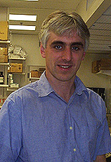
Patricia Becerra received her Ph.D. from the University of Navarra, Spain, in 1979. She did postdoctoral work at NCI and NIAID and then worked at Georgetown University in Washington before returning to NCI. She joined NEI as a visiting scientist in 1991 and became an investigator there in 1994. She is now a senior investigator in the Laboratory of Retinal Cell and Molecular Biology (LRCMB), NEI.
My interests are in the area of protein structure as it relates to function, with a focus on the interactions of components involved in cell differentiation and survival. My research at NEI has applied these interests to relevant systems in the retina.
My lab group has been studying pigment epithelium–derived factor (PEDF), a protein that acts in neuronal differentiation and survival in cells derived from the retina and CNS. PEDF inhibits angiogenesis, and its expression is downregulated over the replicative lifespan of mammalian cells. This interesting factor is secreted by retinal pigment epithelial cells into the interphotoreceptor matrix, where it acts on photoreceptor cells. Its importance in the development, maintenance, and function of the retina and CNS is evident in animal models for inherited and light-induced retinal degeneration, as well as for degeneration of spinal cord motor neurons.
When I joined the LRCMB, little was known of PEDF’s structure, biochemistry, and mechanism of action. Much of our progress in understanding PEDF relied on our development of overexpression systems that yielded recombinant proteins as functionally active neurotrophic factors identical to the native protein and ideal for biochemical, biophysical, and biological studies.
The cDNA sequence for PEDF predicts a unique protein with strong homology to members of the serine protease inhibitor (serpin) superfamily. For this reason, we first studied PEDF as a serpin. We established that PEDF belongs with the subgroup of noninhibitory serpins and that it has characteristics of a substrate rather than an inhibitor of serine proteases. Moreover, a region from its amino-terminus confers the neurotrophic activity to the PEDF polypeptide, not requiring the serpin reactive loop (the structural determinant for serpin inhibition).
We concluded that PEDF’s neurotrophic activity must be mediated by a mechanism independent of serine protease inhibition, and we proposed that during evolution this serpin might have lost its inhibitory activity and gained its neurotrophic function. These findings provided an example of the separation of inhibitory and other activities in a serpin.
Our investigations were next directed toward the hypothesis that PEDF’s neurotrophic activity is mediated by interactions with cell surfaces. Focusing on retinoblastoma and cerebellar granule cells, we prospected for PEDF receptors and found evidence for 1) a saturable, specific, and high-affinity class of receptors on the surface of both cells, with characteristics of an 80-kDa plasma membrane protein, and 2) the amino-terminal region in PEDF that interacts with the receptor. Our work demonstrated that the first step in the biological activity of PEDF is the binding to receptors on the surface of target cells, a significant advance in the elucidation of PEDF’s mechanism of action.
We hope our PEDF research lays the groundwork for the development of therapies for diseases involving defective neuronal differentiation or cell survival, such as retinitis pigmentosa, age-related macular degeneration, and amyotrophic lateral sclerosis.
The reported antiangiogenic effects of PEDF suggest it may also be useful in treating diseases in which new blood vessel formation plays a role, such as diabetic retinopathy, age-related macular degeneration, tumor growth, and rheumatoid arthritis.
The role of a cell-surface receptor in the mechanisms of action of PEDF represents a key aspect of regulation. Along these lines, our first priority is the identification of the PEDF receptor protein, and we are investigating cell-surface receptors in the normal and diseased retina. Consistent with our goal of elucidating the mechanisms of action of PEDF, we are interested in signal transduction and the expression of genes affected by PEDF’s stimuli.
Future work will also explore the development of sustained delivery systems for PEDF in animal models of retinal and CNS diseases.
 |
|
Bill
Copeland
|
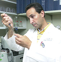
Bill Copeland received his Ph.D. from the University of Texas at Austin in 1988 and did his postdoctoral work at Stanford (Calif.) University before joining the Laboratory of Molecular Genetics of NIEHS in 1993. He is now a senior investigator in the Laboratory of Molecular Genetics, NIEHS.
My major research interest at NIEHS is mitochondrial DNA (mt-DNA) replication and how the structure of replication proteins relates to mt-DNA stability and drug sensitivity. Mitochondria, the subcellular site of energy production, are unique organelles in that they possess a circular chromosome of 16,569 bp coding for 13 polypeptides, 22 tRNAs, and 2 ribosomal RNAs.
Mammalian mt-DNA is 16 times as prone to oxidative damage and evolves 10 to 20 times as fast as nuclear DNA. Point mutations and DNA deletions in the mt-DNA give rise to a wide range of severe diseases. These mutations can occur during DNA replication. DNA polymerase g is responsible for replicating mt-DNA and is composed of a 140-kDa subunit containing catalytic activity and a 55-kDa accessory subunit.
We were the first to clone, overproduce, and characterize the human DNA polymerase g catalytic subunit, p140, and p55 accessory subunit. We discovered that, in addition to its polymerase and exonuclease activities, the human DNA polymerase g catalytic subunit also has dRP lyase activity. This activity is required for base excision repair. This key activity and the fact that DNA polymerase g is the only DNA polymerase in animal cell mitochondria mean that it is necessary in both DNA replication and repair.
We were also the first to determine that the 55-kDa accessory subunit increases the length of DNA synthesized by enhancing the binding of the polymerase to DNA. We are now determining the role of the mt-DNA polymerase with and without the accessory subunit in mt-DNA mutation by studying the fidelity of DNA replication and determining the mutation spectrum produced in vivo.
DNA polymerase g is unique among the cellular replicative DNA polymerases in that it is highly sensitive to anti-HIV nucleoside analogs, such as AZT, dideoxynucleotide, and other nucleoside analogs. These drugs can cause mitochondrial toxicity by producing mitochondrial myopathies such as ragged-red fiber disease in AIDS patients. We are determining what structural elements in polymerase g make this cellular polymerase sensitive to antiviral nucleoside analogs. To accomplish this goal, we are studying the inhibition of DNA polymerase g and several mutant polymerases by antiviral AIDS drugs. We showed that a single tyrosine residue is responsible for the sensitivity of mt-DNA polymerase to dideoxynucleoside analogs.
Accessory proteins are also required for initiation, elongation, and maturation of mt-DNA replication. To understand their role as well as the overall mechanism of replication, we have cloned and overproduced the single-stranded DNA binding protein and helicase. My group is now examining the assembly and communication of these proteins with the DNA polymerase.
We are also studying a nuclear DNA polymerase involved in repair of interstrand crosslinks. Damaging agents such as cisplatin, nitrogen mustard, and other bifunctional alkylating agents cause interstrand crosslinks in DNA that present a difficult problem for replication and must be repaired for faithful replication and viability of the cell. We have cloned and overproduced a DNA polymerase involved in this crosslink repair and named it DNA polymerase q. Human DNA polymerase q is 198 kDa and is homologous to the Drosophila mus308 protein product, a DNA polymerase-helicase involved in repair of interstrand crosslinks.
To fully understand this new human DNA polymerase, the full-length cDNA has been functionally overexpressed in insect cells by a recombinant baculovirus and the active protein purified and characterized. Future work will explore the role of this polymerase in DNA repair and, specifically, how it acts on DNA-containing bulky adducts or crosslinks.
 |
|
Frank
Gherardini
|
Frank Gherardini received his Ph.D. from the University of Illinois, Urbana-Champaign, in 1987 and did a postdoctoral fellowship in the laboratory of Philip Bassford at the University of North Carolina, Chapel Hill. In 1991, he joined the faculty at the University of Georgia, Athens, where he was an associate professor of microbiology before joining the Laboratory of Human Bacterial Pathogenesis at NIAID’s Rocky Mountain Labs in Hamilton, Mont., in July 2001.
My research group focuses on the physiology, biochemistry, gene regulation, and pathogenesis of Borrelia burgdorferi, the causative agent of Lyme disease in humans. The bacterium is transmitted to humans by ticks of the genus Ixoides, and a chronic inflammatory condition develops, causing pathological manifestations in neurologic, cardiac, rheumatologic, and dermatologic tissues. The disease is characterized by early and late stages similar to the disease course in syphilis.
B. burgdorferi faces several environmental and immunological challenges during its infective cycle and must alter (regulate) gene expression to adapt successfully to these conditions. Analysis of the B. burgdorferi genome sequence has revealed that there are very few known regulatory proteins in this bacterium. Conspicuously absent are global regulatory proteins such as CRP, LexA, Fnr, IHF, OxyR, Lrp, and the s factors involved in the heat shock response, s32 and s24. This suggests that compared with other well-characterized pathogenic bacterial systems, the global regulatory systems operating in B. burgdorferi are relatively simple. Clearly, these systems are required for B. burgdorferi to adapt as it encounters very different environments during transfer from an animal reservoir to the tick and then to a human host.
Our research efforts have focused on three important regulatory proteins, PerR, F54, and FS, which control the expression of genes that are critical to the pathogenesis of Lyme disease.
(1) PerR-dependent regulation in B. burgdorferi. Analysis of the genome of B. burgdorferi identified an open reading frame that has 54.3 percent similarity to a protein encoded by perR from Bacillus subtilis. PerR, which requires a metal ion as a cofactor, has been shown to regulate dps, hemA, katA, and mrgA, some of which are involved in an oxidative stress response and response to Mn limitation. Because of the function of PerR from B. subtilis, we have begun to analyze a putative oxidative stress response in B. burgdorferi. B. burgdorferi contains genes encoding an Mn-dependent superoxide dismutase (sodA), a putative flavin-dependent NADH peroxidase (npx) (designated as nox in the B. burgdorferi genome database), and a neutrophil-activating protein (napA) that complements an alkylhydroperoxide reductase mutant of E. coli. Our working hypothesis is that B. burgdorferi cells are using these three proteins to convert toxic O2- to H2O2, and then to H2O, in a two-step process that is regulated by PerR. Because this system would promote the in vivo survival of B. burgdorferi cells when challenged by O2- and H2O2 from host cells, we are particularly interested in this process and how it is regulated.
(2) Regulation of gene expression by F54 and FS. Regulation of gene expression by FS, a member of the F70 family, is responsible for the transcription of a variety of genes encoding virulence factors in Salmonella sp. Salmonella plasmid virulence genes, spvR and spvABCD, are carried on large (50- to 100-kb) plasmids in a variety of Salmonella species, including S. typhimurium, S. choleraesuis, and S. dublin. Loss of these plasmids, of SpvR, SpvC, and SpvD, or of FS, results in loss of virulence and the ability of the cells to multiply in the reticuloendothelial system in mouse models. B. burgdorferi contains rpoS (encoding FS), and our preliminary data indicate that expression of rpoS regulates several plasmid-encoded genes that may be important in the virulence of the bacterium.
We have mapped one transcriptional start site of rpoS from B. burgdorferi to a potential F54-dependent promoter. Furthermore, analysis of the transcription of rpoS in a B. burgdorferi F54-mutant indicates that FS is regulated by F54 during different stages of the infective cycle. Thus F54-holoenzyme is required for FS expression, and both proteins play key roles in the pathogenesis of Lyme disease. We hope that continued elucidation of these genetic and biochemical processes will lead to more effective diagnosis and treatment of Lyme disease.
 |
|
Pu
Paul Liu
|
Pu Paul Liu received his medical degree from Beijing Second Medical College in 1982 and his Ph.D. from the University of Texas M.D. Anderson Cancer Center in Houston in 1991. He did his postdoctoral work at the University of Michigan, Ann Arbor, before joining the Laboratory of Gene Transfer of the then-National Center for Human Genome Research in 1993. He is now a senior investigator and head of the Oncogenesis and Development Section in NHGRI.
The main focus of my research is the genetic control in hematopoiesis and the development of leukemia. Leukemia affects 26,000 Americans each year and represents a significant burden on the health-care system. Hematopoiesis is the process of terminal differentiation of mature blood cells from stem cells, a process controlled by a complex network of proteins regulating cell proliferation and differentiation. Leukemia, a tumor of white blood cells, is an example of abnormal hematopoiesis. In addition, many hematological diseases, such as certain forms of anemia and neutropenia (decreased white blood cells), result from defects in hematopoiesis.
Chromosome 16 inversion is one of the most common chromosome abnormalities in human acute myeloid leukemia (AML). During my postdoctoral training, I found that a fusion gene between CBFB and MYH11 is generated by this inversion. After joining NIH, my lab demonstrated that this CBFB-MYH11 fusion gene, through its encoded fusion protein CBFb-SMMHC, suppresses the normal function of the transcriptional heterodimer CBF (composed of CBFb and AML1) and blocks normal hematopoiesis in transgenic mice.
We showed that this suppression of CBF function by CBFb-SMMHC could be explained by the ability of CBFb-SMMHC to sequester AML1 in the cytoplasm, away from the nucleus, where normally AML1 is located and involved in the regulation of gene expression. Using transgenic mouse models, we demonstrated recently that CBFB-MYH11 is necessary but not sufficient for the development of leukemia and that additional genetic changes are needed for full leukemic transformation. Since this discovery, we have identified such cooperating genetic changes using a retroviral insertional mutagenesis approach in mice.
CBF genes encode transcription factors that associate with other nuclear proteins and regulate expression of target genes. We are now working to identify downstream target genes of CBF oncoproteins, which will lead to better understanding of the process of leukemogenesis and, potentially, to the discovery of new diagnostic and therapeuti targets. In addition, we will test the hypothesis that the other subunit of CBF, AML1, is a leukemia suppressor in transgenic mice. Finally, we will test new therapeutic approaches for AML that specifically counteract the function of CBFB-MYH11 in our established murine leukemia model.
Knowledge of normal hematopoiesis will enhance our understanding of leukemogenic process. To understand the role of Cbfb in hematopoiesis and other developmental processes, we have generated mice with a Cbfb-GFP knock-in fusion. In parallel studies, we are isolating zebrafish mutants defective in their ability to differentiate white blood cells. Our methods include chemical mutagenesis and screening for loss of expression of myeloid-specific markers in fish embryos. We will identify the genes altered in these mutants by genetic mapping and positional/candidate gene cloning. We hope this will yield new insight into the control of myelopoiesis.
 |
|
Alan
Perantoni
|
Alan Perantoni received his Ph.D. from the Catholic University of America, Washington, D.C., in 1983, after having conducted his dissertation research at NCI. He was an assistant professor in the Pathology Department, University of Colorado Medical School in Denver, before returning to NCI in 1992 as a senior staff fellow. He is now a senior investigator in the Developmental Biology Working Group of the Laboratory of Comparative Carcinogenesis, NCI.
My interests are in the area of cell specification and inductive signaling and how they relate to the neoplastic process. I am particularly interested in the mechanisms responsible for the accumulation of aberrant stem cells found in embryonal neoplasms such as the pediatric Wilms’ tumor, which caricatures metanephric development.
Accordingly, I have focused on the characterization of factors that mediate normal metanephric stem cell commitment, that is, the inductive signaling ligands that cause specification; the signaling pathways that direct the differentiation; and the downstream events that regulate and execute morphogenesis—all in an effort to define possible targets of carcinogenesis.
Metanephric development is driven by reciprocal inductive interactions between the epithelial ureteric bud, which is derived from the mesonephric duct and serves as the progenitor of the collecting duct system, and the metanephric mesenchyme (MM), which originates in the adjacent nephrogenic cord, converts to a polarized epithelium under the influence of the ureteric bud, and forms the epithelia of the nephron. I am most interested in this signature event of morphogenesis—epithelial conversion—since it appears to be the point at which differentiation is severely inhibited in the Wilms’ tumor.
The nature of the inductive ligands has remained an enigma for more than 70 years, despite the efforts of several groups to define them. To accomplish this, I established a rat cell line from the natural inductor, the ureteric bud, and purified factors from cell culture medium conditioned by these cells. As a result of such studies, I have identified three growth factors or cytokines that function in combination to induce epithelial conversion with kinetics comparable to those in vivo: fibroblast growth factor–2, leukemia inhibitory factor, and transforming growth factor–b2.
Although these factors can function somewhat independently of one another, causing epithelial tubule formation after several days, they are most effective in combination, yielding tubules in two to three days, as in vivo. Because the factors we identified implicate specific signaling pathways, I am now interested in determining how signaling from these pathways integrates to produce a more profound and accelerated inductive response. To address this question, my group has focused on establishing a stem cell line from rat MM.
In addition, we have developed a model for identification of molecular events specifically associated with epithelial conversion using differential display. This approach has revealed a large series of known and novel sequences that are differentially transcribed with morphogenesis. The list includes transcription (co)factors, cell-cycle regulatory proteins, cell adhesion molecules, and signaling proteins. Of these, CITED1, a CBP/p300-binding transcriptional coactivator, is especially intriguing. It is expressed in the metanephros only in blastemal and stem cells and in blastemal populations of Wilms’ tumors and is downregulated with epithelial conversion. We are currently investigating the possibility that it functions as a gatekeeper for this morphogenetic event.
We hope that these studies lead to a better understanding of the mechanisms responsible for Wilms’ tumor formation and the resulting expansion of metanephric stem cells. But beyond this, we hope the work may help define other blocks in the terminal differentiation of renal cells, such as in renal carcinoma, which originates in tubular epithelia. Furthermore, a comprehensive understanding of the regulation of stem cells responsible for renal tubular and glomerular development may eventually lead to therapies that could invoke mechanisms of tissue regeneration in the treatment of the numerous disorders that damage the tissues of the kidney.
 |
|
Nick
Ryba
|
Nicholas Ryba received his D.Phil. from the University of Oxford in 1985 and did postdoctoral work at the MPI for Biophysical Chemistry in Göttingen, Germany, and at Leeds University, England, before joining the Laboratory of Immunology, NIDR, in 1991. He is now a senior investigator in the Oral Infection and Immunity Branch, NIDCR.
I have always been interested in the biology of sensory perception and since coming to NIH have used molecular approaches to study olfaction, pheromone detection, and taste. Over the last few years, my group has concentrated primarily on understanding the physiological basis of taste sensation and discrimination. This work on taste has involved a wonderful wide-ranging collaboration with Charles Zuker’s group at the University of California in San Diego.
Our sense of taste allows us to distinguish the sweetness of honey from the bitterness of tonic water and the sourness of unripe fruit from the saltiness of seawater. Although taste perception involves many steps, it begins at the surface of taste receptor cells with the recognition of the tastant molecules by taste receptors. This means that the identification of functionally defined taste receptors generates powerful molecular tools to begin to dissect not only the cellular basis of tastant recognition, but also the logic of taste coding. For example, defining the size and diversity of the receptor repertoire provides evidence for how a large number of chemosensory ligands may be recognized, and analysis of the patterns of receptor expression contributes to our understanding of chemosensory discrimination and coding.
Recently, we isolated two novel families of G-protein coupled receptors expressed in subsets of taste receptor cells of the tongue and palate (T1Rs and T2Rs). One of these, the T2Rs, is a family of about 30 different genes that include several functionally validated mammalian bitter taste receptors. Nearly all of the T2R genes are clustered in regions of the genome that have been genetically implicated in controlling responses to diverse bitter tastants in humans and mice, consistent with a role as bitter taste receptors.
Notably, we found that most T2Rs are co-expressed in the same subset of taste receptor cells, suggesting that these cells are capable of responding to a broad array of bitter compounds but not of discriminating between them. This is consistent with what we know about bitter taste, which serves as an important warning of the presence of many chemically unrelated noxious substances.
In contrast to bitter taste, the number of biologically relevant sweet tastants is modest. Thus, it is generally believed that the sweet-receptor family is likely to be quite small. Genetic studies of sweet tasting have identified a single principal locus in mice (Sac) that influences responses to several sweet substances. Very recently, we used transgenic mice to show that the Sac locus encodes a T1R-family receptor and we developed a cell-based reporter system to prove that T1Rs encode functional sweet taste receptors. Intriguingly, we found a complete cellular segregation of T1R and T2R receptors, strongly suggesting that sweet and bitter tastes are encoded by activation of different subsets of taste receptor cells (as might be expected for modalities that influence such opposite behaviors).
In the future we hope to extend these studies to define the logic of taste coding by examining the physiology and connectivity pathways of T1R- and T2R-expressing cells in the various tastebuds and by investigating the effect of genetic ablation, or knockouts, of the different cells and receptor combinations.
 |
|
Nico
Tjandra
|
Nico Tjandra received his Ph.D. in physics from the Carnegie Mellon University, Pittsburgh, in 1993 and did postdoctoral work at the Laboratory of Chemical Physics in NIDDK. He joined the Laboratory of Biophysical Chemistry in NHLBI as a tenure-track scientist in 1997 and is now a senior investigator in that lab.
My interests are in the area of protein structure and dynamics and how they define biological function. We are focusing on proteins in cell signaling and apoptosis. We use high-resolution nuclear magnetic resonance (NMR) to elucidate the structures and dynamics of these proteins.
The interactions between GTP-binding proteins, their receptors, and their effectors are well characterized in the plasma membrane. In contrast, the specific interaction and function of these proteins in the Golgi membranes is still not clear. Human Ga interacting protein (GAIP) is a major component in the regulation of Ga signaling in the Golgi by interacting specifically with the Gai3. We used NMR to find the structure of parts of the GAIP, including the solution structure of the "Regulation of the Ga signaling" (RGS) domain of GAIP. We used NMR for a backbone dynamics study of human GAIP. The dynamics data revealed flexible regions within this protein. Comparison between the NMR structure of GAIP and the X-ray structure of the RGS4-Gai3 complex identified the conformational changes that result from GAIP interaction with the Ga protein that are also consistent with our dynamics data.
We concluded that the overall structure of the whole class of RGS domains has very little variation and that the sequences flanking the RGS domains of these proteins determine what type of signals they will regulate as well as where they are localized.
Bax is a member of the Bcl-2 family of proteins that regulates apoptosis, or cell death. In collaboration with Richard Youle’s laboratory at NINDS, we have determined the solution structure of human Bax by NMR spectroscopy. Our structure identifies an elegant mechanism in which two crucial steps in Bax’s control of apoptosis—dimer formation and insertion into the mitochondrial membrane—are simultaneously regulated.
Our structure also shows that the packing of the COOH-terminal helix of Bax plays an important role in its function. When this helix is packed against the hydrophobic BH3-binding pocket, it eliminates the possibility for dimer formation. The packing of this helix also hinders its exposure, thus inhibiting Bax insertion into the mitochondria membrane. Based on our structure, regulation of Bax in apoptosis must include displacement of the COOH-terminal helix from the hydrophobic pocket.
What is even more surprising is that the structure of Bax, a pro-apoptotic protein, is essentially identical to Bcl-xL, an anti-apoptotic protein, even though they share only 20 percent sequence homology. We found just three flexible regions that substantially differ in the two structures. Identification of these regions would not have been possible without Bax structure. Interestingly, some of these loops that distinguish the two proteins were thought to have no significance in Bax function; therefore, no extensive biological data are available about them.
The high degree of similarity between the two structures has at least two consequences. First, functional models that have been proposed for Bcl-xL might also be applicable to Bax. Second, perhaps other components in the apoptosis pathways or differences in structure of these proteins after insertion into mitochondria membranes might be important in differentiating their functional roles. We believe that our structure will have a great influence on how future experiments in the apoptosis field will be designed and will give us a better understanding of how this important biological process is regulated.
Looking ahead, we hope to examine structures of other protein components that make up the cell signaling of the Golgi as well as the mitochondrial apoptosis pathway to better understand these complex and yet basic cell functions.
 |
|
Darryl
Zeldin
|
Darryl Zeldin received his M.D. degree from Indiana University in Indianapolis in 1986, and completed a residency in internal medicine at Duke University in Durham, N.C., and a fellowship in pulmonary/critical care medicine at Vanderbilt University before joining the Laboratory of Pulmonary Pathobiology of NIEHS in 1994. He is now a senior investigator and head of the Clinical Studies Section at NIEHS.
My research interests are in eicosanoids, with an emphasis on how they function in the heart, kidney, and lung. This work in this area has been driven by three main hypotheses: (1) P450 cytochromes metabolize arachidonic acid to eicosanoids that play critical roles in modulating fundamental biological processes such as cardiac contractile function and vascular tone; (2) environmental or genetic factors that lead to aberrant P450 activity cause altered production of bioactive eicosanoids and result in heart disease and hypertension; and (3) environmental or genetic factors that affect cyclooxygenase (COX) activity lead to altered prostaglandin production and result in lung dysfunction and asthma.
My lab was the first to clone CYP2J2, a human P450 that is abundant in the heart and active in the metabolism of arachidonic acid to epoxyeicosatrienoic acids (EETs). Subsequent studies from our group demonstrated that EETs improve heart contractile function after ischemia, inhibit cardiac L-type calcium channel activity, and beneficially affect cardiac electrical and mechanical properties.
In further work with CYP2J2, we created transgenic mice in which we overexpressed CYP2J2 in heart myocytes. These mice exhibit improved postischemic cardiac function, a shortened cardiac action potential, and, interestingly, enhanced responsiveness to b-adrenergic receptor agonists. These mice are the first in vivo animal model to permit evaluation of P450 functions in heart.
My colleagues and I also demonstrated that CYP2J2-derived eicosanoids decrease cytokine-induced endothelial adhesion molecule expression, an important component in the development of vascular inflammation and arteriosclerosis. This occurs via inhibition of the transcription factor NF-kB. We showed that exposure of endothelial cells to hypoxia and reoxygenation, such as occurs during heart attack and stroke, decreases CYP2J2 expression, but that maintenance of CYP2J2 protein concentrations attenuate hypoxia-reoxygenation–induced cell death. Moreover, we demonstrated that CYP2J2-derived EETs increase tissue plasminogen activator expression and fibrinolytic activity, actions that reduce the formation of intravascular clots. Together, these studies demonstrate that CYP2J2 and its eicosanoid products play critical roles in normal cardiac myocyte and endothelial function and may be involved in limiting damage such as vascular inflammation, hemostasis, and cardiac dysfunction after ischemia.
In 1999, my laboratory reported cloning CYP2J5, a new mouse P450 that is primarily expressed in the kidney, active in the metabolism of arachidonic acid to EETs, and localized to proximal tubules and collecting ducts—sites where EETs are known to affect renal fluid and electrolyte transport and mediate the actions of several hormones, including angiotensin II and arginine vasopressin. We observed that maximal CYP2J5 expression occurs at a critical time during postnatal renal development when abnormalities in renal function are demonstrable in animals that subsequently develop hypertension.
Evidence to support the hypothesis that CYP2J products are involved in the development and/or maintenance of hypertension comes from recent studies in our lab showing upregulation of a CYP2J immunoreactive protein and increased EET biosynthesis in kidneys of spontaneously hypertensive rats. We mapped the Cyp2j locus to a chromosomal area that co-segregates with the hypertensive phenotype in Dahl salt-sensitive rats. Our recently developed Cyp2j5 knockout mice have systemic hypertension de novo and will be instrumental in evaluating the role of this P450 in kidney function, renal eicosanoid metabolism, and blood pressure regulation.
Our combined work on human and mouse CYP2Js has led to a better understanding of the role of cytochromes P450 in the metabolism of arachidonic acid into compounds that modulate critical cardiovascular functions. The opportunities for translational research in this area include development of new drugs for blood pressure control, vascular inflammation, atherosclerosis, and ischemic heart disease. The identification of individuals at increased risk for these disorders due to P450 genetic variation may also lead to new approaches to disease prevention. We hope to pursue some of these areas in the years ahead.
More recently, we have gone on to examine the role of COXs in lung function. Our work in this area is based on the hypothesis that COX-derived eicosanoids are important modulators of the lung immune response to environmental agents and that COX-deficient mice provide a novel model system to study basic immunologic mechanisms in inflammatory lung disease.
My group was the first to report that allergic lung responses are increased in COX-deficient mice. More recently, we have examined the effects of disruption of COX genes on pulmonary responses to inhaled bacterial lipopolysaccharide (LPS). Interestingly, although COX-deficient mice had increased bronchoconstriction after LPS exposure, there were no differences in inflammatory indices between the genotypes. This finding indicates that COX enzymes are important in regulating physiologic but not inflammatory responses to inhaled LPS.
We hope ongoing studies
on the effect of COX-derived eicosanoids on pulmonary immune response to environmental
agents will lead to opportunities for further mechanistic and translational
research. ![]()