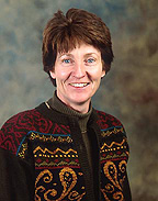
Josephine
Egan
| T H E N I H C A T A L Y S T | J U L Y – A U G U S T 2001 |
|
|
|
| P E O P L E |
RECENTLY TENURED
|
|
Josephine
Egan
|
Josephine Egan received her M.B., B.Ch. (M.D.) in 1979 from the National University of Ireland, Galway. After fellowships in clinical pharmacology (at Baylor College of Medicine in Houston) and endocrinology (the University of Virginia Health Sciences Center, Charlottesville), she joined the NIA in 1990, where she is now chief of the Diabetes Section, Laboratory of Clinical Investigation.
My research focuses on insulin secretion.This means that anything that perturbs insulin secretion is fair game for study—from the most basic level to clinical investigation.
Because type 2 diabetes is the commonest form of diabetes in the elderly, and I work in the NIA, I am especially interested in that disease. But why insulin secretion?
Work in many parts of the world has established beyond doubt that type 2 diabetes is caused by reduced insulin action (insulin resistance) and faulty insulin secretion. In the 1980s and 1990s, there was an explosion in the understanding of how insulin actually activates its receptor, what intracellular processes became activated consequently, and how glucose transport occurs. When it was shown that insulin-mediated glucose transport is decreased in type 2 diabetes, insulin resistance became accepted as the dominant defect of type 2 diabetes. This assumption was regrettable because it is only part of the story.
Obesity, which is the commonest cause of decreased insulin-mediated glucose uptake, is present in 85 percent of type 2 diabetic subjects. But only a minority of obese subjects develop diabetes. This incongruity told us that something besides insulin resistance is needed to cause diabetes. Most obese subjects do not develop diabetes because they become hyperinsulinemic relative to their nonobese counterparts and so can compensate for the insulin resistance. When insulin secretion problems intervene, blood glucose rises and diabetes occurs.
Some of the derangements in insulin secretion in type 2 diabetes have been revealed in clinical studies. They are: 1) absent first-phase insulin secretion (insulin secreted from b-cells in the first few minutes after acute blood glucose elevation), 2) very weak, if any, insulin secretion in response to rising blood glucose—most obvious when glucose is given intravenously, 3) severely diminished 24-hour integrated insulin secretion, and 4) a flattening in the usual pulsatile manner in which insulin is secreted.
In our section, we have used rodent models and human physiology studies to elucidate what lies behind those derangements. We know that insulin secretory capacity depends on both function and mass of b-cells and that b-cells are heterogenous in their response to stimuli. Increasing glucose results in increasing recruitment of secretory b-cells. First-phase insulin secretion comes from a subpopulation of b-cells; in type 2 diabetes this subpopulation is silent.
b-cells that lack glucose competency—the ability to sense changes in blood glucose so that insulin secretion can be adjusted—will not become secretory. We are now gradually peeling back the layers of factors involved in glucose competency, and much of our research has focused on two specific gut peptides, GIP and GLP-1.
Both of these factors are released from the gut in response to food and act on specific receptors on the b-cells to render the b-cells glucose-competent. Activation of their receptors leads to increases in cAMP concentrations, a primary glucose-competency factor. GIP is secreted as soon as food enters the duodenum. We have seen that in type 2 diabetes the b-cells are resistant to GIP, and plasma GIP concentrations rise. No amount of exogenous GIP will induce the receptors to respond. We plan to elucidate the cause of the downregulation of the GIP receptors.
GLP-1 is secreted from the ileum, again after eating, but later than GIP. Its effects are also somewhat downregulated in type 2 diabetes, but, when given in pharmacological doses, it can restore first-phase insulin secretion, increase maximum insulin secretion, and improve pulsatile insulin secretion. Therefore, we have undertaken long-term studies with GLP-1 in treating type 2 diabetic patients. We are developing analogs of GLP-1 to overcome some of the drawbacks—due to its very short half-life of only a few minutes—inherent in using GLP-1 as a pharmacological agent.
We are also exploring the control of the second component required for insulin secretion—sufficient b-cell mass. In type 1 diabetes, as long as 30–50 percent of residual b-cell mass remains, hyperglycemia does not occur. Transplantation experiments have taught us that enough islets must be transplanted in order to restore euglycemia. The capacity of b-cells to synthesize insulin must also be preserved. Infiltrative diseases, such as hemochromatosis and perhaps amyloidosis, can diminish insulin synthesis.
Questions we have been focusing on in this area are: What happens to b-cell mass with age? What regulates the mass? Can the mass be manipulated by pharmacological means? Where do new b-cells come from? Are there endocrine progenitor cells in adult pancreas that can be manipulated? We have shown that GLP-1 also is involved in regulating total b-cell mass and function in rodents. Function is regulated via increasing insulin mRNA and insulin synthesis. We plan to ascertain whether this is so in humans.
Clinically, we are beginning to use data from the Baltimore Longitudinal Study of Aging (BLSA) to examine hormone activities in the natural history of type 2 diabetes. Within our BLSA population are people who have gone from a nondiabetic state to glucose intolerance to frank diabetes. We are asking when GIP concentrations begin to rise—is it at the point of glucose intolerance or only with frank diabetes? What happens to GLP-1 during the progression? We hope to answer these questions—to pinpoint the time in the progression to diabetes when insulin and gut derangements actually lead to diabetes—and thereby elucidate the cause of insulin deficiency.
 |
|
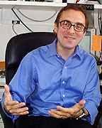
Nicholas
Restifo
|
Nicholas Restifo received his M.D. degree in 1987 from New York University. He was a research fellow at the Memorial Sloan-Kettering Cancer Center in New York before coming to NIH. He is now a principal investigator in the Surgery Branch, NCI.
The focus of our efforts is to develop new immunotherapies for cancer. Our approach is to create new animal models to study the basic immunology of the tumor-host interaction and to develop and test new treatments.
Animal models form the foundation of our current understanding of immunology in general and tumor immunology in particular; preclinical animal data form the core of almost any new therapy. But while animal models are very good for working out mechanistic questions, they are much less predictive of therapeutic benefit in humans. Put another way, animal models are useful to describe what can happen in patients, but they are unreliable at predicting what does happen.
The focus of our mouse work is on the development of anticancer vaccines—vaccines designed to treat, rather than prevent, disease. We have accomplished this experimentally by identifying the mouse homologs of human tumor-associated antigens. We have created and tested recombinant and synthetic anticancer vaccines based on these antigens, examples of which are recombinant viruses, "naked" DNA immunogens, proteins, and peptides.
One of the most interesting and difficult models we work with is based on the B16 mouse melanoma. This tumor was initially discovered as a spontaneously occurring mouse melanoma and is now grown in culture and passaged in syngenic mice. It turns out that B16 expresses all of the homologs of the human melanocyte differentiation antigens that function as tumor antigens.
To elucidate important immunological and therapeutic questions, we use mice that are transgenic for human MHC molecules (such as HLA-A2 and HLA-DR4). We also use knockout mice lacking the antigens we are targeting. This enables us to assess the roles for immunological tolerance to these antigens. Most recently, we have created mice that are transgenic for T-cell receptors, enabling us to study tumor-specific T cells in a variety of activation states.
We are currently using our newly created mouse models to explore an immune strategy in which T lymphocytes are expanded outside the body and "adoptively" transferred to treat patients with cancer. Efforts to date at adoptive immunotherapy have focused on giving killer T lymphocytes or CD8+ T cells. We will soon add CD4+ T cells, which may help killer cells grow, survive, and localize to the tumor site after adoptive transfer.
We also plan to use what some term gene therapy to modify the functions or survival of transferred T cells. The insertion into T cells of genes encoding anti-apoptotic proteins or the gene encoding growth factor interleukin-2 is one possible approach. Alternatively, new reactivities could be conferred on T lymphocytes by inserting genes encoding T-cell receptors specific for tumor-associated antigens.
The immune system can eradicate tumor cells; our goal now is to induce the immune destruction of cancer more consistently. We plan a multipronged approach to the development of new immune-based treatments for patients whose cancers are not currently treatable with traditional chemotherapy, radiation, and surgery.
 |
|
Jeffery
Struewing
|
Jeffery Struewing received his M.D. from Indiana University School of Medicine in Indianapolis in 1985 and his M.S. in preventive medicine from the University of Maryland at Baltimore School of Medicine in 1988. He joined the NCI’s Genetic Epidemiology Branch in 1991 and is now a senior investigator in the Laboratory of Population Genetics.
My research has focused on genetic aspects of breast and ovarian cancer. I have attempted to bridge the gap between epidemiologic studies in humans and more basic molecular and biochemical characterizations of genetic variation.
In doing so, I have progressed from family-based studies of genes with large effects, such as BRCA1 and BRCA2, to more population-based studies of the genetic and environmental determinants of cancer susceptibility.
I began work in this area by recruiting breast and ovarian cancer families for clinical and epidemiologic study in NCI’s Genetic Epidemiology Branch. Our studies helped localize the genes to chromosomal locations, and other groups finally isolated single genes, BRCA1 and BRCA2, that, when mutated, lead to a predisposition to cancer, with the observed dramatic pedigrees.
After obtaining training in molecular genetics, my laboratory studies in breast and ovarian cancer began in 1994 with the analysis of 24 NCI families, 10 of which were found to have BRCA1 mutations. Most of these families had different BRCA1 mutations, but three —all Ashkenazi Jewish—shared the same mutation, designated 185delAG.
In collaboration with Larry Brody of NHGRI, this observation led to our finding that approximately 1 percent of stored DNA samples from Ashkenazi Jews contained the BRCA1 185delAG mutation. For several years, BRCA1 and BRCA2 were studied almost exclusively in high-risk families, in which a very high risk of breast cancer (penetrance) was estimated—85 percent or more by age 70 and nearly 100 percent lifetime. In other words, BRCA1 appeared to be a nearly fully penetrant, autosomal dominant disease gene.
Studies in less restricted populations, however, have shown the situation to be much more complex. Our finding of the 185delAG mutation at a high frequency in Ashkenazi Jews was a prelude to more population-based, genetic epidemiologic studies of this gene. It also gave rise to the possibility of almost immediate commercial testing in this population, despite the fact that it was unknown whether cancer risk estimates from the high-risk families applied to carriers identified from a broader population base.
We therefore quickly designed and implemented a study of the prevalence and penetrance of the 185delAG mutation in the Washington Ashkenazi Jewish population. In more than 5,000 volunteers, we demonstrated a combined 1.2 percent carrier frequency for the 185delAG and 5382insC BRCA1 mutations and a 1.2 percent carrier frequency for 6174delT, the founder mutation of BRCA2 (this gene and mutation were identified after the initiation of the study).
More importantly, using the newly developed kin-cohort method, we estimated that the risk of breast cancer among mutation carriers was 56 percent (40 percent to 73 percent for the 95 percent confidence interval) by age 70—a high risk, but one well below most previous estimates.
Subsequent estimates of penetrance have generally been even lower than our estimate, supporting the idea that the average risks of cancer among BRCA1/2 mutation carriers are much lower than initial estimates from high-risk families.
This discovery opens the door to studies aimed at identifying environmental and genetic factors that modify cancer risk in BRCA1/2 mutation carriers and in the majority of women who do not carry these mutations. This will be a main focus of my future work.
Which genetic loci are likely to be related to cancer susceptibility? The precise biochemical basis for cancer predisposition in BRCA1/2 mutation carriers is unknown, but the protein products of both genes are likely to be involved in DNA double-strand break repair. Variations in genes involved in all aspects of DNA damage recognition and repair, therefore, make ideal candidates as breast cancer susceptibility factors.
We are planning comprehensive analyses of all single nucleotide polymorphisms in DNA repair genes using matrix-assisted laser desorption/ionization–time of flight assays.
 |
|
Irving
Wainer
|
Irving Wainer received his Ph.D. from Cornell University in Ithaca, N.Y., in 1970 and did postdoctoral work at the Institute of Molecular Biology, University of Oregon, Eugene, and Thomas Jefferson University Medical School in Philadelphia. He held positions at the FDA, St. Jude Children’s Research Hospital (Memphis), and McGill (Montreal) and Georgetown (Washington) universities before joining NIA in May 2001 as chief of the Bioanalytical and Drug Discovery Unit.
The research programs in my laboratory include clinical pharmacology and the development of online high-throughput screens for new drug discovery. Our clinical work is primarily focused on how disease state can alter drug metabolism.
We have identified several discordances between genotype and expressed phenotype in patients with advanced cancer and AIDS. For example, patients with advanced cancer or AIDs who also have extensive or fast genotypes for cytochrome P450 2C19 and N-acetyltransferase-2 have displayed poor and slow phenotypes, respectively.
Because these observations were associated with advanced disease, we have initiated studies in patients with terminal syndromes such as cancer cachexia, or wasting. In particular, we have developed a direct measure of a "proteolysis- inducing factor" (PIF) associated with cachexia. PIF is measured in spot urines using capillary electrophoresis (CE). The presence of PIF in urine has been correlated with clinical status and with PIF in tumor biopsies. We have also correlated the presence of PIF in urine with treatment response and clinical relapse. This fall, we will begin a longitudinal study on PIF as a disease marker.
Based on these results, we have initiated a study using CE coupled with mass spectrometry and matrix-assisted laser desorption/ionization–time of flight spectrometry to quantify PIF in tissues and to examine the effect of cachexia on pre- and post-translational expression of hepatic enzymes and transporters. We will also use laser capture microdissection and CE with mass spectrometry or laser-induced fluorescence to study these effects in single cells. In a second line of work, we have developed liquid chromatographic stationary phases containing immobilized receptors, enzymes, and transporters as an online flow system for new drug discovery and characterization of drug candidates.
These columns can range
in size from standard liquid chromatography columns to microcolumns and can
be used to screen complex chemical mixtures, characterize single compounds,
and screen phage libraries. The columns can be used with known targets—for
example, nicotinic, GABA, NMDA, and estrogen receptors; P-glycoprotein and other
ABC transporters; and cytochrome P450 and other enzymes. They can also be used
with orphan receptors and other expressed proteins, and they can be placed online
with mass spectrometers or other instruments that detect structure or activity,
providing real-time data that cannot be obtained using standard microtitration
plates. ![]()