| T H E N I H C A T A L Y S T | M A Y – J U N E 2001 |
|
|
|
| P E O P L E |
RECENTLY TENURED
 |
|
Jay
Chung
|
Jay Chung received his M.D. and Ph.D. from Harvard Medical School in Boston in 1988. After his residency in internal medicine at Brigham and Women’s Hospital, Boston, he joined NIDDK in 1990 as a clinical endocrinology fellow and in 1991 began doing research as a postdoctoral fellow in the Laboratory of Molecular Biology. In 1994, he moved to the Laboratory of Biochemical Genetics in NHLBI, where he is now a senior investigator.
My group is interested in understanding how the behavior of transcription factors is regulated in vivo by other transcription factors and signaling pathways. Although a great deal is known about transcription factor–DNA interactions in vitro, very little is known about how transcriptional factors are recruited in living cells.
To visualize transcription factor-DNA interaction in living cells, we developed the PIN*POINT (protein position identification with nuclease tail) method. This method is based on the idea of fusing the nuclease domain from the restriction endonuclease Fok1 to the transcription factor of interest and expressing the fusion protein in the cell.
Because the nuclease domain does not have sequence specificity of its own, the nuclease domain fused to the transcription factor cleaves DNA near where the transcription factor portion of the fusion protein binds. Detection of these cleavage sites with molecular biology techniques permits the visualization of the recruitment of the transcription factor to a region of DNA in vivo.
By applying PIN*POINT to understand how b-globin gene expression is regulated in vivo, we addressed several fundamental questions regarding transcription factor recruitment. We have discovered that the proximal promoter can also influence the recruitment of transcription factor EKLF to the distant regulator of b-globin expression (LCR, the locus control region). EKLF binds to a specific DNA sequence, the CACCC box, in vitro, and it has therefore been assumed that the EKLF-CACCC box interaction controls EKLF recruitment in vivo. Although both b-globin and g-globin promoters contain the CACCC box, EKLF activates the b-globin promoter but not the g-globin promoter. Our findings demonstrate that a novel suppressor that binds to the g-globin promoter but not to the b-globin promoter suppresses EKLF recruitment to the g-globin promoter. This finding provides evidence that proteins bound to the promoter have strong influence on recruitment of other proteins to the promoter.
In erythroid cells, the chromatin over the b-globin gene is in an "open" conformation but is in a "closed" conformation in other tissues. How the chromatin is remodeled in such a tissue-specific way is completely unknown. One possibility is that chromatin-remodeling complexes such as the BRG1 complex open the chromatin over the b-globin promoter in erythroid cells. However, the chromatin-remodeling complexes generally do not have DNA sequence specificity, and it is therefore not known whether these complexes are targeted to specific sites such as the promoter region.
Our findings indicate that EKLF and the general transcriptional machinery help recruit the BRG1 complex to the b-globin promoter, confirming that chromatin-remodeling complexes can be targeted to a specific site in vivo. This work will be important not only for understanding transcriptional mechanisms but may have implications in drug development for diseases such as sickle cell anemia.
Thyroid hormone receptor activates the transcription of genes with a TRE (thyroid hormone responsive element) in the presence of thyroid hormone. Using PIN*POINT, we have demonstrated that thyroid hormone triggers physical interaction between TRE and promoter.
More recently, our group has begun a new line of investigation in the area of DNA damage signaling. We cloned a kinase called hCds1, which plays an important role in DNA damage response. We discovered that one of the substrates of hCds1 is the BRCA1 tumor suppressor. We have discovered that hCds1 colocalizes with BRCA1 in nuclear foci and phosphorylates serine 988 of BRCA1 in vivo. Phosphorylation of serine 988 is increased after DNA damage and causes dispersion of BRCA1 to sites of its action. Mutation of serine 988 to alanine, which is not phosphorylated, destroys BRCA1’s ability to confer DNA damage resistance to BRCA1-mutated cells, suggesting that serine 988 phosphorylation is important for BRCA1 function in DNA damage response. We have identified the consensus sequence for hCds1 substrates, which we are using to identify in vivo substrates of hCds1. We are now developing transgenic mouse models to extend these studies.
We are currently integrating our interests in transcription regulation and signal pathways by focusing on the regulation of transcription factor activity by various signal pathways. In the near future, we will use high-throughput functional genomics approaches in genetically tractable model organisms to help us discover disease-related transcription factors and signal pathway components in humans.
We are halfway through the analysis of a library of yeast strains in which each strain contains a single mu-tation in one of 6,000 different genes and will begin high-throughput gene knock-down analyses using RNAi in Drosophila cells and Coenorhabditis elegans. Our hope is to take full advantage of the genomics and proteomics approaches to answer mechanistic questions regarding transcription and signal transduction that have implications for human diseases and their treatments.
 |
|
Victor
Pike
|
Victor Pike received his Ph.D. from the University of Birmingham (U.K.) in 1976 and did postdoctoral work there before joining the Medical Research Council Cyclotron Unit in London in 1978 and becoming the head of its Chemistry and Engineering Group in 1996, with concurrent honorary appointments at Imperial College of Science, Technology and Medicine in London and the University of Surrey. In February 2001, he became chief of the PET Radiopharmaceutical Sciences Section in the Molecular Imaging Branch, NIMH.
My principal interests are in all chemical aspects of the design, synthesis, and biological evaluation of novel radioactive probes for use in pharmacological and clinical research with positron-emission tomography (PET). PET is now recognized as a powerful molecular imaging technique for unraveling the biochemistry underlying various clinical disorders and for evaluating the efficacy of existing or proposed treatments. In particular, my work has focused on the challenge of radiochemistry with short-lived cyclotron-produced carbon-11 (t1/2 = 20 min) and fluorine-18 (t1/2 =110 min). This interest began at the MRC Cyclotron Unit, with the establishment of one of the world’s first PET centers.
My first success was the development of a rapid automated procedure for the preparation of carbon-11–labeled acetate for the study of myocardial energy metabolism. This tracer can be used for the positive identification of transient myocardial ischemia in human subjects. After more than two decades, this tracer remains in widespread clinical use at several PET centers. At this time, I also labeled the antibiotic erythromycin A with carbon-11 in order to measure directly the antibiotic concentration at its site of action in human pneumonic lung, a pioneering example of the use of a PET tracer in drug research and development.
My group also developed a new class of labeling agents, [11C]acid chlorides, which opened up the potential to prepare a wider range of labeled compounds; an early example is [11C]di-prenorphine, one of the first radioligands for imaging brain opiate receptors and for investigating the involvement of these receptors in neuropsychiatric conditions. This radioligand continues to be used for clinical research. My group also successfully developed radioligands for heart and lung adrenoceptors.
The pharmaceutical industry has appreciated the capability of PET to assist in drug discovery and development, and I helped develop several tracers for this purpose. An interesting example was 18F-labeled HFA 134a, which was used to show that HFA 134a is safe for inhalation by human subjects. HFA 134a is now produced in enormous quantities to replace ozone-depleting chlorofluorocarbons in their large-scale applications and also as a drug aerosol propellant. Drug particles (for example, fluticasone propionate) administered by inhalation have also been labeled with positron-emitters to measure their regional lung deposition with PET. These measurements are crucial to a better understanding and optimization of drug-inhalation therapies.
The ability of PET to give precise data—the occupancy of a specific population of receptors by a given dose of drug—can be of immense benefit in drug discovery and development. However, such measurements depend on the availability of a selective radioligand for the receptor in question. My group has been instrumental in developing the first effective radioligand for brain serotonin type 1A (5-HT1A) receptors, [11C]WAY-100635. This radioligand has widened possibilities to examine the role of the serotonergic system in various neuropsychiatric disorders, including anxiety, depression, and schizophrenia. This radioligand is now used in clinical research worldwide and also has been widely used for receptor occupancy studies with established or candidate drugs. My collaborators and I received the Marie Curie Award and Springer Prize for our contributions to radioligand development.
Extensive medicinal chemistry and advances in radiochemistry have been at the core of my past success. I aim to continue work in these areas in support of radiotracer and radioligand development in the new Molecular Imaging Branch by developing probes for brain biochemistry of animal and human subjects to better understand neuropsychiatric disorders.
 |
|
Rashmi
Sinha
|
Rashmi Sinha received her Ph.D. in nutritional sciences from the University of Maryland, College Park, in 1987. She joined NCI’s Laboratory of Cellular Carcinogenesis and Tumor Promotion that year, moved to the Epidemiology and Biostatistics Program in 1992, and is now a senior investigator in the Nutritional Epidemiology Branch of the Division of Cancer Epidemiology and Genetics.
My research focuses on the complex role of diet in cancer etiology, especially regarding carcinogens such as heterocyclic amines (HCAs) and aromatic hydrocarbons (PAHs) that are produced in meat cooked at high temperatures. Early epidemiologic studies found a suggestive association between meat-cooking techniques and cancer risk, but did not differentiate factors that influenced the production of HCAs and PAHs. For example, roast beef and steak were included within the same meat category, despite widely dissimilar cooking methods and very different levels of HCA formation. Following up on these initial leads, I designed an interdisciplinary research program to investigate the roles in human cancer etiology of mutagens and animal carcinogens formed during meat cooking.
First, I needed tools to estimate HCA and PAH intake in an accurate and reliable manner; I took two approaches to secure them–improving questionnaire-based measures of intake and evaluating biological markers of HCAs and PAHs. To improve intake estimates, I developed databases similar to those used for other dietary components (such as fats, proteins, and vitamins) that could be with a food frequency questionnaire (FFQ). In collaboration with several research laboratories, I measured mutagenic activity, HCAs (such as MeIQx, PhIP, and DiMeIQx), and PAHs (such as BaP) in meat samples cooked by different methods to varying levels of doneness.
Results from this study underscored the need to include questions on type or cut of meat, cooking technique, and doneness levels for individual meat items in order to capture the variability in HCA and PAH content. Using this information, I designed and validated a FFQ that takes account of these details. I also carried out a large metabolic study to evaluate HCA biological markers of internal exposure that could be used in etiologic studies and to assess the influence of polymorphic metabolic enzymes on these biomarkers.
I have used both questionnaire information and biomarkers in case-control studies of colorectal adenomas and lung, breast, and stomach cancers. I found an increased risk of colorectal adenoma associated with high intake of red meat. Most of this risk was attributable to high levels of doneness (well or very well done) and/or to high-temperature cooking techniques such as grilling. Linking the FFQ information to the mutagen-carcinogen database, I evaluated the effect on risk of mutagenic activity and exposure to several HCAs and to BaP. Mutagenic activity is an integrative measure of all mutagens contained in meat. I found an increased risk associated with higher levels of meat-derived mutagenic activity; intake of MeIQx, a highly mutagenic HCA; and possibly PhIP. I am now investigating several polymorphic enzymes involved in metabolizing these compounds. I am also evaluating the relationship of BaP to colorectal adenoma risk using questionnaire data on diet and cooking and biologic measures.
Other studies in which I have collaborated also lend support to a causative role for HCAs in human cancer. For example, red meat, especially fried and/or well-done red meat, was associated with an increased risk of lung cancer in a population-based case-control study of Missouri women, and MeIQx was associated with elevated risk in both nonsmokers and moderate smokers. In a case-control study in Nebraska, intake of well-done red meat, a surrogate for HCA, and grilled red meat, a proxy for both HCAs and PAHs, were related to stomach cancer. And in a collaborative case-control study of breast cancer nested in the Iowa Women’s Health Study, where well-done red meat was associated with increased risk of breast cancer, I found that high intake of PhIP, especially, was related to elevated risk.
Furthermore, the association of specific HCAs with different tumor sites–such as MeIQx and PhIP with colorectal adenomas, MeIQx with lung cancer, and PhIP with breast cancer–parallels to a certain extent the organ specificity of animal carcinogenicity studies. In rodent models, several HCAs produce colon tumors, MeIQx causes lung tumors, and PhIP leads to mammary tumors.
To extend my initial observations, I have established collaborations with multiple intramural and extramural investigators. I have contributed key components to dietary questionnaires on meat cooking in numerous case-control studies and in several cohort studies as well, for example, the Nurses’ Health Study; the Health Professionals Follow-up Study; the NCI Prostate, Lung, Colorectal and Ovarian Cancer Screening Trial; and the Agricultural Health Study. These studies should help clarify the role that HCAs play in the etiology of several types of cancer in the United States. Should a consistent association between cancer and meat cooking mutagens be established, we can then make public health recommendations on the safest way to prepare meat to eliminate bacterial contamination and to minimize possible carcinogens.
 |
|
Peter
Sun
|
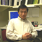
Peter Sun received his Ph.D. from the Institute of Molecular Biology, University of Oregon, Eugene, in 1990 and did postdoctoral work at NIDDK. He joined NIAID in 1994 and is now a senior investigator in the Laboratory of Immunogenetics.
My research focus has been on the structure and function of immunoreceptors. In particular, we study how the receptors recognize their ligands and initiate activation or inhibitory signals. We primarily choose to use the X-ray crystallography technique to define three-dimensional molecular structures of receptors and their ligand complexes.
By doing so, we can visualize the receptor-ligand molecular interface and thus map out the hot spots on the receptor—the regions critical for ligand recognition. In practice, knowing the shape of a receptor-ligand interface and the residues involved will help us not only in the diagnosis of diseases caused by the impairment of receptor function, but also in the design of potential new drugs that are capable of inhibiting the function of these receptors.
Recent advances in the molecular and cellular biology of innate immune functions have opened a new, exciting frontier for structural biology—to reveal the receptor activation mechanism involved in innate immunity.
Following the cloning of several natural killer (NK) cell surface receptors by others, we began to study their ligand recognition by X-ray crystallography and solution-binding methods. At the center of our interest is the mechanism by which NK cells distinguish between self and nonself and thereby direct their cytolytic killing against virally infected rather than healthy host cells.
Two families of inhibitory receptors, the killer immunoglobulin receptors (KIR) and the C-type lectin-like receptors, have been identified on human NK cells to interact with class I MHC antigens and thus may be critical in mediating this self vs. nonself recognition.
Members of C-type lectin-like CD94/NKG2 receptors recognize a nonclassical class I molecule–HLA-E–bound with peptides derived from the signal peptide of other classical class I MHC molecules; in contrast, KIR receptors recognize classical class I HLA molecules.
In an effort to elucidate the molecular interface between KIR receptors and their HLA ligands, we have determined the crystal structures of an HLA-Cw3–recognizing KIR and its complex with the HLA molecule. This is the first structure of a KIR-HLA complex, and it illustrates the resemblance between KIR and T-cell receptor recognition of HLA molecules. It also reveals the contribution of the class I peptide to KIR recognition and the nature of allotypic specificity of the receptor.
Through mutations and binding studies, we also demonstrated the importance of interface residues in KIR-HLA recognition.
Another receptor involved in NK cell–mediated innate immunity is an Fc receptor, FcgIII. Fcg receptors are expressed on the surface of macrophages, mast cells, and NK cells to mediate opsonization of antibody-coated pathogens and antibody-directed cellular cytotoxicity.
They are also implicated in certain autoantibody-mediated autoimmune diseases. In an effort to define how immunoglobulins bind and activate humoral immune systems through their Fc receptors, we crystallized and solved the X-ray structure of FcgIII in complex with an Fc fragment of IgG1.
The co-crystal structure revealed an unexpected binding of one receptor to the dimeric Fc ligand. This mode of receptor binding, although very different from other known ligands of Fc, explains the role of the antigen–specifically the need for immune complex formation–in receptor activation. Based on our crystal structure, we explored the possibility of using small molecular ligands that are capable of inhibiting the receptor function as potential useful therapeutic reagents.
We have designed four
peptides and investigated their ability to bind to receptor FcgRIII. Using solution-binding
experiments, we demonstrated that these peptides bind specifically to FcgRIII
with 1/20 to 1/10 the affinity of IgG1 and are able to inhibit Fc binding to
the receptor. The results of these peptide studies have opened new ways of designing
therapeutic compounds. ![]()