Joshua
Farber
| T H E N I H C A T A L Y S T | J A N U A R Y – F E B R U A R Y 2001 |
|
|
|
| P E O P L E |
RECENTLY TENURED
|
|
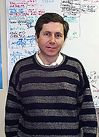
Joshua
Farber
|
Joshua Farber received his M.D. degree from the Johns Hopkins University School of Medicine, Baltimore, in 1977 and completed residency and fellowship training in infectious diseases at the Johns Hopkins Hospital. He did postdoctoral work in the laboratories of Earl Stadtman (Laboratory of Biochemistry, NHLBI) and Daniel Nathans (Department of Molecular Biology and Genetics, Johns Hopkins University School of Medicine). He joined NIH in 1993 and is now a senior investigator in the NIAID Laboratory of Clinical Investigation.
My clinical training in infectious diseases and my basic science training in molecular biology led me to search for novel genes induced in macrophages in response to factors produced by activated T cells. I presumed that such genes would encode proteins important in defense against infection and inflammation, and I was particularly interested in identifying secreted proteins because either the proteins themselves or small-molecule agonists or antagonists of their receptors might be useful therapeutically. In my screening using material made from mouse macrophages, I discovered genes encoding two proteins, Mig and CRG-2/IP-10, that belong to a family of secreted proteins now known as chemokines. These discoveries served as the starting point for my work at NIH.
The chemokines are now recognized as a family of more than 40 chemotactic cytokines that are of critical importance in regulating the trafficking of leukocytes throughout the body as part of leukocyte development and differentiation, homeostasis, and host defense. Chemokines signal through seven transmembrane-domain G-protein–coupled receptors, of which 18 have been identified in humans. Signals produced by these receptors are necessary for the activation of integrins on the surfaces of leukocytes, which is in turn required for white-cell adherence to endothelium and egress from the vasculature into lymphoid organs and tissue. These signals are also presumed to be important for the positioning of leukocytes within tissues after leaving the circulation.
Mig, IP-10, and a third chemokine, I-TAC, were found to share a receptor, CXCR3. Besides their roles in leukocyte physiology, chemokine receptors—particularly CCR5 and CXCR4—were found to function with CD4 as obligatory co-receptors for the entry of HIV and SIVs into cells, discoveries that provided major insights in HIV and SIV biology and disease and have created the possibility for new therapeutics.
In characterizing the Mig protein, members of my laboratory found that this chemokine targeted T cells and, in particular, T cells that had been recently activated. Analysis of experimental infections in mice revealed widespread induction of Mig and CRG-2/IP-10 in response to production of IFNg. We presumed that Mig and CRG-2/IP-10 were important for recruiting activated T cells as well as other CXCR3-expressing cells, such as natural killer cells, to peripheral tissues for host defense. While experiments in our and other laboratories have supported this presumption, experiments with Mig knockout mice that we created revealed an unexpected role for Mig in the optimal production of antibody against a bacterial pathogen. These and other recent studies of ours using human tonsils have suggested that a pro-inflammatory chemokine such as Mig may have a role within lymphoid organs in optimizing interactions among T cells, B cells, and dendritic cells.
Besides our work on chemokines, my laboratory also discovered two chemokine receptors expressed on lymphocytes that are now known as CCR6 and CXCR6, and we cloned and characterized two forms of a third lymphocyte chemokine receptor, CCR9. We found that CCR6—the receptor for Mip-3a, a chemokine induced by pro-inflammatory cytokines at epithelial surfaces—was expressed on subsets of memory CD4+ T cells that home in on the skin and the intestine. Of particular interest, CCR6 was also expressed on both naïve and memory B cells, and activation of B cells through antigen receptors led to a significant increase in receptor activity without changing the number of receptors per cell. Together with data of ours for other chemokine receptors on T cells, this observation suggests novel mechanisms for regulating chemokine receptor expression on activated lymphocytes.
Understanding these mechanisms is one focus of my laboratory’s current work. We reported that CCR9 is a receptor for TECK, a chemokine expressed constitutively by the epithelia of the thymus and small intestine. CCR9 is expressed on thymocytes and specifically on the subsets of memory T cells of the intestine. We found that alternative splicing of the CCR9 mRNA led to two forms that vary at their NH-termini, and that these forms, CCR9A and CCR9B, respond to different concentrations of TECK. This represents a new way of extending the range of concentrations over which a cell can respond to a chemokine ligand.
Finally, my laboratory has worked on understanding the role of the chemokine system in AIDS. We discovered a receptor that we named STRL33, now called CXCR6, that, together with our collaborators in NIAID and the FDA, we found functioned along with CD4 as a co-receptor for diverse strains of HIV and SIV. And we collaborated on the first studies to show that envelope glycoproteins of HIV and SIV can signal through the primary HIV and SIV co-receptor, CCR5.
Taken together, our studies and those of many other labs suggest that particular chemokine receptors function to direct the migration of specific lymphocyte subsets, defined by the cells’ states of development, activation, and differentiation, as well as the compartments onto which the cells home. We seek more detailed understanding of the roles of the individual chemokine receptor-ligand groups in host defense, immunopathology, and AIDS in order to reveal how the chemokine system functions as a whole in lymphocyte biology. We expect that by manipulating the system—such as by using chemokine receptor antagonists now under development—we will better understand how to treat both AIDS and immune-mediated disease.
 |
|
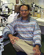
Ashok
Kulkarni
|
Ashok Kulkarni received his Ph.D. from the Maharaja Sayajirao University of Baroda, India, in 1980 and did postdoctoral work at Columbia University in New York and in the Developmental and Metabolic Neurology Branch, NINDS, before joining NIDCR in 1995. He is now a senior investigator and head of the Functional Genomics Unit, NIDCR, and director of the NIDCR Gene Targeting Facility.
My interest is in functional genomics as it relates to in vivo gene function, with a focus on candidate genes in development and disease. In my early work, I generated and characterized transforming growth factor-ß1 (TGF-ß1) knockout mice. These mice were shown to develop multifocal inflammation with similarities to human autoimmune disorders. Further analysis of these mice revealed critical roles of TGF-ß1 in embryonic development, hematopoiesis, inflammation, tooth mineralization, and carcinogenesis.
My lab used gene targeting to generate a much-needed animal model for Fabry disease, a painful and fatal X-linked lipid-storage disease. We disrupted the a-galactosidase A gene in mouse embryonic stem cells to generate a line deficient in this enzyme. These mice develop subclinical symptoms similar to those in Fabry disease patients. With collaborators, we demonstrated the effectiveness of bone marrow transplantation, gene therapy, and lipid deprivation to ameliorate metabolic defects in Fabry mice. We are continuing work with this mouse model to develop therapeutic approaches to Fabry disease.
Another focus of my research is molecular mechanisms underlying neuronal phosphorylation and its role in neurodegenerative diseases such as amyotrophic lateral sclerosis (ALS) and Alzheimer’s disease. We first generated cyclin-dependent kinase 5 (Cdk5) null mice. These mice show abnormal neuronal migration, cerebellar defoliation, and abnormal motor neurons. Although this mouse model revealed unique roles of Cdk5 in neuronal migration, it was of limited value in studying the aging nervous system because of its neonatal fatality. To circumvent this problem, we developed Cdk5 conditional knockout mice in which Cdk5 is disrupted after neuronal migration is complete. These mice display locomotor and postural abnormalities along with neuromuscular defects that mimic some of the symptoms found in ALS patients.
We have also used gene targeting to generate mouse models for hereditary dental disorders, such as amelogenesis imperfecta (AI) and dentinogenesis imperfecta (DGI). Mice bearing a targeted mutation in the amelogenin gene, which is implicated in AI, have discolored and disfigured teeth. These defects are similar to those seen in AI patients, and these mice will be valuable for understanding the role of amelogenin in tooth development and designing treatments for this disorder. We have also targeted overexpression of TGF-ß1 to teeth using a tooth-specific promoter. Mice overexpressing TGF-ß1 in teeth develop discolored and fractured teeth, which resemble the tooth phenotype seen in DGI. We are currently analyzing the role of TGF-ß1 in tooth development and disease using proteomics and genomics.
Our overall goal is to use functional genomics to identify specific roles of genes in the molecular processes that underlie development and disease. Doing so will also help us unravel functions of the numerous genes being identified in human and mouse genome studies.
 |
|
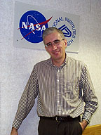
Leonid
Margolis
|
Leonid Margolis received his Ph.D. from Moscow State University, where he also earned an advanced doctoral degree in 1985. He progressed through the academic ranks there to full professor and lab chief. With the beginning of "perestroika" and collapse of the Soviet Union, he worked as a visiting professor at University College, London, and Johns Hopkins University in Baltimore. In 1994, he became a Fogarty Scholar-in-Residence at NIH. He joined NICHD’s Laboratory of Cellular and Molecular Biophysics in 1995, where he now heads the Intercellular Interactions Section; he is also deputy director of the NASA/NIH Center for Three-Dimensional Tissue Culture.
In a broad sense, I would like to understand the mechanisms of normal and pathological cell behavior in a native microenvironment of real tissue in vitro. The biology of any given cell depends on a complex system of local contacts with cellular and noncellular structures within the tissue.
Meanwhile, most of our knowledge in cell biology comes from experiments on isolated cells that do not adequately represent the complexity of cell-cell interaction. Specifically, my current interest is to understand tissue HIV pathogenesis.
Critical events in HIV disease occur in lymphoid tissue, where HIV infects cells via surface molecules, CD4, and one of the co-receptors, CXCR4 or CCR5. Infection leads to CD4+ T lymphocyte loss, deterioration of cell-cell interactions, and immunodeficiency. To study HIV tissue pathogenesis, we developed a new system of human lymphoid tissue infected with HIV-1 under controlled conditions ex vivo. The goal was to delineate the role of viral co-receptors in HIV pathogenesis.
There are three important unanswered questions I want to answer: 1) why HIV infection is transmitted by viruses using the CCR5 co-receptor, 2) why later in the course of HIV disease CXCR4-utilizing variants often evolve, and 3) why this switch of "co-receptor tropism" coincides with rapid CD4+ T cell depletion and development of immunodeficiency.
Over the last few years, together with members of my unit—in particular, Jean-Charles Grivel, Svetlana Glushakova, and Wendy Fitzgerald—and encouraged by LCMB Chief Joshua Zimmerberg, we have focused on answering the last of these three questions. Using chimeric isogenic viruses that differ only by the sequences in the viral envelope protein that determine co-receptor recognition, we demonstrated that HIV-1’s use of CXCR4 is sufficient to induce severe T lymphocyte depletion.
We delineated the mechanism of this phenomenon, explaining why CCR5-utilizing HIV-1 variants deplete far fewer CD4+ T lymphocytes than CXCR4-utilizing viruses—namely, because fewer T lymphocytes express CCR5 than CXCR4. Surprisingly, some of the "dual-tropic" viruses that in transfected cell lines recognize both co-receptors (and which comprise the majority of HIV isolates), in human lymphoid tissue use only one co-receptor. In this setting again, CXCR4 usage is associated with severe CD4+ T lymphocyte depletion because its prevalence makes the cells cognate targets for these viruses.
Furthermore, we found that ex vivo human lymphoid tissue retains immune function and responds to antigen challenge by producing specific antibodies. CXCR4- but not CCR5-utilizing HIV strains suppress this immune function in ex vivo tissues. We are now trying to understand the mechanism for such a dramatically different immune response in tissues infected with HIV-1 that differ only in co-receptor tropism.
I am now focusing more on the first two of our main questions regarding HIV transmission and the co-receptor switch. I think they should be addressed at the highest level of tissue complexity, involving cell-cell interactions and cell trafficking. I expect to use the NASA bioreactor—available through the NICHD-based NASA/NIH Center for Three-Dimensional Tissue Culture—to try to combine various human tissues with lymph to create a multicom-partment "artificial patient." In this system, we will test various hypotheses regarding virus transmission as well as various antiviral drugs and possibly vaccines.
My dream is to develop new techniques to monitor the interactions of a cell—how it establishes and disrupts cell-cell contacts and changes its metabolism in response.
Although my current focus is predominantly on HIV, several ongoing projects address mechanisms of tissue pathogenesis by other human pathogens, such as parasites, bacteria, and other viruses. I think that our studies are important for developing new ways to understand, prevent, and treat the diseases caused by these agents, as well as for development of new diagnostics. I believe that in the 21st century, "biology" will increasingly become "tissue biology," emphasizing the cell—not as a separate entity, but as an element of a multicellular structure. My research is aimed at this goal.
 |
|
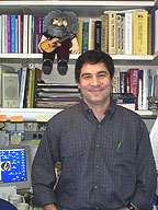
Paul
Roche
|
Paul Roche received his Ph.D. from Duke University in Durham, N.C., in 1988. He did postdoctoral studies with Peter Cresswell at Duke and with Eric Long in the Laboratory of Immunogenetics in NIAID. In 1994 he joined the Experimental Immunology Branch of NCI, where he is now a senior investigator.
My research interest is in understanding basic mechanisms of protein trafficking in lymphocytes. Lymphocytes are major effectors of immunity, recognizing and ultimately eliminating foreign pathogens from the body.
My group has two distinct areas of interest: elucidating the basic mechanisms of intracellular granule exocytosis and studying the cell biology of antigen processing and presentation to T lymphocytes. We have approached our work using traditional techniques of molecular cell biology, and we are currently using confocal microscopy to address these issues in living cells.
Like all eukaryotic cells, lymphocytes rely on intracellular transport vesicles to shuttle membranes and cargo proteins from one compartment to another along the constitutive and regulated secretory pathways. These vesicles must dock and ultimately fuse with specific target membranes, and our goal is to understand the molecular mechanisms by which this specificity is achieved.
In neurons, a class of proteins known as SNAREs form a multiprotein complex that catalyzes the fusion of synaptic vesicles with the presynaptic plasma membrane, thereby allowing neurotransmitter to be released from intracellular stores into the synaptic cleft.
When I joined the Experimental Immunology Branch, the SNAREs that catalyze similar granule-plasma membrane fusion events in nonneuronal cells were not known. We therefore set out to identify the proteins necessary for secretion in immune cells. Using the yeast two-hybrid system, we isolated a ubiquitously expressed SNARE, termed SNAP-23, that mediates membrane-membrane fusion events in a wide variety of eukaryotic cells including lymphocytes.
SNAP-23 and other SNARE subunits behave as integral membrane proteins, yet they do not insert into membranes using the "traditional" co-translational translocation machinery. We have shown that partial SNARE complexes containing SNAP-23 actually assemble in the cytosol and traffic to membranes post-translationally. Furthermore, we have cloned a novel kinase, termed SNARE-kinase, or SNAK, that phosphorylates cytosolic SNAP-23 and promotes assembly of SNARE complexes. Our current focus is on whether stimulating exocytosis in secretory cells alters SNARE complex assembly and whether regulated assembly-disassembly of SNAREs is required for granule exocytosis in lymphocytes. We are examining exocytosis by video microscopy. This allows us to observe the behavior of individual fluorescent secretory granules in real time.
I have also had a long-standing interest in the cell biology of antigen processing and presentation to T cells. During immune surveillance, foreign pathogens are engulfed and degraded by antigen-presenting cells (APCs). Peptide fragments of these antigens are then presented on the cell surface of the APCs for recognition by antigen-specific T lymphocytes. Most of our work has been examining the biosynthesis, assembly, and intracellular trafficking of peptide-binding major histocompatibility complex (MHC) class II molecules. For example, we have found that newly synthesized MHC class II molecules are phosphorylated by protein kinase C in human APCs. This phosphorylation regulates the kinetics of intracellular transport of the class II molecules to endocytic peptide-loading compartments.
We have recently become very interested in the behavior of class II molecules on the surface of APCs. We have identified a novel role for plasma membrane lipid microdomains in concentrating the class II–peptide complexes necessary for T-cell activation. We are examining the components at the plasma membrane interface of APCs with T cells (the so-called "immunological synapse"). We are specifically interested in identifying the molecular signals that lead to the recruitment of T-cell–activating ligands (such as class II molecules) to this site.
It is likely that these two interests of the lab will converge, because some plasma membrane SNAREs themselves reside in lipid microdomains and may play a role in the targeting of intracellular class II—peptide complexes to specific regions of the plasma membrane. Although both SNARE function in protein traffic and the cell biology of T-cell activation are areas of intense research, more questions are left unanswered than answered, keeping our interest and excitement as high as ever.
 |
|
Alexander
Wilson
|
Alexander Wilson received his doctoral degree in medical genetics from Indiana University, Indianapolis, in 1980. His postdoctoral training was in statistical genetics in the Department of Biometry and Genetics at the Louisiana State University Medical Center, New Orleans, where he rose to the rank of full professor in 1993 before joining NIH in 1995. He is currently a senior investigator and head of the Genometrics Section in the NHGRI Inherited Disease Research Branch and an adjunct full professor in the Department of Epidemiology at the Johns Hopkins University School of Public Health in Baltimore.
During the past 20 years, my research efforts have focused on using sophisticated statistical methods to identify genetic effects that may be responsible for phenotypic variation in quantitative traits.
My theoretical and methodological work has focused on developing new methodologies to identify genetic effects and, using computer simulation, investigating the statistical properties of methods of genetic analysis for quantitative traits. I’m probably best known for developing a theoretical method that can be used to adjust for the effects of identified genetic loci, so that modifier loci can subsequently be identified. Before coming to NIH, I was one of the principal designers of the Statistical Analysis for Genetic Epidemiology (S.A.G.E.) software package, one of the most widely used statistical packages for the genetic analysis of family data.
Since coming to NIH in 1995, I’ve helped to establish the joint NIH/Johns Hopkins University Center for Inherited Disease Research (CIDR) in Baltimore and have authored or coauthored proposals to obtain over 650,000 genotypes from CIDR for studies searching for genetic loci involved in the phenotypic variation of scoliosis, obesity, cranio-lenticulo-sutural dysplasia, and hypertension.
Most recently, my work has focused on developing alternative methods for multipoint linkage analysis. Lynn Goldin of NCI and I have developed a method based on moving averages to incorporate information from neighboring markers. And, Alexa Sorant, also of NHGRI, and I have proposed a method that uses composite markers derived from bi-allelic single-nucleotide polymorphic (SNP) markers.
We’ve shown that single-locus and multilocus marker systems are mathematically equivalent under certain critical assumptions and that with the appropriate statistical methods, composite markers based on SNP markers for linkage analysis can be nearly as informative as the traditional short tandem repeat markers.
My work in the future
will continue to focus on the identification of genetic loci for quantitative
traits such as hypertension and hypercalcuria, as well as on the development
of statistical methods that can be used to tease the genetic effects out of
these complex disorders. ![]()