Jorge
Carrasquillo
| T H E N I H C A T A L Y S T | J A N U A R Y – F E B R U A R Y 2001 |
|
|
|
Translational ResearchJORGE
CARRASQUILLO:
|
text and photos by Margaret Coulombe |
|
|
Jorge
Carrasquillo
|
The first FDA-approved radiolabeled antibody for imaging was born at NIH, and Jorge Carrasquillo was the sire of the radionuclide that was coupled to the antibody.
The antibody itself was discovered in Jeff Schlom’s NCI Laboratory of Tumor Immunology and Biology. But it was from the ensuing collaborative clinical work between Schlom’s and Carrasquillo’s teams that there emerged in 1992 a new product to uncover metastatic colorectal and ovarian carcinoma (OncoScint, Cytogen Corp., Princeton, N.J.).
"It is fascinating to take something from its basic concept into the lab and then into the clinic," says Carrasquillo, deputy chief of Nuclear Medicine at the Clinical Center. "My main interest is translational research."
And in his nearly two decades of research at NIH, where he came with his mentor in 1983 to establish a radioimmunodetection-radioimmunotherapy program—in the Nuclear Medicine (NM) Department—Carrasquillo has been instrumental in the development of more than 40 clinical research protocols involving antibodies and radioisotopes.
As reflected in the partnership with Schlom, NCI scientists have been the main co-investigators in these projects and have served as the physicians to the patients enrolled in CC clinical trials in this "brand new area of research"—nuclear medicine in diagnosis and therapy.
Carrasquillo’s radioimmunotherapy brewery resides at the Building 21 Nuclear Pharmacy, where NM chemist Chang Paik performs the preliminary labeling and testing of research reagents destined for clinical trials. Carrasquillo and his team also occupy a 4th floor lab in Building 10, where they assay products to ensure their quality and function and perform pharmacokinetic analyses on patient samples.
To support an IND (investigational new drug) submission to the FDA, the NM team typically characterizes and tests the desired radiolabeled antibody both in vitro and in animals. Then Phase I clinical trials are designed and implemented with one or more (usually) NCI collaborators.
During a trial, the NM team takes serial blood samples, urine specimens, and often bone marrow or tumor biopsies to evaluate the distribution of the monoclonal antibody in the body. The pharmacokinetic data, together with the imaging data, are used to calculate dosimetry and help guide changes in protocol.
Home Runs
With Tom Waldmann, chief of the NCI Metabolism Branch, the NM team has performed preclinical evaluations and clinical analyses of pharmocokinetics and dosimetry of anti-Tac monoclonals (directed against the IL-2Ra receptor) in patients with adult T-cell leukemia [Treatment of Tac-Expressing Cutaneous T-Cell Lymphoma and Adult T-Cell Leukemia with Yttrium-90 Radiolabeled Anti-Tac]. Initial results, including two complete remissions, were promising. Ongoing clinical trials are focusing on using humanized anti-Tac for therapy.
Carrasquillo points to Phase I and II trials as the most fulfilling segment of his work. "The most satisfying part is getting something into the clinic that has potential to help patients. Anti-Tac [see "Anti-Tac Takes Off in Three Directions," The NIH Catalyst, March–April 1998] is an example of a successful outcome, with good patient responses. Unfortunately, it’s not often that you hit a home run like that," he adds. However, Carrasquillo also has some runs batted in—end products in which he clearly had a direct influence if not a direct hand,
He points to two radiolabeled immunotherapy products now nearing the market as treatments for non-Hodgkin’s lymphoma—BexxarTM (Coulter Corporation, Miami, Fla.) and ZevalinTM (IDEC Pharmaceuticals Corporation, San Diego, Calif.). The former couples iodine-131 to a monoclonal antibody, and the latter links an antibody to yttrium-90. "At NIH," he notes, "we performed the first successful studies targeting lymphomas with indium-labeled antibodies. It’s building this kind of knowledge base that allows development of such products and reagents by others."
Loaded Bases
Collaborations with Ira Pastan, chief of the Laboratory of Molecular Biology, NCI, and his team have been particularly fruitful. One study aims to "develop chemical methods to radiolabel monoclonal antibodies and fragments to construct scintigraphic imaging agents that detect hematologic malignancies that express IL-2a receptors." The goal is to label "genetically synthesized single-chain disulfide stabilized variable region fragments (scdsFv) of anti-Tac with Tc-99m for tumor imaging.
"In this investigation, we realized that because of their small size, scdsFv fragments had high renal uptake. This led us to a different line of research that resulted in animal studies that suggested optimal ways to block this uptake by using amino acid infusions or by chemically modifying the Fv fragments," Carrasquillo recalls. The two teams also worked together on preclinical studies with indium-111–radiolabeled immuno-toxins.
A collaboration with Pastan in progress since 1992 involves B3 antibody, which Pastan developed and characterized and which Pastan and Carrasquillo have been exploring as the basis for radioimmunotherapy in colon and breast cancer. B3, says Carrasquillo, is an "interesting" antibody that recognizes Lewis Y antigen, which is expressed in high concentrations in various adenocarcinomas.
"Using a xenograft tumor model, my group demonstrated excellent targeting of radiolabeled B3 monoclonal antibody," he says. The finding warranted the filing of an IND with the Food and Drug Administration and resulted in approval of a Phase I clinical trial conducted by Pastan and his NCI colleague Lee Pai.
A follow-up trial, in collaboration with Pai and NCI’s Michael Bishop, is testing a higher radiation dosage with bone marrow support and is ongoing. Carrasquillo continues to design improvements—such as pretargeting—to incorporate into future clinical trials.
In 1998, Carrasquillo and Pastan applied for and received a Bench to Bedside Award to study pretargeting in the treatment of epithelial cancers with radiolabeled monoclonal B3 antibody.
In conventional antibody therapy, radioactive material is coupled to the antibody at the outset. But because most antibodies are fairly large and circulate slowly, surrounding tissues and organs may be damaged by the time the radionuclide finds its mark. The pretargeting strategy investigated by Carrasquillo separates the tumor targeting from the radionuclide delivery.
To do this, the antibody is coupled to the nonradioactive "sidearm" streptavidin, a reagent that recognizes biotin—the small, speedy molecule to which the radionuclide is attached. The antibody package is delivered first and can take its time localizing to the tumor without inflicting damage on surrounding tissues and organs; once tumor localization is achieved, the radionuclide is sped on its way to the tumor by its biotin chauffeur.
"If you attach the radioactivity to biotin, a relatively small molecule, it will get out of the circulation quickly and bind to the streptavidin to which the antibody is attached. What does not attach to the target antibody is quickly eliminated from the system," Carrasquillo explains, noting that the work could not have been done without the Bench to Bedside Award, an award established in 1998 by the NIH Clinical Research Revitalization Committee to encourage intramural collaboration between clinical and basic researchers at NIH.
Carrasquillo is now studying the pretargeting approach in the xenograft setting, in which animal models have yielded results promising enough to pursue reagents for clinical studies, he says.
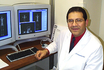
PET Projects
Carrasquillo is particularly enthusiastic about ways to capitalize on the CC’s positron emission tomography (PET) facility, under the direction of Bill Eckelman, to enhance current options in tumor diagnosis and cancer therapy monitoring.
Radioactive materials that give off a single gamma ray when they decay are those traditionally used in nuclear medicine, he notes. Those called positron emitters, on the other hand, release a positively charged electron when they decay, which, in turn, gives off two 511KeV gamma rays, 180 degrees apart. The PET scanner can detect these gamma rays and make exquisite images of their distribution, Carrasquillo says, enabling researchers to "label biological compounds and trace physiologic events to answer clinical questions.
"Very exciting."
Among these positron emitters are carbon-11, oxygen-15, nitrogen-13, and fluorine-18.
Carrasquillo describes several ongoing PET protocols that rely on trace amounts of these positron emitters to measure blood flow and blood volume. Tiny amounts, he notes, citing the "tracer principle," do not perturb the system but allow tracing of the physiologic processes of interest.
Blood flow, blood volume, and glucose metabolism in tumors are the measures of interest in studies undertaken in collaboration with Steve Libutti (NCI) and Steve Bacharach (Nuclear Medicine) to evaluate the effects of anti-angiogenic agents. "Patients are being treated with different antiangiogenic agents, such as anti-VEGF [vascular endothelial growth factor] on various NIH protocols," Carrasquillo elaborates. Using PET imaging techniques, he says, the investigators can assess whether these reagents result in relevant changes in blood flow, blood volume, or glucose metabolism. "PET thus may serve as a surrogate marker of what is happening at the tumor level."
Most recently, Carrasquillo
has applied his PET activities to collaborative work with NIAID’s Anthony
Fauci and Douglas
Brust using fluorodeoxyglucose (FDG) as a measure of glucose metabolism
and, by extension, a possible indirect marker of sites of HIV infection and
active viral replication. The research applies the tumor-imaging properties
of the positron emitter fluorine-18 to the search for HIV reservoirs. The radiolabeled
FDG reagent is taken up by cells with high glucose metabolism and becomes trapped
inside them, resulting in an unusually high signal that flags the possibility
of viral activity. ![]()
 |
As
a resident in nuclear cardiology at the University of Seattle, Jorge Carrasquillo
anticipated a conventional clinical practice, but his work with Steve
Larson, then chief of nuclear medicine at the Seattle Veterans Affairs
Hospital and a pioneer in radiolabeled antibody research, changed his
outlook. Instead, he came with Larson to NIH in 1983 to establish a program
in radioimmunodetection and radioimmunotherapy. Carrasquillo became the
head of the Nuclear Medicine Antibody Program in 1985. He is deputy chief
and one of five nuclear medicine physicians in the CC Nuclear Medicine
Department.
|