
Jeff
Green
| T H E N I H C A T A L Y S T | N O V E M B E R – D E C E M B E R 2000 |
|
|
|
| P E O P L E |
RECENTLY TENURED
|
|
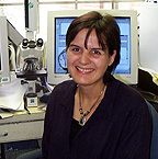
Jeff
Green
|
Jeff Green received his M.D. in 1981 from McGill University in Montreal and residency training in pediatrics at the Children’s Hospital of Philadelphia. He joined the NIH Clinical Center in 1984 as a clinical genetics fellow and subsequently became an NCI biotechnology postdoctoral fellow in the laboratory of the late George Khoury. After an appointment as an investigator in the Laboratory of Molecular Oncology at FCRDC, he joined the Laboratory of Cell Regulation and Carcinogenesis, NCI, in 1997, where he has served as the head of the Transgenic Oncogenesis Group.
The use of transgenic animals for modeling human diseases has been the primary focus of the work in my laboratory. My early interests involved the phenotypic and molecular characterization of transgenic mice carrying the human T-cell lymphotrophic virus (HTLV-1)-tax gene. The tax mice were shown to develop perineural tumors with similarities to human neurofibromas, as well as inflammatory lesions in the salivary and lacrimal glands leading to abnormalities similar to those found in human Sjögren’s syndrome. Further analyses of these transgenic animals also demonstrated in vivo that the tax gene can upregulate important growth regulators, including granulocyte macrophage colony stimulating factor, the interleukin-2 receptor, and nerve growth factor.
With my growing interest in hormone-related malignant disease, I became intent on developing a transgenic model for human prostate cancer. To reach this goal, our lab designed a targeting vector using the C3(1) component of the rat prostate in 5' flanking region. This led to the first transgenic model for prostate cancer that was driven by the expression of large and small T-antigen (Tag) from the SV40 early region. Interestingly, female transgenic mice carrying the same transgene develop mammary cancer that exhibits many important features observed in human breast cancer. Although Tag is not etiologically involved in human prostate or breast cancer, its mechanism of transformation involves the inactivation of two important tumor suppressor genes, Rb and p53. The loss of these tumor suppressors has been implicated in many human cancers, including prostate and breast cancer. Thus, Tag-induced transgenic oncogenesis may involve relevant pathways for human cancer development.
Transgenic prostate and mammary cancer lesions in these mice develop over a very predictable time course. We have taken advantage of this fact to define histopathological and molecular changes that occur at particular stages of tumor formation. The lesions that develop in the prostate appear strikingly similar to human prostate intraepithelial neoplasia and progress to invasive carcinomas. A significant number of these lesions develop ras mutations, as occurs in a subset of human prostate cancers. Although most transgenic mammary tumors do not harbor ras mutations, amplification of ki-ras occurs during tumor progression. We have demonstrated the functional significance of this finding by crossing the C3(1)/Tag mice with ki-ras knockout mice. The resulting mice show delayed tumor development. We have also demonstrated that expression of the transgene in the mammary gland is not responsive to estrogen and, as with human breast cancer, expression of estrogen receptor a is lost during tumor progression.
Our work has also demonstrated that there is a biphasic alteration in the apoptotic response in the transformed cells during the development of mammary cancer in the C3(1)/Tag mice: Rates of apoptosis rise during the preinvasive phase, but fall during the transition to invasive carcinomas. Our lab has demonstrated that the bax gene appears to be a critical regulator of the early pro-apoptotic response in the mammary tumors—C3(1)/Tag mice lacking bax have an accelerated rate of tumor formation.
Currently, I serve as the principal investigator of the NCI Mouse Models of Mammary Cancer Collective, which is part of the national NCI Mouse Models of Human Cancer Consortium. The NCI Mouse Collective is a community of 25 highly interactive NIH investigators seeking to advance the understanding of breast cancer through animal models.
With support from the Collective, my lab is expanding research using cDNA microarray technologies to define genetic pathways and identify new genes in mammary and prostate tumor development. We are comparing the expression profiles of tumors from several major transgenic mouse models of mammary cancer with those of human breast cancer, as part of the validation process for mouse models of human disease. In addition, we are continuing to use transgenic models to test several therapeutic approaches to mammary and prostate cancer including chemoprevention and antiangiogenesis agents. We are using array technologies to uncover mechanisms of action for these agents.
Our goals in this work and for the future emphasize development of new transgenic models for prostate and mammary cancer and the development of new targeting vectors and strategies to improve heterologous gene expression in vivo.
 |
|
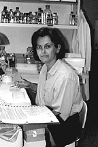
Vanessa
Hirsch
|
Vanessa Hirsch obtained a D.V.M. in 1977 and a Masters of Veterinary Science in pathology in 1981 from the University of Saskatchewan in Saskatoon. She became a diplomate of the American College of Veterinary Pathologists in 1984 and obtained a Doctor of Science degree at the Harvard School of Public Health in Boston in 1988. After four years as a research assistant professor at Georgetown University in Washington, D.C., she joined the Laboratory of Infectious Diseases in 1992 and is now a senior investigator in the Laboratory of Molecular Microbiology, NIAID.
My research focuses on the pathogenesis of AIDS and the development of a vaccine for HIV-1, using simian immunodeficiency virus (SIV) infection of monkeys as an animal model. SIV induces an immunodeficiency syndrome in macaque monkeys that is remarkably similar to that in HIV-infected humans.
Although HIV-1 infection of humans is fatal, the disease course is highly variable, ranging from asymptomatic survival for more than 15 years to progression to AIDS within two years of infection. The level at which plasma viremia stabilizes after primary infection is a highly significant prognostic indicator of subsequent disease course. This suggests that host immune mechanisms are critical in the control of viremia. Studies in my lab have shown that SIV-infected macaques also exhibit variable disease course and that viral load is a strong predictor of disease progression.
My lab is investigating host and viral factors that contribute to the variable disease course in SIV infection of macaques. We recently demonstrated that the susceptibility of peripheral CD4+ T cells to viral infection in culture is highly predictive of primary viremia in these animals after their inoculation with SIV. The relative proportion of CD4+ T cells, their level of activation, or the level of expression of the major SIV co-receptor, CCR5, does not explain the major differences in susceptibility. This result suggests there are other host cell factors that contribute to the establishment of persistent viral replication and progression to AIDS.
I am also investigating the origins of primate lentiviruses. It is important to remember that SIV infection of macaques, an Asian monkey, is artificial. SIVs originate in primates of African origin, including sooty mangabeys (SIVsm) and African green monkeys (SIVagm). My lab characterized the first SIVsm isolate, SIV strains from three species of African green monkeys, the sole SIV from Sykes’ monkeys (SIVsyk), and SIV from L’Hoest monkeys and suntailed monkeys (SIVlhoest and SIVsun). A close genetic relationship between SIV from the latter two monkeys, despite their long-term (over 10,000 years) geographic separation, supports the hypothesis that the primate lentiviruses co-evolved in African monkeys over the last million years. Interestingly, SIVlhoest and SIVsun are genetically most closely related to SIVmnd from mandrills, despite the fact that mandrills and L’Hoest monkeys are only distantly related. Recently, we characterized SIV from redcapped mangabeys (Cercocebus torquatus torquatus) and drill (Mandillus leucophaeus) and mandrill monkeys (Mandillus sphinx). We have found that many of these SIV isolates induce AIDS in Asian monkeys.
Despite serologic evidence of widespread SIV infection in African monkeys, AIDS-like disease is observed only in macaques. SIVsm infection of sooty mangabey monkeys is asymptomatic, whereas inoculation of this same virus strain into macaques results in AIDS. My lab demonstrated a similar phenomenon with SIVagm in African green monkeys and macaques. This species-specificity of virulence allows us to examine underlying mechanisms of attenuation in the natural host species and is a focus of my pathogenesis studies.
We recently evaluated plasma viral load and expression of SIV in tissues of naturally and experimentally infected African green monkeys. A wide range in viral load was observed among healthy African green monkeys, ranging from undetectable to concentrations similar to those observed in HIV-infected humans or SIV-infected macaques. Therefore, containment of viremia is an unlikely explanation for nonpathogenicity of SIVagm in its natural host. A major focus of future studies will be virologic and immunologic characterization of experimental SIVagm infection of African green monkeys vs. the susceptible pigtailed macaque host. Determining how host species coexist with their naturally occurring lentiviral infections will aid us in understanding the pathogenesis of HIV infection in humans.
The development of a vaccine for AIDS is the other focus of my research. Although anti-retroviral therapy has had a significant effect on the survival of HIV-infected patients, treatment is costly and prone to the emergence of drug-resistant viral mutants. Thus the development of safe, realistic, and effective vaccine strategies is essential. Unfortunately, many of the vaccine approaches tested thus far have resulted in only partial protection from infection in primate models of AIDS.
My lab has been developing a vaccine using live viral vectors to prime a cell-mediated immune response. Using a highly attenuated, modified vaccinia virus Ankara (MVA), we demonstrated that vaccination with MVA expressing SIV genes resulted in significant modulation of viremia and improved survival when animals were challenged with SIV.
In one study of MVA expressing SIV Gag-pol proteins, rhesus macaques received four immunizations with the MVA recombinant virus or nonrecombinant MVA as a control. Gag-specific cytotoxic T lymphocyte (CTL) responses were detected in all experimentally immunized macaques, with levels peaking after the second immunization. After challenge with pathogenic SIV, all macaques became infected; however, viral load was lower in MVA-gag-pol-immunized macaques than in the control macaques. The level at which CTL stabilized after resolution of primary viremia correlated inversely with plasma viral load set point. Most importantly, the magnitude of reduction in viremia was predicted by the magnitude of the vaccine-elicited CTL response prior to SIV challenge.
In summary, these studies demonstrate that recombinant MVA-SIV as a sole immunogen generates a robust CTL response in macaques that mediates protection from high levels of viremia after SIV challenge. The effectiveness of recombinant MVA in the macaque suggests that MVA-based vaccines warrant evaluation for preventing AIDS in humans.
 |
|
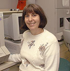
Olli
Kallioniemi
|
Olli Kallioniemi received his M.D. in 1985 and Ph.D. in 1988 from the University of Tampere in Finland. After residency training in laboratory medicine, he did postdoctoral work at the University of California, San Francisco, and then returned to the University of Tampere, where he became a professor of cancer genetics before joining NHGRI in 1996. He is now a senior investigator in the Cancer Genetics Branch and head of the Translational Genomics Section.
My interest is in the molecular basis of human breast and prostate cancer and strategies and technologies to translate basic research findings into clinical applications. Our research focuses on the discovery of inherited germ-line alterations, as well as somatic genetic events and gene expression changes involved in tumor progression.
In the area of genetic predisposition, I have collaborated with investigators in Scandinavia on genetic epidemiological studies of breast and prostate cancer. Using genetic linkage analysis of cancer families, we recently identified a putative third major locus, at 13q21-q22, for breast cancer susceptibility. Our laboratory was also involved in the identification of a candidate susceptibility locus for human prostate cancer on the X chromosome (HPC-X).
In 1992, we developed the comparative genomic hybridization (CGH) technique to identify chromosomal regions that are altered in cancer. At NHGRI, we are focusing on the identification of genes that are affected by genomic alterations, particularly high-level DNA amplifications that often take place in genetically unstable cancer cells. In collaboration with other investigators, we have identified several genes whose increased expression as a result of gene amplification may drive cancer progression and the development of therapy resistance. These include androgen receptor gene amplification in prostate cancer, and AIB1 and the ribosomal protein S6 kinase, which are amplified in breast cancer. We are now using a combination of high-resolution DNA chip technologies, such as CGH and cDNA microarray analyses, to rapidly identify amplified and overexpressed genes throughout the genome. Such genes may contribute to the clonal progression of cancer and provide targets for therapy.
In the postgenomic era, translating gene discoveries into clinical applications will be an increasingly important challenge. We recently developed the tissue microarray (TMA) technology to conduct rapid, large-scale analyses of promising candidate genes in hundreds or thousands of clinical tumor specimens. Up to 1,000 tiny "tissue spots" originating from different patients can be arrayed on a single microscope slide. We are applying the TMA slides to study the role of novel genes and proteins in thousands of tissue specimens from different cancer types, associating molecular findings with clinical and treatment outcome information. Such "genome-scale translational research" will be important to identify the most promising diagnostic and therapeutic gene targets from the genomic data now becoming available.
 |
|
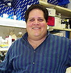
Thomas
Kristie
|
Thomas Kristie received his Ph.D. from the Committee on Virology at the University of Chicago in 1986 and did postdoctoral work with Phillip Sharp at the Center for Cancer Research, Massachusetts Institute of Technology. He joined the NIAID Laboratory of Viral Diseases in 1993 and is now a senior investigator in the Molecular Genetics Section.
My laboratory is interested in the molecular biology of herpes simplex virus (HSV), specifically focusing on the biochemical mechanisms of viral gene expression and transcriptional control of the viral lytic-latent cycle.
HSV is a human pathogen affecting a large percentage of the population. After a primary infection, the virus remains latent in the sensory neurons of the individual until complex stimuli such as stress, hormonal alterations, or tissue damage reactivate viral replication. During the lytic cycle, the immediate early (IE) genes of HSV are controlled by a complex viral regulatory enhancer that provides the framework for the assembly of macromolecular complexes. Our focus has been on determining the components, interactions, and functions of the various polypeptides involved.
As a postdoctoral fellow, I worked on identifying and purifying C1 factor, a critical host cell component of the enhancer complex. After arriving at NIH, I began cloning and characterizing this protein. C1 factor is a unique transcription factor produced as a 230- kDa protein and is proteolytically processed into a family of polypeptides. In a series of studies, my laboratory has defined the interactions of the C1 factor with each primary component of the HSV enhancer complex and demonstrated that it is both the coordinator of the enhanceasome assembly and a transcriptional coactivator. We have also isolated a relatively large bank of cellular proteins that interact with various domains of C1. We are now characterizing these to determine the normal functions of the C1 factor in mammalian cells.
One of the most interesting and critical questions in herpesvirus biology is the mechanism for establishment of and reactivation from the latent state. Considering the cell-specificity of latency and the cell stimuli that reactivate the virus, we think cellular factors probably play the most significant role in the lytic-latent switch. It is also likely that the regulation of the IE genes would be a primary target in reactivation. Models for the reactivation of HSV from the latent state propose either that 1) reactivation proceeds through a viral gene expression pattern distinct from the lytic phase or that 2) reactivation is due to coordinated activation of transcription of the five IE genes by cellular transcription factors.
My laboratory has demonstrated that although the C1 protein is localized in the nucleus of most cell types, it is uniquely sequestered in the cytoplasm of sensory neurons. In the mouse model, conditions that reactivate the virus from latency also result in rapid relocation of C1 to the nucleus. This observation and our work on C1 functions led us to propose that the C1 factor is a critical component of the signaling mechanism that initiates viral reactivation.
We are now using biochemical and genetic approaches to find how C1 factor is sequestered as well as the signaling pathway(s) for activation of C1 transport. In addition to determining the role of C1-interacting proteins, we have been developing animal models, including C1 domain-specific knockout mice to aid analysis of the roles of the C1 factor in the regulation of cellular and viral processes.
A second area of interest in the laboratory is the unusual specific proteolytic processing of the C1 factor. The precursor protein is cleaved at a series of 20-amino-acid repeats in the central domain to generate a family of poly-peptides. The product polypeptides do not segregate, however, suggesting that proteolysis is a unique mechanism for regulating the activity of the protein. We have determined that the C1 factor itself contains an autocatalytic activity that is responsible for processing of the reiterations. We are now focusing on demonstrating our hypothesis, namely that processing regulates the protein-protein interactions and, therefore, the functional activity of the C1 factor.
Our interest in this area led to the development of a genetic screen for the isolation and characterization of novel site-specific proteases. Site-specific proteolysis plays significant roles in the regulation of many basic normal and disease processes, including:
![]() The control of cholesterol metabolism by proteolytic regulation of transcription
factor SREB
The control of cholesterol metabolism by proteolytic regulation of transcription
factor SREB
![]() The cleavage of amyloid precursor protein (APP), which may affect the development
of Alzheimer’s disease
The cleavage of amyloid precursor protein (APP), which may affect the development
of Alzheimer’s disease
![]() The intricate pathway of proteolytic targeting resulting in cell apoptosis
The intricate pathway of proteolytic targeting resulting in cell apoptosis
Using a model protease, my laboratory developed a system capable of selectively isolating a rare protease of modest activity from a complex cDNA pool. We are now involved in several collaborative efforts to identify and isolate novel site-specific proteases in a variety of biological systems.
 |
|
Alison
McBride
|
Alison McBride received a Ph.D. from the Imperial Cancer Research Fund and University of London, U.K., in 1986. She was a postdoctoral fellow and investigator with Peter Howley in NCI and joined NIAID in 1994. She is now a senior investigator in the Laboratory of Viral Diseases, NIAID.
I am interested in the strategies that viruses have developed to manipulate key host cell factors at different stages of the virus life cycle. Such studies have helped characterize many fundamental regulatory pathways in eukaryotic cells, such as DNA replication, gene expression, cell cycle control, and signal transduction.
Since joining NIH, I have studied the papillomaviruses. These are small viruses that infect and replicate in the stratified layers of the epithelia and cause benign warts, or papillomas. In some cases these lesions can progress to malignant carcinomas, the most notable of which is cervical cancer. The papillomaviruses also have an interesting biology: The virus infects basal epithelial cells and maintains its genome as an episome within these persistently infected cells. Viral DNA amplification and capsid antigen production occur only in the differentiated layers of a stratified epithelium. The papillomaviruses encode oncogenes that interfere with host cell growth control to enable the virus to replicate in nondividing, differentiated cells.
My research has focused on the molecular mechanisms by which two papillomavirus proteins control the viral life cycle. The viral E1 protein is required to initiate episomal viral DNA replication. The E2 proteins regulate viral transcription, DNA replication, and genome segregation. My colleagues and I have defined the structure, function, and interactions of these proteins.
A few years ago we identified an additional function for the E2 protein. We showed that the E2 protein and viral genomes are bound to mitotic chromosomes in dividing cells. E2 links the genomes to the host chromosomes to ensure that they are not lost as the cells divide, and that they are segregated equally to daughter cells. This finding defines the mechanism by which episomal viruses maintain and segregate their genomes in persistently infected cells. We are currently trying to identify the chromosomal factor to which E2 binds and to determine whether it is a common target of other episomal viruses.
We have also found phosphorylation of E2 regulates its degradation by the proteasome and modulates episomal viral genome copy number. Our current hypothesis is that E2 phosphorylation is cell cycle—specific and regulates genome segregation by determining the amount of E2 bound to mitotic chromosomes. High concentrations of E2 protein can be observed in the differentiated cells of a papilloma that amplify viral DNA, and we are testing whether E2 has yet another role in regulating vegetative viral DNA replication.
In addition to these molecular approaches, we have developed two biological systems with which to study the role of the E1 and E2 proteins. The study of the complete papillomavirus life cycle is difficult because it requires keratinocyte differentiation. We have developed an animal model in which organotypic rafts are generated from bovine or human keratinocytes and are grafted onto immune-compromised mice. When transfected with viral DNA, the grafts develop into papilloma-like lesions that synthesize infectious viral particles. This allows genetic analyses of the viral functions required for the lytic cycle. It also allows us to study DNA segregation and persistence in an animal model.
Certain papillomaviruses are associated with the development of cervical cancer, and inactivation of E1 and E2 gene functions (by viral genome integration) is thought to play a role in malignant progression. Because E1 and E2 are crucial for viral DNA replication, it has been difficult to determine whether it is the absence of these gene products or the integration event per se that is important in carcinogenesis. To study this process, we have developed a system that allows analysis of the effect of mutations in E1 and E2 on biological indicators of malignant progression, in the absence of integration.
We hope that our study of the papillomavirus E1 and E2 proteins will continue to provide important insights into the pathogenesis of papillomavirus infection. This knowledge will be useful in designing strategies to intervene in the viral life cycle and could lead to prevention or treatment of papillomavirus-associated diseases. Our studies will also provide insights into mechanisms of transcriptional regulation and differentiation-dependent regulation of epithelial gene expression. A detailed understanding of the mechanism of papillomavirus DNA replication and segregation could lead to the development of a new generation of persistent extrachromosomal vectors that may have applications for gene therapy and genetic immunization.
 |
|
Daniel
Pine
|
Daniel Pine received his M.D. degree from the University of Chicago-Pritzker School of Medicine and did his postgraduate training at New York’s Columbia University before becoming a faculty member in the College of Physicians and Surgeons there. He is now head of the Section on Developmental Psychopathology and Affective Neuroscience, in the Program on Mood and Anxiety Disorders, NIMH.
My research interests pursue two complementary avenues. First, I am an active investigator in pediatric psychopharmacology. Second, I am testing hypotheses on brain-behavior associations in childhood psychopathology.
My interest in pediatric psychopharmacology developed while I was a member of the Department of Psychiatry at Columbia University. The effectiveness of medications with selective serotonin reuptake inhibiting activity (SSRIs) in adult anxiety disorders led our group to conduct an open trial of SSRIs for childhood anxiety disorders. Results from this study in turn led to our participation in a multisite, randomized, controlled trial of SSRIs in 128 children with a pediatric anxiety disorder. This study established the robust efficacy and safety of SSRIs in children with anxiety disorders.
In light of potential pharmacologic parallels between pediatric and adult anxiety disorders, I have examined the psychobiology of pediatric anxiety. Following work in adult panic disorder, my initial studies found that children with respiratory illnesses, such as asthma associated with dyspnea, faced a higher risk of separation anxiety disorder than did children with other illnesses. My subsequent work, funded by extramural grants from NIMH, demonstrated that children with separation anxiety disorder exhibit the same set of respiratory abnormalities that are classically found in adult panic disorder. These include heightened dyspnea, chaotic breathing, and heightened respiratory response to CO2 inhalation.
Because SSRIs ameliorate such breathing abnormalities in panic disorder, I designed a study to test the hypothesis that SSRIs ameliorate these abnormalities in separation anxiety disorder. Through related psychophysiological projects conducted within the context of this study, I am examining the hypothesis, based on animal studies, that antidepressants exert more consistent effects on innate than learned aspects of fear. We are planning studies in the NIMH intramural program to examine the effects of SSRIs on learned and innate fears in children and adolescents.
My prior studies found that only a subset of children with separation anxiety disorder exhibits respiratory characteristics of panic—characteristics that also identify adults with familial forms of panic disorder. Hence, this subgroup of children may manifest a potentially heritable, respiratory-based susceptibility to panic disorder, a hypothesis I have pursued through other NIMH-funded projects.
Supporting this hypothesis, I conducted a longitudinal study showing that most adult panic attacks are preceded by a childhood history of separation anxiety disorder, though most children with separation anxiety disorder do not develop panic. To further test the hypothesis, I developed a portable apparatus for assessing CO2 sensitivity in children. Our group has used this apparatus to begin comparing respiratory parameters in children of normal parents and parents with three types of disorders: panic, depressive, and anxiety disorders other than panic. In further support of the central hypothesis, the study is based on a sample in which there is a strong familial association between adult panic disorder and childhood separation anxiety disorder.
Beyond studies on anxiety disorders, I have extended to children other adult work on brain-behavior associations. For example, based on the association between adult anxiety and growth hormone deficiencies, I documented a longitudinal association between anxiety in young girls and growth deficits from childhood into adulthood.
Similarly, adult studies document associations among stress-induced anxiety, the hypothalamic-pituitary-adrenal axis (HPA), and hippocampal function. I demonstrated spatial memory deficits and elevated cortisol in children with anxiety disorders. Because spatial deficits, HPA activity, and some forms of anxiety could each relate to hippocampal dysfunction, I developed fMRI methods for assessing hippocampal activity across development using a spatial navigation paradigm. With these methods, I am now testing the hypothesis that enhanced HPA activity in childhood anxiety is associated with both reduced memory-based navigation ability and reduced hippocampal activation during navigation. We also plan to pursue this hypothesis in our intramural lab.
Finally, studies among adults find that abnormalities in impulse control and emotion regulation each show unique associations with distinct brain asymmetry profiles in posterior regions engaged by perceptual tasks. I documented parallel associations with disorders of impulse control and emotional regulation in children, using cognitive and electrophysiological indices of posterior brain asymmetry.
This work in turn led to a series of fMRI studies examining developmental variations in brain regions engaged by emotional perception tasks. In each of these projects, and in proposed projects in the NIMH intramural program, I plan to continue illustrating how findings from adult psychiatry inform research on treatments and risk factors for childhood psychiatric disorders.
 |
|
Patricia
Rosa
|
Patricia Rosa obtained her doctorate in 1980 from the Institute of Molecular Biology at the University of Oregon in Eugene. After postdocs at Washington University in St. Louis and the Research Institute of Scripps Clinic in La Jolla, Calif., Rosa joined NIAID’s Laboratory of Microbial Structure and Function, Rocky Mountain Laboratories, in Hamilton, Mont. She is now a senior investigator at RML’s Laboratory of Human Bacterial Pathogenesis.
In a broad sense, I am interested in how the phenotype of a cell at any particular time reflects both its genetic structure and the environment in which it is located. This interest probably began with an epigenetics course that I took as an undergraduate and has taken various forms in graduate and postdoctoral studies. Since coming to Rocky Mountain Labs, my research has focused on Borrelia burgdorferi, the spirochete that causes Lyme disease. Features of this relatively simple bacterium and its infectious cycle present a good system in which to study the adaptive responses of a bacterial pathogen to its vector and host environments. All steps of this infectious cycle can be reproduced in the laboratory, making it accessible to scientific investigation.
Maintenance of B. burgdorferi in nature requires efficient transmission between the tick vector and the mammalian host. Like many bacterial pathogens, B. burgdorferi must cope with a changing array of environmental conditions to persist, proliferate, and be transmitted between hosts. My lab and others have found good evidence for differential gene expression by borreliae in these disparate settings. Yet, it is unknown how B. burgdorferi senses its environment and orchestrates appropriate adaptive responses. What environmental signals define the tick vs. the mammal and induce the appropriate bacterial response? How does B. burgdorferi sense these signals and transduce them? Which proteins are made in different places or at different stages of the infectious cycle, and what are their functions? What features of the spirochete or the infection are pertinent to pathogenicity? Our broad objective is to use a molecular genetic approach to answer these questions and elucidate the mechanisms of adaptation and variation in B. burgdorferi over the course of the infectious cycle.
Our current knowledge of B. burgdorferi at the molecular level represents an interesting paradox; we have the sequence of the entire genome, but we know very little about the functions of most of the encoded proteins. Although we have identified several B. burgdorferi genes whose expression or putative function suggest that they are important in the infectious cycle, we do not know the actual biological roles or significance of their protein products.
Ongoing and future studies are designed to test the roles of these genes and their products in the infectious cycle and to identify additional genes that allow spirochetes to adapt, persist, and be transmitted between ticks and mammals. We also want to understand regulation of these genes and their roles in pathogenesis. A major thrust of our work is to analyze the phenotypes of specific gene mutants in the context of the natural infectious cycle.
B. burgdorferi is structurally and genetically quite dissimilar from other bacteria, having a small linear chromosome and a large number of linear and circular plasmids. How these DNA molecules replicate, why the genome is segmented, and the functional significance of this unique structure are not well understood and are another focus of research in my lab. The practical consequence of B. burgdorferi’s odd genome is that genetic tools and methods developed for more typical bacteria do not work in B. burgdorferi, so we have developed basic tools with which to undertake genetic studies in this bacterium.
We have recently succeeded in inactivating genes in pathogenic strains of B. burgdorferi. This represents a significant advance because until now, no one had been able to introduce mutations into a wild-type background, impeding analysis of the phenotype of mutant spirochetes in the infectious cycle.
Although the genetic system of B. burgdorferi is rudimentary and far from elegant, it still provides a powerful tool with which to explore the unique structure of the borreliae genome and to study the adaptive responses of a bacterial pathogen to its vector and host environments.
 |
|
Ellen
Sidransky
|
Ellen Sidransky received her M.D. from Tulane University in New Orleans in 1981. She trained in pediatrics at Children’s Memorial Hospital—Northwestern University in Chicago, Ill., and in clinical genetics at the NIH Interinstitute Genetics Training Program. She joined the Clinical Neuroscience Branch at NIMH in 1988 and is now a senior investigator leading the Unit on Clinical Genetics.
My laboratory integrates basic and clinical research in a "bench-to-bedside" approach, focusing on Gaucher disease, the most common of the sphingolipidoses. I use Gaucher disease, which has a broad spectrum of symptoms, as a prototype for other disorders affecting the nervous system. Most of these inherited disorders are characterized by a wide range of presentations, yet the factors contributing to this heterogeneity are often elusive. My clinical, biochemical, and molecular studies of humans and animals are designed to understand this heterogeneity and to improve therapy.
My principal research aim is to understand genotype-phenotype relationships in Gaucher disease and other inherited disorders affecting the nervous system. Our clinical and molecular studies of more than 200 patients have shown that there is significant genotypic heterogeneity among clinically similar patients. Moreover, patients with the same genotype can have different disease manifestations.
Our recent work revealed that the region surrounding the human glucocerebrosidase gene on chromosome 1q21 is particularly gene-rich, with seven genes and two pseudogenes within 75 kb of sequence. We found that in a significant number of patients, mutant alleles arise by recombination or gene conversion occurring between the glucocerebrosidase gene and its nearby pseudogene. We discovered that these recombinations can occur at a variety of sites throughout the gene and by different mechanisms of recombination. In addition to recombination events within and around the glucocerebrosidase locus, the actions of contiguous genes, environmental factors, and modifying genes may contribute to the phenotypes observed. We are actively exploring these possibilities via transcriptional studies of the contiguous genes in the region, transcriptional arrays and mouse models.
Our active clinical service investigates the natural history and spectrum of manifestations in Gaucher disease. We focus on patients with atypical or extreme phenotypes, because they may reflect the involvement of other genes or modifiers. Two fascinating phenotypes I am investigating include Gaucher patients with parkinsonian symptoms and patients with hydrocephalus and cardiovascular involvement. Genotypic analyses reveal that patients in the first group have many different Gaucher mutations, while the latter appear to share a common genotype. In addition, a knockout mouse model of Gaucher disease generated with a null allele led me to recognize an analogous human phenotype—a neonatal lethal form of Gaucher disease. Affected patients die with hydrops fetalis in utero or shortly after birth. Since our original description of this phenotype, dozens of additional cases have been identified.
I also discovered that null allele mice and severely affected human patients have skin ultrastructural abnormalities and altered epidermal barrier function secondary to the deficiency of epidermal glucocerebrosidase. This skin abnormality may serve as a valuable clinical tool, enabling early differentiation of different Gaucher phenotypes.
Another challenge has been to understand the development of neuronopathic Gaucher disease. Because this and other aspects of the pathology of Gaucher disease are not adequately explained by glucocerebroside accumulation, we are also exploring the significance of an alternate substrate, glucosylsphingosine, that we found elevated in brains of patients and mice with neuronopathic Gaucher disease.
In working to improve treatment for Gaucher disease, I participate in a collaborative effort to test chemical modifications of recombinantly produced glucocerebrosidase. Modifications include using molecules such as polyethylene glycol to prolong circulation of the enzyme. This may permit lower dosage and drug cost, reduce antigenicity, and make it easier and more convenient to administer the enzyme to patients. We are also investigating more efficient production of the enzyme from new transgenic sources, as well as the use of inhibitors of glycolipid synthesis.
Many of the approaches used in our studies of Gaucher disease will also be applied to another group of inherited disorders—congenital disorders of glycosylation (CDG)—in collaboration with Donna Krasnewich of NHGRI. These disorders involve defective assembly of NH2-linked oligosaccharides. Patients with CDG show diverse symptoms, including cerebellar abnormalities, malformations of facial features, gastrointestinal disease, and profound delays in psychomotor development. CDG provides an opportunity to investigate the elusive correlation between clinical manifestation and the molecular mechanism of the metabolic defect.
The results of our genotype-phenotype studies provide evidence that even well-characterized Mendelian disorders are not necessarily "simple." I believe that investigations into the mechanisms contributing to the heterogeneity encountered in these disorders will ultimately enable us to discover the "magic in the web of it." Moreover, the strategies, experience, and knowledge gained through this work may be helpful in understanding and treating other hereditary diseases, including more "complex" neurologic and psychiatric illnesses.
 |
|
Lee
Weinstein
|
Lee Weinstein received his M.D. from New York’s Columbia University College of Physicians and Surgeons in 1983 and did a medicine residency at Montefiore Hospital in New York before joining the Metabolic Diseases Branch of NIDDK in 1986. He is now a senior investigator in that branch.
I am interested in G protein signaling and hormone action. My research at NIDDK has examined the role of G protein genetic defects in hormonal disorders. Our work has focused on the heterotrimeric G protein (Gs) that couples receptors to the enzyme adenylyl cyclase, which catalyzes the generation of intracellular cyclic adenosine monophosphate (cAMP). The Gs a-subunit (Gsa) is ubiquitously expressed and is required for the intracellular cAMP response to hormones and other extracellular signaling molecules.
In my early work at NIH, my colleagues and I showed that mutations that produce a constitutively active Gsa protein are present in a mosaic distribution in patients with the McCune-Albright syndrome, which is characterized by hyperpigmented skin lesions, focal skeletal lesions (fibrous dysplasia), and endocrine hyperfunction. This observation provided the impetus for ongoing studies at NIH on the role of Gs pathways in osteoblast differentiation. We also showed that heterozygous inactivating mutations of the same gene lead to Albright hereditary osteodystrophy (AHO), characterized by obesity, skeletal defects, and, in some cases, mental deficits. Interestingly, maternal transmission of these mutations also leads to multihormone resistance (pseudohypoparathyroidism type Ia, PHPIa) whereas paternal transmission does not (pseudopseudohypoparathyroidism, PPHP).
Using a Gsa knockout mouse model, we have shown that Gsa is imprinted in a tissue-specific manner—maternally expressed in some tissues (such as renal proximal tubules, the site of parathyroid hormone action) but biallelically expressed in most other tissues. This provides a likely explanation for the variable clinical presentation in AHO patients (PHPIa vs. PPHP). We also showed that Gsa plays a major role in energy and glucose metabolism. Specifically, maternal vs. paternal transmission of a heterozygous knockout leads to opposite effects on energy metabolism and fat accumulation. However, all heterozygous mice have increased sensitivity to insulin in vivo.
We now appreciate that the Gsa gene (GNAS1) has multiple alternative promoters. We have recently identified the fourth one of these that is imprinted, and we believe this promoter region is critical for the tissue-specific imprinting of Gsa. In patients with renal resistance to parathyroid hormone in the absence of AHO (PHPIb), this region has a paternal-specific imprinting pattern in both parental alleles. Therefore, PHPIb is caused by a GNAS1 imprinting defect that presumably leads to decreased Gsa expression in renal proximal tubules. Analysis of this region provides a useful diagnostic tool for the evaluation of patients who present with parathyroid hormone resistance.
In
the future, we hope to determine the mechanisms by which Gsa
is imprinted in a tissue-specific manner and by which imprinting of GNAS1
is established and maintained. We also plan to study the physiological roles
of Gsa in greater detail by examining mice with loss
of Gsa in specific tissues. We hope these studies
will provide important insights into the mechanism of genomic imprinting and
the role of Gs signaling in metabolism and hormone action. ![]()