| T H E N I H C A T A L Y S T | J U L Y – A U G U S T 2000 |
|
|
|
| P E O P L E |
RECENTLY TENURED
 |
|
Norman
Coleman
|
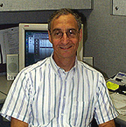
C. Norman Coleman received his M.D. from Yale University in New Haven, Conn., and trained in internal medicine at UCSF, medical oncology at NCI, and radiation oncology at Stanford (Calif.) University, where he served on the faculty for seven years. Before returning to NIH as director of the Radiation Oncology Sciences Program (ROSP), he was the Fuller-ACS Professor and Chairman of the Joint Center for Radiation Therapy at Harvard Medical School from 1985 to 1999.
My foremost objective at NIH is to develop the ROSP, which is a program that combines expertise among the intra- and extramural branches relating to radiation oncology—specifically, the Radiation Oncology Branch, Radiation Biology Branch, and Radiation Research Program.
The field of radiation oncology deals with all tumor types, and the efficacy of radiation therapy depends on the response of both the tumor and the normal tissue within the radiation field. To exploit new advances in imaging, technology, and molecular biology, the ROSP program will collaborate extensively with other cancer specialists within NIH, surrounding universities, the region, and other research centers.
Radiation delivery requires definition of the target and normal tissue and the assessment of their response to treatment. To that end, our technical research and development work includes anatomical and functional imaging, real-time imaging (ultrasound or MRI) during radiation to assure beam targeting, optical sensors for dose calculation, and dose-calculation modeling of radiation-tissue interaction at the cellular and subcellular level
We are now planning a Technology Development and Assessment Center within the Radiation Oncology Branch. One goal of the new center will be to develop implantable micro- and nanosensors to measure biological processes before, during, and after a range of treatments and to assess rapidly the molecular and cellular response to treatment by tumor and normal tissue.
The biological aim of the ROSP is to understand the tissue, cellular, and molecular response of tumors to radiation alone and in combination with radiation-modifying agents. We also want to develop improved therapeutics based on tumor genotype and phenotype. The programs that we are establishing toward these goals include a microarray facility at the NCI’s Division of Clinical Sciences Advanced Technology Center in Rockville, Md., and an in vitro and in vivo facility at FCRDC to study and assess radiation-drug interactions.
My primary personal research interest is in developing new therapeutics based on molecular targeting. Although radiation is typically described as an average dose to a tissue, at the end of its path in tissue, the energy is delivered in dense packets. Furthermore, radiation can be focused to tissue via external beam, implanted sources, and systemically administered isotopes. Thus, our conceptual model is that radiation is "focused biology" that can perturb a wide range of cellular targets. This perturbation can be exploited with molecular therapeutics. Moreover, because many of the newly developed cancer treatments—such as antiangiogenesis agents or signal transduction modifiers—are cytostatic and not cytocidal, radiation may be essential to make the agents effective in killing tumor cells.
My research interest over the past decade has been targeting the tumor microenvironment, including tumor hypoxia, which is present in about half the tumors studied. This not only leads to radiation resistance, but the microenvironmental stress and hypoxia itself can alter cellular phenotype, for example, the induction of HIF-1–responsive genes. Microenvironmental perturbation within tumors, such as an abnormal redox state, may alter protein structure and function.
James Mitchell’s group in the Radiation Biology Branch is advancing this work by developing techniques to image hypoxia in real-time that would help guide therapeutics. My own laboratory interest has turned to the role of the cyclooxygenase inhibitors as enhancers of tumor cell killing. We have demonstrated that the nonspecific nonsteroidal anti-inflammatory drug ibuprofen is an effective radiation sensitizer in vitro and in vivo. We are investigating possible mechanism(s) for this effect, including perturbation of arachadonic acid metabolism, inhibition of NFkB, which is constitutively active in a number of cancers, and possibly through the action of PPAR (peroxisome proliferator-activated receptors) family.
ROSP is now recruiting basic molecular biology researchers, physicists, and engineers for the biomolecular sensor program and clinicians interested in translational research. We welcome inquiries and look forward to collaborations with colleagues at NIH.
 |
|
Barney
Graham
|
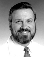
Barney Graham received his M.D. from the University of Kansas, Kansas City, in 1979 and completed clinical training and a fellowship in infectious diseases at Vanderbilt University in Nashville, Tenn., in 1986; in 1991, he completed his Ph.D. in microbiology and immunology at Vanderbilt, where he is currently a professor of medicine in the Division of Infectious Diseases and an associate professor of microbiology and immunology. He will join the NIH Vaccine Research Center as the director of clinical trials, commuting on a regular basis as soon as the building is ready and settling in next spring.*
My primary research interests are viral pathogenesis and vaccine development. I have focused on two major areas of work, AIDS vaccine evaluation in clinical trials and pathogenesis of respiratory syncytial virus (RSV) in a murine model. My specific recent interest in this area is in the role of RhoA, a small GTPase, in RSV entry, morphogenesis, and pathogenesis. I am also interested in science education and expanding research opportunities for African-Americans and other underrepresented minorities. In this regard, I am a member of the Development Committee for National Medical Fellowships, Inc., and a mentor in the Fellowship Program in Academic Medicine for Minority Students sponsored by Bristol-Myers Squibb.
Since 1987 I have been involved in the development of the AIDS Vaccine Evaluation Group, recently renamed the HIV Vaccine Clinical Trials Network (HVTN). This group has collectively conducted more than 40 Phase I and II clinical trials of candidate AIDS vaccines. I chaired the initial human studies evaluating recombinant Vaccinia virus vectors, including studies combining a live recombinant virus with a subsequent subunit protein booster.
In addition, I chaired studies evaluating novel adjuvants for subunit envelope antigens, and multivalent peptide vaccines combining both B- and T-cell epitopes. I also chaired the studies evaluating subjects who developed breakthrough HIV-1 infection despite prior vaccination, and I served in leadership positions for all the major subcommittees in the HVTN. I am particularly interested in the use of cytokines, chemokines, and other natural immuno-modulators as vaccine adjuvants, and in the evaluation of vaccine approaches that combine immunogens, delivery vehicles, and routes of administration.
My laboratory focus is to define basic mechanisms of RSV pathogenesis and to apply this knowledge to vaccine development. RSV is an important cause of respiratory disease that has received high priority for vaccine development. Previous vaccine candidates have failed in clinical trials, and a formalin-inactivated whole virus (FI-RSV) preparation was associated with vaccine-enhanced illness.
After early studies characterizing a murine model of RSV infection and defining the role of T cells and antibody in primary RSV infection, we addressed the pathogenesis of the FI-RSV vaccine-enhanced disease. My laboratory demonstrated that priming with inactivated RSV induces a type 2 T-helper lymphocyte (Th2) response (dominant IL-4 expression), whereas priming with live RSV induces a Th1-like response (dominant IFN-g expression) in mice following RSV challenge.
We have subsequently used the murine RSV model to evaluate new vaccine approaches and to investigate how immunization determines the composition of immune responses. Specific projects include defining the mechanisms by which: 1) the RSV G glycoprotein induces IL-5 and eosinophilia, 2) IL-4 modulates the cytolytic mechanism of CD8+ cytolytic T cells, 3) RSV-induced immune responses interact with allergic inflammation to cause airway hyperrespon-siveness, 4) co-administered antigens influence RSV-induced immune responses and lung pathology, and 5) integrins mediate lymphocyte trafficking and activation during RSV infection. The studies specifically aim to answer questions about in vivo phenomena, taking advantage of a variety of knockout and transgenic mouse strains and assays designed to avoid in vitro artifacts.
A recent discovery in my laboratory of a cellular ligand for the RSV F glycoprotein has led to new projects in the areas of RSV-induced membrane fusion and viral entry and the role of intracellular signaling pathways triggered by the RSV F interaction in virus morphogenesis, immunity, and disease.
Our belief is that understanding basic mechanisms of antiviral immunity and viral pathogenesis will lead to novel and more effective approaches to vaccine development. Both laboratory and clinical studies yield essential information that must be integrated to inform the process of vaccine development.
* The September–October issue of The NIH Catalyst will include a story on new VRC recruits and programs. A ceremony to mark the official opening of the VRC is planned for October.
 |
|
Michael
Krause
|
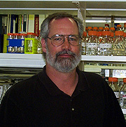
Michael Krause received his Ph.D. from the University of Colorado in 1986 and did postdoctoral work with the late Harold Weintraub at the Fred Hutchinson Cancer Research Center in Seattle. He joined the NIDDK Laboratory of Molecular Biology in 1993 and is now a senior investigator heading the Section on Developmental Biology.
I am interested in transcriptional regulation of cell fate during development. I use the nematode Caenorhabditis elegans as an experimental system.
C. elegans adults have only 959 somatic cells and develop from an essentially invariant cell lineage over the course of three days. The nearly complete genome sequence of C. elegans became available last year, revealing about 19,000 genes. Recent techniques, using traditional mutagens and/or molecular approaches, make it possible to quickly create loss-of-function phenotypes for most genes. The combination of relatively simple anatomy, defined cell lineage, complete genomic information, and powerful forward and reverse genetics makes this organism an ideal system in which to study the transcriptional regulation of cell fate.
My lab focuses primarily on the regulation of muscle cell fates in C. elegans, including the early events of mesodermal patterning that underlie the formation of the correct type, number, and position of muscle cells. Some of the muscle types in C. elegans can be correlated with muscle types found in mammals and many of the transcription factors involved in mammalian myogenesis have homologs in C. elegans. The simple anatomy of C. elegans allows us to study the function of these transcription factors in a single cell, sometimes revealing functions obscured in more complex animal systems.
A landmark discovery in the field of myogenic cell fate was the identification of the protein MyoD. MyoD is a basic helix-loop-helix (bHLH)–type transcription factor restricted primarily to skeletal muscle cell precursors; MyoD is not present in cardiac or smooth muscle. MyoD activates transcription of multiple downstream genes to cause proliferating cells to exit from the cell cycle and initiate the skeletal muscle program of differentiation. MyoD is one of four highly related proteins that together make up a family of myogenic factors in mammals. The identification of MyoD validated the notion of "master" regulatory genes capable of initiating whole programs of differentiation and marked the beginning of an intense research effort on skeletal muscle differentiation.
I arrived at the NIH shortly after identifying the only C. elegans homolog of MyoD (CeMyoD) and having shown that the expression profile of CeMyoD was consistent with its playing a key role in myogenesis. In collaboration with Andy Fire’s group at the Carnegie Institution of Washington in Baltimore, we used CeMyoD knockouts to show that skeletal muscle–like cells in the nematode (body wall muscle) can be formed in the absence of CeMyoD activity. This observation is surprising, because mammalian tissue culture studies and mouse knockout experiments demonstrate that some members of the mammalian MyoD family are essential for skeletal myogenesis. Although CeMyoD is not essential for body wall muscle formation in C. elegans, it is required for these cells to function; homozygous CeMyoD-null animals cannot move normally and die shortly after the embryo hatches.
Through work in my lab at NIH, we have now shown that the CeMyoD finding is just one in a series of observations revealing both conservation and divergence in the function of several highly conserved, muscle-associated transcription factors in C. elegans. Through further dissection of the functions of these factors we have found that embryonic and postembryonic periods of myogeneis in C. elegans use different regulatory pathways. These results have implications beyond the nematode and help to explain some of the dramatic differences between C. elegans and other organisms in the function of these evolutionarily conserved factors.
More recently we have turned our attention to another bHLH transcription factor, Twist, which acts early in the myogenic pathway. We have identified mutants in Twist that reveal that it is required for patterning a subset of the muscles in C. elegans. Twist also functions in a subset of mesodermal tissues during mammalian development, and Twist mutations in humans result in Saethre-Chotzen syndrome, which affects craniofacial development. We are interested in studying CeTwist in more detail to identify interacting factors and downstream target genes.
Together with other labs, we have begun to identify the transcriptional hierarchy regulating postembryonic muscle cell fates in C. elegans. Future work is aimed at adding additional detail to the regulatory network of genes and defining pathways functioning in other muscle cell types. It should be possible to describe in complete molecular detail how each muscle cell of C. elegans arises, beginning with fertilization of the egg. By defining developmental paradigms, this will be an important milestone, not only for myogenesis but for developmental biology in general.
 |
|
Chris
McBain
|
Chris McBain received his Ph.D. from the University of Cambridge, England, in 1988 and did postdoctoral work at the University of North Carolina and Duke University, both at Chapel Hill, before joining the Laboratory of Cellular and Molecular Neurophysiology of NICHD in 1993. He is now a senior investigator heading the Section on Cellular and Synaptic Physiology.
It is becoming clear that to begin to understand the coordinated activity of large ensembles of neurons, we must first understand the nature of transmission between individual pre- and postsynaptic elements within a circuit and each and every neural element involved. My interests are in elucidating the precise nature of excitatory and inhibitory synaptic transmission between specific identified neural populations within the hippocampal and cortical formations.
For the hippocampal neuronal network, the net flow of information is strongly modulated by the action of the local-circuit GABAergic inhibitory interneurons, whose cell bodies are distributed throughout all layers of the hippocampus and comprise about 10 to 15 percent of the total neuronal population. The importance of these inhibitory neurons is underscored by the number of animal models of epilepsy that result from manipulation or loss of these inhibitory pathways.
My research has found that, rather than act as GABAergic counterparts of glutamatergic excitable cells, interneurons possess both intrinsic and extrinsic properties that set them apart from principal cells of the cortical formation. This divergent cell population can be distinguished by the molecular identity of receptors and channels they express, by their mechanisms of short- and long-term synaptic plasticity, and by signal transduction mechanisms associated with both ligand- and voltage-gated receptors.
Work in my lab has focused primarily on three areas of research. The first is the nature of synaptic plasticity regulating hippocampal inhibitory interneuron excitability. We found that, in contrast to principal cells, interneurons do not possess conventional forms of N–methyl-d-aspartate (NMDA)–receptor postsynaptic long-term plasticity, but are influenced indirectly by a "passively propagated" form of plasticity occurring within a polysynaptic pathway.
In addition, we demonstrated cell-specific expression of presynaptic forms of plasticity at synapses formed by the axons of dentate gyrus granule cells, the so-called mossy fibers. Target specificity of presynaptic plasticity results from the segregated expression of cAMP-dependent signaling cascades. This signaling pathway is targeted to presynaptic axons associated with principal cells but is absent at the same synapses made onto inhibitory interneurons. Expression of glutamate receptor proteins is similarly segregated on the postsynaptic side of interneuron synapses. Within a single cell the a-amino-3-hydroxy-5-methyl-4-isoxazolepropionic acid (AMPA)–preferring class of glutamate receptors, which permit the entry of calcium ions, are expressed exclusively at mossy fiber synapses, while Ca-impermeable AMPA receptors are associated with inputs arising from the CA3 pyramidal neurons. Such receptor and axonal targeting act to increase the computational power of both individual neurons and their associated network. Future experiments will investigate the mechanisms responsible for receptor targeting to specific synapses.
The second area of interest in our lab emerged as we realized that to fully understand the role played by an individual cell within a neural network it was also important to elucidate the intrinsic properties of each cell type. These intrinsic properties largely dictate how the cell integrates and responds to the thousands of synaptic signals arriving on its dendritic processes. To this end we investigated the role, the developmental expression, and subcellular localization of a variety of potassium channel subtypes (namely, Kv2.1, Kv4.2, Kv3.1, and Kv3.2) expressed in interneuron subpopulations.
We found that the temporal and spatial overlap of each K channel subunit dictates the firing pattern and integrative properties of each interneuron subpopulation. Interestingly, a picture is emerging that members of the Kv3 family are downstream targets for neurotransmitters and modulators (for example, histamine) that activate protein kinase A (PKA) cascades. PKA phosphorylation of Kv3 channels reduces currents through these channels, consequently setting the upper limit of fast spiking within inhibitory interneurons, which, in turn, pace high-frequency oscillations within the hippocampal network.
Our third area of interest emerged as a natural extension of our research into hippocampal function. Collaborative studies with Tony Wynshaw-Boris (NHGRI, now UCSD) investigated an animal model of the neurological disorder type I lissencephaly. Type I lissencephaly is a neuronal migration disorder that affects 1 in 40,000 children annually in the United States. Seizures are universal in people with type I lissencephaly, but precisely how a deficiency of the LIS1 protein disrupts normal brain development and precipitates seizures is not known.
By combining molecular, immunohistochemistry, and electrophysiological approaches, we demonstrated severe neuronal dysplasia and heterotopia throughout the hippocampal and cortical formations of both human lissencephalic hippocampus and mice containing a deletion of the Lis1 gene. Mechanisms of synaptic transmission were severely disrupted, revealing hyperexcitability of the hippocampus and a predisposition for electrographic seizure activity. These data are the first to pinpoint the neuronal basis of seizures associated with lissencephaly within the hippocampus. We will continue to use clinically relevant animal models to investigate the intrinsic properties, synaptic transmission, and receptor targeting in defined neuronal networks.
 |
|
Toru
Miki
|
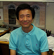
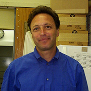
Toru Miki received his Ph.D. from Kyoto University, Japan, in 1979. He taught at Yamaguchi University School of Medicine, Ube, before joining the Laboratory of Cellular and Molecular Biology, NCI, in 1985. He is now chief of the Molecular Tumor Biology Section in the Basic Research Laboratory, NCI.
My research interests are in molecular mechanisms that convert normal cells to cancerous cells. To explore this field, I have developed an efficient expression cloning system and used it to isolate cDNAs that can convert normal fibroblasts into morphologically transformed cells. In an earlier study, we introduced an epithelial cell expression library into NIH3T3 cells and identified three morphologically distinct foci of transformed cells, from which we rescued three transforming cDNA clones designated ECT (epithelial cell transforming genes).
We soon identified ECT1 as a receptor of keratinocyte growth factor (KGF). KGF is related to fibroblast growth factors (FGFs), but specifically targets epithelial cells. Several research groups were in the process of isolating FGF receptors at that time, and structural comparison of KGF receptor and FGF receptor–2 (FGFR2) revealed a difference only in a small region of the ligand-binding domain, suggesting that these receptors are encoded by alternative transcripts of a single gene. This finding established that ligand binding specificity can be determined by alternative splicing.
We also isolated a constitutively activated FGFR2 from rat osteosarcoma cells. Acquisition of a new sequence, designated FRAG1, at the COOH-terminus of FGFR2 played a major role in the activation by overriding the negative regulatory effect of the normal ligand-binding domain. Finally, we found that FGFR2 isoforms with an acidic amino acid stretch were heavily modified with glycosaminoglycans. The modified receptor exhibits sustained activation of MAP kinase and stimulation of DNA synthesis. This modification event is also regulated by alternative splicing, because the modification site is encoded by an alternative exon.
In contrast to ECT1, which exhibits structural homology to several protein kinases, ECT2 initially lacked structural information. However, our continued attempts to isolate novel transforming genes from different cell types, as well as accumulation of new protein sequences in databases, led us to new members of this group of oncogenes, which share structural motifs of the guanine nucleotide exchange factors for the Rho family GTPases. While these oncogenes, ECT2, OST, TIM, NET1, and NTS, share common motifs, they also display distinct features. OST comprises multiple isoforms, catalyzes guanine nucleotide exchange on RhoA and Cdc42, and associates with Rac1 in its GTP-bound form. OST also regulates the JNK/SAPK MAP kinase pathway and induces actin reorganization in fibroblasts. NET1 is one of the genes induced with oxidative stress. NTS, an exchange factor for Rac1, is a major nucleolar antigen expressed in proliferating, but not resting, cells.
Most of my recent interests are in the biological functions of ECT2. Besides the Rho exchange factor homology domain, ECT2 also contains domains related to several molecules involved in cell-cycle checkpoint control and repair. We have recently found that ECT2 is a critical molecule regulating cell division. ECT2 functions as a guanine nucleotide exchange factor for Rho GTPases and is phosphorylated specifically in G2 and M phases. Interestingly, this phosphorylation is required for the exchange activity of ECT2. Unlike other exchange factors, ECT2 localizes in the nucleus in interphase, but spreads to the entire cell after nuclear membrane breakdown. During cell division, ECT2 localizes at the cleavage furrow and then the midbody. Inhibition of ECT2 activity in cultured cells either by microinjection of anti-ECT2 antibody or expression of dominant-negative isoforms strongly inhibits cytokinesis. ECT2 also localizes in spindle microtubules during mitosis.
We have recently cloned Xenopus ECT2 and are studying its function in mitosis using a Xenopus in vitro cell-cycle control system. We are also increasing our attempts to clarify the role of Rho exchange factors in cell-cycle control.
 |
|
Bob
Seder
|
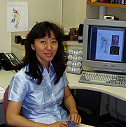
Bob Seder received his M.D. from Tufts University in Boston in 1986 and completed a residency in internal medicine at Cornell University–New York Hospital. He came to NIH in 1989 as a postdoctoral fellow in the Laboratory of Immunology, NIAID, and is currently chief of the Clinical Immunology Section in the Laboratory of Clinical Investigation, NIAID.
Cellular immunity plays an important role in protecting hosts from a variety of infectious pathogens. These include fungal (such as Histoplasma capsulatum), parasitic (Leishmania major, Toxoplasma gondii), mycobacterial ( Mycobacterium tuberculosis, M. avium complex), and bacterial (Listeria monocytogenes) pathogens. Over the past 15 years, the increase in opportunistic infections in individuals infected with HIV has underscored the importance of the cellular immune response in mediating protection against some of these pathogens. Despite a greater understanding of the factors regulating immune responses against these pathogens, patients often died from these infections due to an inadequate immune response. The worldwide pandemic of M. tuberculosis infection further attests to the need for understanding protective primary and memory cellular immune responses, which could lead to effective vaccines.
Protective immunity to many intracellular pathogens requires a complex sequence of events involving cells of the innate and acquired immune response. While innate immune response does provide some protection, acquired cellular immune response appears essential for sterilizing immunity and maintaining effective memory responses. Cytokines, such as IL-12, operate as a critical link between innate and acquired immunity. Their effect on the immune response overall and specifically on CD4+ T-cell differentiation has been the major focus of my career and current laboratory.
The importance of CD4+ T cells in regulating immune responses was highlighted by the seminal observation that CD4+ T-cell clones could be segregated into specific subsets—Th1 and Th2—based on their pattern of cytokine production. This distinction and its importance in vivo provided a framework to examine the factors involved in the generation of these subsets. In my postdoctoral lab, we showed that cytokines such as IL-12 and IL-4 were potent regulators of Th1 and Th2 differentiation, respectively. Based on these observations, my current laboratory is using a variety of experimental infectious disease models to further understand the role of cytokines in immune regulation in vivo. To this end, we have studied three human pathogens (H. capsulatum, M. tuberculosis, L. major) in vivo. We also use these models to study cytokines as the basis for new therapies and vaccines.
Our recent work applies our understanding of how cytokines alter specific immune responses to a daunting challenge—the development of vaccines against intracellular infection. The great success of all existing vaccines depends on long-lived humoral immune response via antibodies. These responses to bacteria and viruses are readily achieved by many different vaccine formulations. By contrast, there are no uniformly effective vaccines for infections such as tuberculosis, malaria, and HIV, which now account for a majority of the deaths worldwide from infection. For all of these infections, a cellular immune response mediated by CD4 and/or CD8+ T cells is required for protective immunity.
We have used the mouse model of Leishmania major infection, which requires Th1 responses, to study how long-term cellular immunity can be induced in vivo. Our results so far have shown that plasmid DNA vaccination encoding a specific leishmanial antigen induces long-lived protective responses in mice. This work has led us to explore whether this approach can work in nonhuman primates. If successful, we will follow up these studies in humans to determine safety and efficacy. We hope that this model will also help guide a rational approach for vaccination against M. tuberculosis, which requires a similar type of immune response.
 |
|
Wei
Yang
|
Wei Yang received her Ph.D. from Columbia University in New York in 1991 and did postdoctoral work at Yale University in New Haven, Conn. In 1995, she joined the Laboratory of Molecular Biology of NIDDK, where she is now a senior investigator.
I have been interested in protein-DNA interactions for quite some time. Trained to be a biochemist and X-ray crystallographer in graduate school, I became fascinated by DNA recombination during my postdoc period at Yale University. At that time, I determined the crystal structure of a site-specific recombinase, gd-resolve, complexed with a 34-bp substrate DNA. Since joining NIH, my group has widened the research target from DNA recombination to mismatch repair and has taken a combinatorial approach, including X-ray crystallography, biochemistry, and mutagenesis, to study mechanisms of both processes.
DNA repair is an essential biological process in all living organisms. Mistakes in base pairing occur during DNA replication and are routinely repaired. MutS, MutL, and MutH are necessary and sufficient to initiate methyl-dependent mismatch repair in Escherichia coli. MutS possesses an ATPase activity and initiates repair by recognizing DNA containing mispaired or unpaired nucleotides. After binding to a mismatch, MutS recruits MutL to mediate the activation of MutH endonuclease, which cleaves the newly synthesized strand 5' to a transiently unmodified d(GATC) sequence. Both MutS and MutL also play essential roles in the subsequent removal and resynthesis of the daughter strand. The mismatch repair proteins MutS and MutL are conserved from prokaryotes to humans, and defective human homologues, for example, MSH, PMS, and MLH1 proteins, have been directly implicated in hereditary nonpolyposis colon cancers (HNPCC) and other familial and sporadic cancers.
During the past four and a half years, we have determined the crystal structures of MutH, a conserved 40-kD fragment of MutL (LN40), and LN40 in complex with an ATP analog and with the product ADP. Most recently, in collaboration with Peggy Hsieh’s group (GBB, NIDDK), we have determined crystal structures of Thermus aquaticus MutS alone, its complex with DNA, and a ternary complex of MutS, DNA, and ADPMg2+. Based on these structures, we identified the active site residues of MutH and the MutS ATPase, and we confirmed the functional importance of these residues by mutagenesis. The crystal structures of MutS and its complex with DNA revealed the mode of mismatch recognition, which is based on the instability of heteroduplex DNA at mispaired or unpaired bases. We also uncovered an ATP-ase activity intrinsic to MutL that previous investigators had concluded did not exist.
Using hydrodynamic methods and crystallography, we have shown that binding of ATP triggers a structural trans-formation of MutL. Finally, we have developed an in vitro assay for initiation of mismatch repair. Combining our structural, mutagenesis, and biochemical results, we have proposed a proof-reading mechanism of mismatch recognition by MutS—similar to that found in protein synthesis and DNA replication, but the first example identified in DNA repair processes. Our studies of mismatch repair proteins also have shed light on defects of mutations in HNPCC kindreds.
The other project my research group has been focused on is V(D)J DNA recombination, which is a site-specific event essential for the assembly of the immunoglobulin (Ig) and T-cell receptor (TCR) genes in the vertebrate immune system. The Ig and TCR genes are encoded in separate V, D, and J segments. Each segment has multiple copies with slight variations in sequence. By combinatorial means of joining V, D, and J segments into Ig and TCR genes and modifying the junctions between segments in somatic cells, immune systems are able to make millions of different Igs and TCRs from a limited set of coding sequences.
The V(D)J recombination
is initiated by the combined action of two lymphoid-specific proteins, RAG1
and RAG2, which introduce double-strand breaks at the borders of the V, D, and
J segments. The second stage of V(D)J recombination is to process and rejoin
the cleaved coding ends of V, D, and J segments to form the mature Ig and TCR
genes. Several proteins involved in the general repair of DNA double-strand
breaks, including the catalytic subunit of DNA-dependent protein kinase, Ku
protein, XRCC4 and ligase IV, are required for the second stage of V(D)J recombination.
Mistakes at any stage of the V(D)J can cause cancers, growth and developmental
defects, and premature death in humans. We are studying almost all of the involved
proteins and in collaboration with Martin
Gellert’s group (LMB,
NIDDK) have succeeded in determining the crystal structure of XRCC4. We are
continuing to unravel the structure and function of macromolecular complexes
involved in DNA repair and recombination. ![]()