| T H E N I H C A T A L Y S T | M A Y – J U N E 2000 |
|
|
|
| P E O P L E |
RECENTLY TENURED
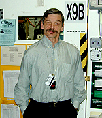
Zbigniew Dauter obtained his Ph.D. from the Technical University of Gdansk, Poland, in 1975. He lectured on crystallography at that university, then conducted research on structures of biologically active small and macromolecules at the University of York, England. He also worked at the synchrotron outstation of the European Molecular Biology Laboratory in Hamburg, Germany, before coming to the United States and assuming his current position as chief of the new Synchrotron Radiation Research Section within the Program of Structural Biology of the NCI in Frederick. His group is located at the synchrotron of Brookhaven National Laboratory on Long Island, N.Y.
|
My interests have always been in the elucidation of the structures of biologically active compounds—at first, smaller organic ones, such as antibiotics and anti-tumor acridine derivatives, and for the past 18 years, macromolecules. I specialize in the application of the unique properties of the synchrotron X-radiation to diffraction studies of macromolecular crystals, in particular, atomic-resolution structural analyses and use of anomalous scattering effects.
The enormous intensity of X-rays produced by the synchrotron makes it possible to record diffraction data on protein crystals at atomic resolution—about 1 Å. This provides a wealth of structural information with the accuracy in the range of 0.01 Å, comparable with the structures of small molecules. This leads to improved libraries of protein stereochemistry and provides better target values of geometrical parameters for validation of protein models refined at lower resolution.
Many hydrogen atoms become directly visible in electron density maps generated with the synchotron. This is important because hydrogen atoms are very often crucially involved in the reaction mechanism of enzymes. Atomic resolution also allows investigation of the protonation states of charged groups within the protein. I have participated in several studies of macromolecular structures at atomic resolution of proteins ranging from small metalloproteins (rubredoxins and ferredoxins) to larger enzymes. The 1.0-Å model of 75-kDa alcohol dehydrogenase complexes has led to revision of the classic mechanism of this enzyme.
Another property of synchrotron radiation is its tunability, which makes it possible to capitalize on anomalous dispersion effects of heavier elements inherently present or introduced into proteins. This is the basis of the popular multiwavelength anomalous dispersion method of solving crystal structures. We recently devised a novel modification of this approach, based on incorporation of anomalously scattering bromide or iodide ions into protein crystals. We incorporate the ions into the protein crystals with a flash dip in the appropriate cryosolution. This approach may be particularly useful for high-throughput projects, such as structural genomics. Several new structures have been recently solved with this approach. For example, the structure of the yeast hypothetical protein yhp9 containing the GAF domain, with a dimer of 2 times 18 kDa in the asymmetric unit, has been solved from NaBr-soaked crystals by Jim Hurley and his team at NIDDK, and the results are being prepared for publication in Cell.
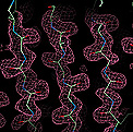 |
Also, the structure of the human acyl-protein thio-esterase of 56 kDa (see figure for a piece of the initial electron density map with a fragment of a b-sheet) was recently solved from brominated crystals using single-wavelength data in the laboratory of Zygmunt Derewenda of the University of Virginia in Charlottesville (the protein came from Teresa Jones of NIDDK). It will be published in Nature Structural Biology.
The applications we work with demand diffraction data that are as accurate as possible. Optimization of data collection procedures is my particular specialty. By recording very accurate diffraction data on lysozyme crystals and by using very small anomalous signals of sulfur and chlorine atoms, we have shown lysozyme binds several halide anions at the surface.
In addition to pursuing my own research interests, I am the facility manager for the macromolecular crystallography synchrotron beam line X9B at Brookhaven. I supervise data-collection facilities that are available to all intramural NIH laboratories (at present about 12 groups) that need to use synchrotron radiation for crystallographic data collection. To obtain more information about access to this beam line, contact me by e-mail or at (631)-344-7367. I would be glad to collaborate with those interested in crystal structures of their macromolecules.
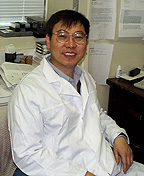
Chuxia Deng received his Ph.D. from the University of Utah in Salt Lake City in 1992 and did postdoctoral work in Philip Leder’s laboratory at Harvard Medical School in Boston before joining the NIDDK Laboratory of Biochemistry and Metabolism in 1995. He is now a senior investigator in the Mammalian Genetics Section, Genetics of Development and Disease Branch, NIDDK.
 |
I am interested in studying human skeletal dysplasias and breast cancer using mouse models. A particularly exciting focus of current research in my laboratory are mechanisms of BRCA1-associated tumorigenesis. About half of familial breast cancer cases and 90 percent of combined familial breast and ovarian cancers are associated with mutations in the BRCA1 gene.
The lack of a suitable animal model has made it difficult to identify how BRCA1 mutations affect the timing and process of tumor development. Speculations that BRCA1 is not a simple tumor suppressor and that BRCA1-associated tumor formation is not straightforward are fueled by the fact that mice heterozygous for BRCA1-null mutations do not develop tumors, while mice homozygous for mutations die early in embryonic life and display cellular proliferation defects.
To plumb the molecular mechanisms through which BRCA1 represses tumor formation, we introduced a series of mutations—including a null mutation, an isoform mutation, and a conditional mutation—into the mouse BRCA1 locus. Mutational analyses at both cellular and whole animal levels demonstrated that the primary function of BRCA1 is to maintain genome integrity through its control over the G2-M cell cycle checkpoint and centrosome duplication. BRCA1 mutations result in genetic instability (DNA damage and chromosomal aneuploidy), which then activates cellular protection mechanisms, including cell-cycle checkpoints and programmed cell death, to eliminate the mutant cells. This is why BRCA1 mutant cells fail to grow in culture. On the other hand, the genetic instability in BRCA1 mutant cells theoretically increases mutation rates of all genes, including tumor suppressors and oncogenes, and this increase ultimately overcomes the proliferation defects caused by the BRCA1 loss and results in tumor formation. Studies in our animal model, whose BRCA1 is specifically mutated in mammary epithelium, indicate that this is the case.
Analyzing mammary tumors from the BRCA1-conditional knockout mice, we noticed that two-thirds of the tumors exhibited alterations in p53, a potent tumor suppressor that is mutated in more than 50 percent of all human cancers. This observation suggests that p53 tumor suppressor gene is involved in BRCA1-associated tumorigenesis. To directly test whether inactivation of p53 contributes to the BRCA1-associated tumorigenesis, we deleted one wild-type allele of p53 in the mice with mammary epithelium-specific inactivation of BRCA1. We found that the remaining wild-type allele of p53 was quickly mutated, and the mammary tumor formation was dramatically accelerated. These results demonstrate that disruption of BRCA1 allows p53 (and other unidentified genes) to mutate more readily and leads to tumor formation. We plan to use the BRCA1-conditional mutant mice to further study molecular aberrations arising from BRCA1 deficiency, to identify genetic modifiers and exogenous factors that influence the onset of tumor formation, and to validate potential therapeutic strategies.
We have also generated mouse models mimicking achondroplasia, Pfeiffer syndrome, and thanatophoric dysplasia I and II. Our research is elucidating the role of fibroblast growth factor receptors (FGFRs) and TGFb/Smads signals in mammalian development and skeletal formation and is furthering our understanding of the mechanisms underlying dwarfism. FGFRs are membrane-spanning tyrosine kinases that serve as high-affinity receptors for at least 22 growth factors. It has been shown that missense mutations in three out of four known FGF receptors (FGFR1-3) are responsible for at least nine human inherited skeletal dysplasias, including the FGFR3-associated achondroplasia, which is the most common form of dwarfism. Using our mouse model, we demonstrated that a loss-of-function mutation of FGFR3 resulted in faster and prolonged growth of long bones of the arms and legs—phenotypes that are opposite to those displayed in human achondroplasia patients—suggesting that the human diseases are caused by gain of function, or a constitutive activation of FGFR. This hypothesis has been supported by other investigators, as well as by further studies in our own lab. Our work has also demonstrated that the activated FGFRs retard long-bone growth by activating Stats (signal transducer and activator of transcription) and cell cycle inhibitors. We are currently searching for potent inhibitors of FGF/FGFR signals and downstream modifiers in order to develop effective therapeutic approaches for these skeletal dysplasias.
Mustafa Dosemeci received his Ph.D. in occupational health from the Hacettepe University, Turkey, in 1982 and did postdoctoral work on exposure assessment at the London School of Hygiene and Tropical Medicine in the United Kingdom before joining the NCI Environmental Epidemiology Branch in 1986. He is now a senior investigator in the Occupational Epidemiology Branch, NCI.
 |
My research career has focused on assessing exposure to occupational risk factors in cancer epidemiology. My research activities at NCI fall into four areas: 1) exposure assessment in occupational cancer epidemiology and exposure-related methodological issues; 2) large interdisciplinary case-control studies that examine the interaction of genetic susceptibility and occupational and environmental exposures in cancer risk; 3) evaluation of cancer risks in large surveillance studies; and 4) other scientific activities, including chairing the Exposure Assessment Working Group of the Division of Cancer Epidemiology and Genetics.
I have assessed various occupational and environmental exposures for numerous case-control, cohort, and cross-sectional biomarker studies conducted either in the branch or in other research institutes around the world. Cohort and case-control approaches have their own advantages and disadvantages. Assessing exposure retrospectively in cohort studies is time consuming and requires careful evaluation of substantial historical exposure information. Because of the availability of monitoring data, exposure assessment in cohort studies typically is considered superior to estimates in case-control studies where we usually lack monitoring data.
In most cohort studies, however, assessment of exposure is specific only to the job title level, in contrast to case-control studies, in which the availability of subject-specific exposure information allows evaluation of between-worker variability within the same job category. Between-worker variability reflects both genetic variability and differences in external exposures to environmental and occupational cancer risk factors; my recent research activities have focused on the estimation of the internal dose of occupational and environmental risk factors in cancer epidemiology.
I have completed quantitative assessments of historical exposure to benzene for 75,000 workers and to silica for 68,000 workers in cohort studies conducted in China and am currently conducting quantitative exposure assessments for three major cohort studies: the Agricultural Health Study, the Diesel Exhaust Cohort Study, and the Shanghai Women’s Cohort Study.
I have also conducted subject-specific assessments of exposure to various solvents, particulates, and industrial chemicals in seven case-control studies and have developed job exposure matrices (JEMs) for more than 40 chemical and physical hazards. In three cross-sectional biomarker studies conducted in China and India, we have assessed exposures to benzene, butadiene, and benzidine and their relationship to biological markers. I have also carried out studies to validate the exposure assessment methodologies we’ve used in our cohort and surveillance studies. Moreover, based on the results of studies simulating exposure misclassification, we have been able to develop specific recommendations for industrial hygienists to reduce the ef fects of misclassification on risk estimates.
Two large interdisciplinary case-control studies are under way to evaluate bladder and lung cancer risks using external and internal doses of occupational and environmental exposures. The first, based in Spain, is a study of bladder cancer that will involve 1,500 cases and 1,500 controls from 18 hospitals. Using a state-of-the-art, computer-assisted technique, information will be obtained by personal interview on occupational, environmental, clinical, and dietary risk factors; blood samples will be collected to determine genetic susceptibility markers.
The second study examines lung cancer risk factors in Russia and is autopsy based. In the pilot phase of the study, we are identifying 500 lung cancer cases and 500 control subjects from 88 hospitals with a high autopsy rate. For this study, I have been collecting work and residential histories and measurement data on more than 100 occupational and environmental hazards and am also obtaining normal and tumor tissue samples to identify genetic susceptibility markers.
I also coordinate three large mortality and cancer linkage databases: One uses death certificate data from 24 states and includes occupational information for about 7.2 million individuals who died between 1984 and 1996; another, a Swedish cancer and environmental linkage database, contains occupational information from the 1960 and 1970 censuses; and the third, the Shanghai Cancer Registry, uses JEMs geared to working conditions in China to evaluate the risk of specific cancers in relation to occupational categories or exposures. These activities afford opportunities to collaborate with intramural and extramural investigators in various hypothesis-generating studies.
Maribeth Eiden received her Ph.D. in genetics from the George Washington University in Washington, D.C., in 1985. She is now chief of the Unit on Molecular Virology in the Laboratory of Molecular and Cellular Regulation, NIMH.
 |
I am interested in the molecular mechanisms of retroviral entry into mammalian cells. Viral entry is a multistage process that involves a series of interactions between viral envelope proteins and cell surface receptors, and later viral core proteins and intracellular proteins, resulting in delivery of the nucleocapsid to intracellular compartments appropriate to activation of reverse transcriptase and synthesis of proviral DNA.
My studies on molecular determinants of retroviral entry began during my doctoral work with Marv Reitz in the Laboratory of Tumor Cell Biology, NCI. My research there characterized the gibbon ape leukemia virus (GALV), the first known retrovirus to be associated with a primate leukemia.
The simplicity of the GALV particle—a virion made up of only three components (genome, core, and envelope)—makes it amenable to use in in the construction of hybrid retroviral vectors. A hybrid vector contains genome, core, and envelope components derived from different viruses.
My NIMH lab has focused on the use of GALV vectors to probe cellular requirements for viral entry. To accomplish this, we constructed a series of GALV retroviral vectors. These vectors, like wild-type GALV, are capable of infecting appropriate target cells. Unlike their wild-type counterparts, however, they fail to replicate after infection. The first GALV vectors were produced using PG13 packaging cells. PG13 cells are murine NIH3T3 cells that have been engineered to stably express GALV envelope in combination with murine leukemia virus (MLV) core proteins. The hybrid vectors produced from PG13 cells contain MLV core and genome components in combination with GALV envelope proteins. These hybrid vectors were demonstrated to be capable of infecting several target cells that were resistant to or inefficiently infected by retroviral vectors bearing MLV envelope proteins. We later cloned a full-length, biologically active GALV genome from which we were able to construct a GALV-based packageable genome. Vectors produced in human 293T cells containing this modified GALV genome in combination with GALV core and envelope components are more efficient gene-transfer vehicles than either MLV-based vectors or PG13-produced GALV hybrid vectors for a variety of animal cell types, including human.
Working with Wayne Anderson’s laboratory in NCI, we determined that the GALV receptor normally functions as a type III phosphate transporter. Type III Pi transporters are present on most cell types, where they absorb Pi from interstitial fluid during normal cellular processes such as cellular metabolism, signal transduction, and nucleic acid and lipid synthesis. We and others later discovered that type III Pi transporters serve as receptors, not only for GALV but also for feline leukemia virus type B, two murine leukemia viruses, and simian sarcoma-associated virus. This finding allowed us to investigate specific features of viral entry shared by GALV and other retroviruses that also use this receptor-transporter family for entry into mammalian cells.
At this writing, it appears that GALV entry requires not only primary binding of the GALV envelope, but also secondary receptor interactions that allow the initial virus-receptor interaction to progress through a series of additional steps that culminate in the release of the nucleocapsid into the cytosol of the infected cell.
My lab has steadily increased its facility in the use of retroviral vectors to infect cells as we have constructed and tested new hybrid vectors. I expect this will lead us to the discovery of additional stages in the multistep process of viral entry. I also expect that we will identify new intracellular protein actors in the process, as well as develop increasingly efficient viral vectors.
Susan Pierce received her Ph.D. from the University of Pennsylvania in Philadelphia in 1977 and was the William and Gayle Cook Professor of the Biological Sciences in the Department of Biochemistry, Molecular Biology and Cell Biology at Northwestern University in Evanston, Ill., before joining NIAID as chief of the Laboratory of Immunogenetics in December 1999.
 |
The long-standing interest of my laboratory is in the molecular mechanisms by which B lymphocytes are stimulated by the encounter with a foreign antigen to proliferate and differentiate into antibody-secreting plasma cells and memory B cells.
It is well established that activation of B cells requires the binding of foreign antigen to the B-cell antigen receptor (BCR). We now understand that the BCR plays a dual role in B-cell activation by T-cell-dependent antigens: It initiates signal transduction cascades and it transports antigens to an intracellular compartment in which peptide–MHC class II complexes are assembled. The subsequent expression of the peptide–class II complexes on the B-cell surface allows the B cell to engage antigen-specific helper T cells, a critical event in B-cell activation.
We have focused our efforts over the last several years on delineating the cellular and molecular mechanisms underlying the signaling and antigen-targeting functions of the BCR and the means by which these functions are regulated. We hope that understanding these critical BCR processes and their regulation at a molecular level will lead to new strategies for vaccine design to stimulate antibody responses to both pathogens and tumor cells and for therapeutics to block or modify B-cell responses in allergy, autoimmunity, and transplantation.
Evidence from our lab and others indicates that the signaling and antigen-transport functions of the BCR are not independent and that correct targeting of the BCR to the class II peptide–loading compartment requires signaling. Indeed, BCR signaling dictates the correct targeting of antigen, the rate of transport of the BCR, and the efficiency of the assembly of peptide class II complexes.
In addition, B-cell co-receptors that influence BCR signaling in vivo and in vitro—including the FcgRIIB, CD19/CD20, and CD40—also influence antigen targeting. Thus, the B cell integrates information from a variety of signaling receptors to regulate its BCR signaling and antigen-processing function and, in turn, its interactions with helper T cells.
In addition to the B cell signaling receptors, antigen processing in B cells is regulated by several other factors, including the developmental state of the B cell, induction of the stress response, and viral infection.
The observation that the signaling and targeting functions of the BCR are interrelated raised the question of how these two functions are coordinated. Recent advances in cell biology have led to the description of sphingolipid- and cholesterol-rich membrane micro-domains, or lipid rafts, within the plasma membrane that act as platforms for receptor signaling and trafficking.
We have recently shown that upon cross-linking, the BCR is translocated into lipid rafts in which the Src family kinase Lyn is concentrated and the phosphatase CD45 is excluded. The BCR is phosphorylated in the raft and subsequently targeted to the peptide-loading compartment.
We believe the translocation of the BCR into lipid rafts, although previously unappreciated, could be a control point in BCR function and thus a very significant step in the BCR signaling and antigen processing pathways. Indeed, our recent results indicate that the lipid rafts function to coordinate the activity of the BCR and other B-cell coreceptors and to modulate BCR function during development and during infection by Epstein-Barr virus.
In the future, we plan
to use a combination of biochemical and genetic tools to characterize the components
of the lipid rafts, their relationship to the BCR signaling pathway and antigen-targeting
pathways, and their function in immature and memory B cells. ![]()