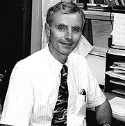
Darrell
Abernethy
| T H E N I H C A T A L Y S T | S E P T E M B E R – O C T O B E R 1999 |
|
|
|
| P E O P L E |
RECENTLY TENURED
|
|
Darrell
Abernethy
|
Darrell Abernethy received his M.D. and Ph.D. in pharmacology from the University of Kansas School of Medicine in Kansas City in 1976. Following a residency program at Jackson Memorial Hospital in Miami, Fla., and a postdoctoral fellowship at Massachusetts General Hospital in Boston, he moved up the academic ranks at the medical schools at Tufts University in Boston, Baylor University in Houston, Brown University in Providence, R.I., and Georgetown University in Washington before assuming his current position earlier this year as clinical director and chief of the Laboratory of Clinical Investigation at the NIA.
My research has focused on the mechanisms of peripheral distribution of drugs, drug disposition in the context of obesity, and the pharmacokinetic and pharmacodynamic relationships of cardiovascular drugs in aging.
Recent studies center on the role of genetic polymorphisms that influence responses to cardiovascular drugs. We are exploring both phenotypic and genotypic changes that contribute to altered phenotype, as well as nongenotypic splice variant transcriptional changes that result in phenotypic changes.
These studies currently involve endo-thelial nitric oxide synthase (eNOS) and the L-type calcium channel.
We and other investigators have previously noted marked differences in forearm vascular relaxation due to alterations in endothelial-derived relaxing factor(s) in aged and in African-American subjects. The basis for this finding is a current line of investigation, with the hypothesis that polymorphic variants of eNOS make a major contribution to this variability over and above environmental and disease-based factors.
In laboratory studies, we have found expression of functionally different splice variants of the a1 subunit of the L-type calcium channel in young vs old human vascular smooth muscle cell lines. We are now exploring the basis for this observation in the laboratory and are pursuing its clinical correlates by characterizing the expression pattern of these splice variants in human surgical specimens.
 |
|
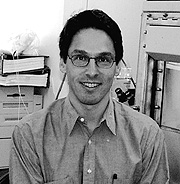
Robert
Kotin
|
Robert Kotin received his Ph.D. in microbiology from Rutgers University in New Brunswick, N.J., and the University of Medicine and Dentistry of New Jersey in Newark in 1986. After a postdoctoral fellowship at Cornell University Medical College in New York, he worked at Lederle Laboratories in Pearl River, N.Y., and Genetic Therapy, Inc., in Gaithersburg, Md., where he developed recombinant adeno-associated virus vectors. In 1994, he joined the the Molecular Hematology Branch in NHLBI, where he is now a senior investigator in the Laboratory of Molecular Hematology.
My research interests are focused on the molecular biology of adeno-associated virus (AAV), a human parvovirus. AAV is a deceptively simple virus with only two open reading frames—one encoding the structural proteins and the other the nonstructural, or Rep, proteins. The AAV genome consists of a linear single strand of DNA of approximately 4,700 nucleotides. One of the unique aspects of AAV is that it requires co-infection with a helper virus, such as adenovirus or herpesvirus, for productive infection. The only AAV gene product necessary for replication is the Rep protein(s). Helper virus infection renders the host cell permissive for AAV gene expression and replication, rather then complementing a genetic defect in AAV. In the absence of helper virus co-infection, the Rep protein represses AAV gene expression and the AAV DNA integrates into the cellular genome.
My early work examined the structure and organization of the AAV proviral DNA. Using the flanking cellular DNA as probes for genomic Southern blots, I discovered that the viral DNA integrated into a specific locus on human chromosome 19. Site-specific (or targeted) integration is still a very exciting discovery that, so far, is unique to AAV. This mechanism of targeted integration, however, remains obscure. A correlation between targeted integration and Rep protein expression is evident, but the known activities of Rep are not sufficient to explain targeted integration.
Our investigations of Rep have revealed several interesting activities that we think play an important role in AAV integration and virus replication and may explain some of the effects of Rep on the host cell.
Much of our research is driven by the desire to use recombinant AAV as a gene transfer vector. Understanding the functions of the AAV Rep proteins and how these proteins interact with the cell is central to advancing this goal. A major limitation for producing recombinant AAV results from the transient production methods used. Cells expressing Rep proteins display aberrant growth rates and phenotype. High concentrations of Rep appear to be cytotoxic to the cell, and it is not possible to produce a cell line that constitutively expresses Rep proteins. My laboratory has recently discovered that Rep binds and inhibits the cyclic AMP-dependent protein kinases, protein kinase A and protein kinase X (PrKX). The cellular role of PrKX is unknown, and we are currently investigating the molecular biology of this potentially important protein kinase.
The use of recombinant virus for gene transfer is a compelling idea for which the technology and applications have yet to be fully developed. The interaction between AAV and the host cell is complex, and a greater understanding of this system will be necessary before investigators can obtain transgene expression with therapeutic effect. Current AAV gene transfer vectors are based on AAV serotype 2; we have produced recombinant AAV type 4 and type 5. These three serotypes exhibit overlapping, but distinct, tissue tropism. Characterizing tissue specificity will enable investigators to select the appropriate vector for a given application.
In summary, my laboratory is making good progress on two fronts with AAV. First, we’ve been able to determine some of the ways that AAV interacts with cells—such as the interactions between Rep and PrKX and Rep and cellular DNA–which yield insights into viral and cell biology. Second, we’ve made strides in developing AAV as a gene transfer vector with great potential therapeutic value. AAV continues to surprise us in the ingenuity with which it manipulates the cell. I think this is very impressive for a simple, nonpathogenic virus.
 |
|
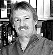
Stuart
Le Grice
|
Stuart Le Grice received his Ph.D. in biochemistry at the University of Manchester, United Kingdom, in 1976 After postdoc-toral research in the UK, Germany, and the United States, he served as a senior scientist with Hoffmann-La Roche, Basel, Switzerland. In 1990, he joined Case Western Reserve University, Cleveland, where he directed the Center for AIDS Research (CFAR) from 1994 to 1999. He is now chief of the Resistance Mechanisms Laboratory in the HIV Drug Resistance Program at the Frederick Cancer Research and Development Center, NCI.
My primary research interests are in protein structure and function in the regulation of gene expression. Studies of transcription in gram-positive and gram-negative prokaryotes laid the foundation for my current research. In these bacteria, multiple forms of sigma factors interact with the host RNA polymerase to control gene expression. We used chemical and enzymatic footprinting techniques to investigate RNA polymerase/promoter interactions in binary (polymerase/promoter) and ternary (polymerase/promoter/nascent RNA) complexes. We also studied cessation of transcription at sequence-dependent terminators to explore barriers to the use of heterologous signals in prokaryotes.
These studies have allowed me to move into retrovirology and apply similar concepts to investigate mechanisms of reverse transcription in HIV and related lentiviruses, the focus of my research since 1987. My studies were aided significantly by the development of metal chelate technologies for purification of polyhistidine-extended proteins at Hoffmann-La Roche, where I reported the first single-step purification of p66/p51 HIV-1 reverse transcriptase (RT). The p66 and p51 subunits of this enzyme are derived from the same gene, but p51 undergoes COOH-terminal processing by the viral protease. Understanding the role each subunit plays within the parental heterodimer required preparing enzymes containing point mutations in a single subunit. A reconstitution protocol I developed made this possible and was first applied to prepare selectively deuterated p66/p51 heterodimers ([H]-p66/[D]-p51 and [D]-p66/[H]-p51) for analysis by neutron diffraction. Subsequently, I have been engaged in a program of subunit-selective mutagenesis and was the first to illustrate that the p51 subunit of p66/p51 RT was catalytically inactive; that is, all enzymatic functions reside within the p66 subunit. This mutagenesis strategy has been adopted by several laboratories studying structurally related lentiviral RTs.
The multifunctional nature of RT provides both its DNA polymerase and ribonuclease H (RNase H) activities as targets for development of antiretroviral agents. Thus, a second theme of my research is RNase H activity as it pertains to nonspecific and highly specific hydrolysis of the RNA-DNA replication intermediate. I was first to demonstrate that eliminating RNase H activity inhibited HIV replication in culture and that host enzymes could not complement an impaired retroviral function, thus suggesting this activity as a bona fide therapeutic target. In 1990, I showed that RNase H activity could be subdivided into polymerization-dependent and polymerization-independent modes, although their relevance was not immediately clear. Further research revealed mutants that retained exclusively polymerization-dependent RNase H activity. Eliminating the latter function significantly impaired DNA strand transfer, a process in retroviruses whereby nascent DNA is relocated between templates.
Despite such successful in vitro mutagenesis, the structural basis for phenotypes of many mutants prepared in the laboratory remains unclear. I am now developing bioconjugate strategies for probing retroviral replication complexes in solution and at high resolution. This approach entails site-specific tethering of photoactive, fluorescent, nucleolytic, and proteolytic probes to RT or its nucleic acid substrates. These probes will allow us to investigate contact between individual subdomains, "communication" between the p66 and p51 subunits, and potential interactions between RT and other viral proteins. Preliminary experiments have defined contacts between the extreme COOH-terminus of the RNase H domain and duplex DNA that cannot be detected through crystallographic analysis. The same approach indicates that RT accommodates duplex DNA and RNA-DNA hybrids differently. The fast-emerging field of bioconjugate chemistry provides the ideal complement to crystallographic approaches. The combined power of these disciplines should reveal features of the retroviral polymerase that lend themselves to therapeutic intervention
 |
|
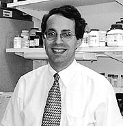
Robert
Star
|
Robert Star received his M.D. from Harvard University-MIT Program in Health Sciences and Technology in Cambridge and Boston in 1980, did postdoctoral work at NIH, and then moved to the University of Texas Southwestern Medical Center in Dallas in 1987. He has returned to NIH as a senior investigator in NIDDK to form the Renal Diagnostic and Therapeutic Unit. He is also a Senior Scientific Advisor to the Division of Kidney, Urologic and Hematologic Diseases.
I am interested in translational research for the diagnosis and treatment of renal disease. My initial bench studies at UT Southwestern wandered from renal transport mechanisms to mathematical modeling to nitric oxide; throughout, however, I attempted to translate my bench discoveries to solve clinically relevant problems. For example, I found that urea movement in the terminal part of the kidney is limited by the apical membrane. Reasoning that trapping of urea within cells might explain some symptoms in dialysis patients, we studied urea movement during dialysis. While incorrect, this led to the development of an easy method to measure the dialysis dose based on the simple concept of urea removal. We then showed that several widely used methods to quantify dialysis dose were inaccurate.
Most recently, we have been studying diagnostic and therapeutic approaches to acute renal failure, a condition that lacks a specific treatment and carries a 50 percent mortality. Again, serendipity played a role. While studying nitric oxide in the kidney, my colleagues and I discovered that nanomolar concentrations of the pituitary hormone a-melanocyte stimulating hormone (a-MSH) inhibited nitric oxide synthesis (NOS) in macrophages. Unlike standard NOS inhibitors that directly inhibit the enzyme, a-MSH inhibited the induction of the inducible NOS (iNOS). a-MSH darkens skin color in amphibians, but, more importantly, decreases fever and has profound anti-inflammatory effects in acute, chronic, and systemic inflammation. a-MSH is increased by systemic stress and is produced at sites of inflammation. We found that a-MSH is produced by inflammatory cells, indicating that a-MSH is an anti-inflammatory cytokine.
We worked on its mechanism of action in cultured and isolated cells, but switched to animal studies to search for clinically relevant uses. We found that a-MSH protected against endotoxin-induced liver damage; unlike many other agents, a-MSH was effective even when given 30 minutes after endotoxin. We then tested a-MSH in a renal ischemia-reperfusion model of acute renal failure, a model in which injury is caused by inflammatory and cytotoxic (NO) injury cascades, both of which are inhibited by a-MSH. We discovered that a-MSH inhibits renal ischemia reperfusion injury. Again, unlike many other agents tested in animals, a-MSH was still effective even when started six hours after injury. We then investigated its mechanisms of action. As expected, a-MSH inhibited both inflammatory (neutrophil, chemokines, adhesion molecules) and cytotoxic (NO) pathways; however, we found it also simulated an endogenous protective mechanism involving IL-10.
Based on its multiple effects and mechanisms of action, we conducted a Phase I dose-escalation trial to establish a-MSH safety in humans. a-MSH was safe; however, we unexpectedly found that a-MSH increased blood pressure. This "side effect" could be useful in treating patients with ischemic acute renal failure and adds another mechanism of action of a-MSH.
Human acute renal failure is difficult to diagnose, because currently available tests are insensitive and do not detect renal damage for several days. We are, therefore, pursuing new diagnostic tests for acute renal failure and have developed and validated a bedside method for measuring glomerular filtration rate that takes only 45 minutes—five times as fast as the current method. We are using a variety of biochemical and molecular techniques—akin to a renal CPK test—to search for molecules that specifically appear in the injured kidney or the urine it produces. For example, we are using laser-capture microdissection to study gene expression in specific portions of the injured nephron. We are using this technique to identify genes that are differentially expressed in different portions of the kidney in response to ischemia. We hope that some of these genes may point to novel protective agents and that some may serve as markers for renal injury. We plan to use advanced imaging techniques to measure renal function, inflammation, and fibrosis.
 |
|
Gisela
Storz
|
Gisela Storz received her Ph.D. in biochemistry from the University of California, Berkeley, in 1988 and did postdoctoral work at NCI and Massachusetts General Hospital in Boston before joining the Cell Biology and Metabolism Branch of NICHD in 1991.
My main interest is in how cells sense and protect themselves against oxidative stress. Reactive oxygen species—such as superoxide, hydrogen peroxide, and hydroxyl radical—have the potential to damage almost all cell components, including DNA, lipid membranes, and proteins. Oxygen radicals have been implicated as causative agents in several degenerative diseases.
Most organisms have an adaptive response to defend against oxidants. For example, treatment of bacterial or yeast cells with low doses of hydrogen peroxide induces expression of a distinct group of proteins, the decreased expression of other proteins, and resistance to killing by subsequent higher doses of hydrogen peroxide. My group at NIH has focused on this adaptation.
The Escherichia coli and Salmonella typhimurium responses to hydrogen peroxide involve at least two regulators, OxyR and OxyS. The expression of nine hydrogen peroxide–inducible proteins is controlled by the OxyR protein, the first transcriptional regulator found to be directly sensitive to the redox environment of a cell. Studies of the DNA-binding properties of oxidized and reduced OxyR showed that the two forms have different binding specificities, allowing OxyR to act as an activator after oxidative stress and as a repressor during normal growth.
We recently discovered that OxyR is activated by the formation of a disulfide bond and is deactivated by enzymatic reduction by glutaredoxin. Because the formation of a disulfide bond in the reducing environment of the E. coli cytosol was unexpected, we are now examining the nature of the exquisite redox sensitivity of OxyR. Our studies should give new insight into cysteine chemistry and the regulation of other redox-sensitive proteins.
Hydrogen peroxide treatment also leads to the induction of a novel 109-nucleotide, untranslated RNA regulator denoted OxyS. OxyS is conserved between E. coli, S. typhimurium, and Shigella flexneri. We found that the OxyS RNA activates and represses the expression of as many as 40 genes in E. coli, and we identified eight targets. This unusual RNA also acts as an antimutator, reducing chemically induced mutagenesis. Thus, we propose that OxyS integrates adaptation to hydrogen peroxide with other cellular stress responses and helps to protect cells against oxidative damage.
We are now using mutational studies of the OxyS RNA and its target sequences to investigate the mechanism of the regulation by this RNA. In addition, we are conducting genetic and biochemical assays to identify and characterize proteins that interact with OxyS. We have also begun to examine the functions of other small RNAs of unknown function in E. coli.
Compared with the bacterial response to hydrogen peroxide, relatively little is known about the cellular mechanisms used by higher cells to sense and protect against oxidative damage. To initiate studies of the oxidative stress response in eukaryotes, we constructed isogenic Saccharomyces cerevisiae strains carrying mutations affecting known signal transduction pathways.
We are now comparing the
oxidant sensitivities and whole genome expression patterns of these mutants
and are genetically screening for yeast mutants with altered sensitivities to
hydrogen peroxide in order to identify new components of the eukaryotic response
to oxidative stress. ![]()