| T H E N I H C A T A L Y S T | M A R C H – A P R I L 1999 |
|
|
|
| C O M M E N T A R Y |
|
by
Michael J. Iadarola,
Ph.D. |
CREATING A GENE THERAPY
FOR
CHRONIC PAIN AND SPINAL
CORD DISORDERS
 |
|
Alan
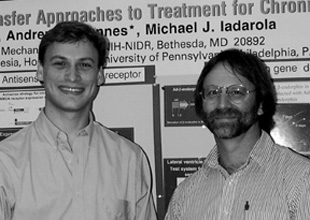
Finegold (left) and Michael Iadarola
|
This research demonstrates a new treatment strategy for chronic pain. It is currently in transition from the lab bench to the patient bedside, as we prepare for a first clinical trial in human subjects. What follows is a personal account of how the research evolved and where it can go in the future. The "paracrine paradigm" we developed is applicable in a general fashion to therapy for chronic neurological disorders.
Pain: Study It or Treat It?
This work began in the summer of 1993 with a small program in therapeutics—small because I was able to carve out only limited time over two summers with an HHMI high school student, Susan Lee (who has since gone on to Harvard Medical School in Boston). I had always been a basic bench scientist, and my lab had been studying synaptic-induced gene regulation in the spinal cord in models of persistent peripheral inflammation. I had discovered that persistent pain up-regulates the opioid peptide dynorphin in the dorsal spinal cord, the first synaptic processing station for pain—first observing this with a radioimmunoassay for dynorphin peptide and later measuring the corresponding mRNA increases, performing studies to localize the spinal neurons involved, and eventually examining seven base pairs of enhancer sequence in the promoter.
The transition to translational research was sparked through our weekly laboratory meetings. What was then the Neurobiology and Anesthesiology Branch contained both basic and clinical research groups, and the clinical group sometimes presented patients with chronic pain problems. This was my first exposure to patients with chronic neuropathic pain disorders, and it was a real eye-opener. Chronic neuropathic pain is notoriously difficult to control with currently available drugs and procedures, and the subjects we were seeing exemplified this clinical state of the art. Often, what had begun as relatively minor nerve damage after a traumatic injury progressed to a severe chronic pain disorder. Patients experienced high levels of spontaneous pain and mechanical allodynia (pain from a normally nonpainful stimulus). Just brushing the skin in the neuropathic zone was enough to cause them excruciating pain. This exposure stimulated us to begin exploring new treatments for pain, in addition to studying the molecular neurobiology of pain.
First Steps
In choosing among treatment approaches, we wanted to do something new and to use some of the molecular methods that we had expertise in and control over within our own lab. At the time, we were performing transient transfections to investigate those seven base pairs in the dynorphin promoter. Moreover, there was real excitement over the beginnings of gene therapy, much of which was occurring here on the NIH campus. So the idea of adapting techniques of gene transfer to pain treatment seemed like a natural extension of our current program. Still uncertain as to the exact gene to use in pain treatment, we nonetheless needed to assess the basic process of in vivo gene transfer.
That first summer, we asked whether plasmids could transfect neurons or glia in the spinal cord in vivo or in primary cultures. Plasmids were sweeping the literature, and reports had appeared suggesting simple systemic injections were effective at transducing cells. Plasmids were certainly convenient, although I had my doubts about how effective they would be in nonmitotic cells of the nervous system.
|
NOTHING WORKED VERY WELL, EITHER IN VIVO OR IN THE PRIMARY CULTURE SYSTEM, WHERE THE GLIAL CELLS ACTED AS INCREDIBLE SPONGES FOR DNA, NO MATTER WHAT FORM IT WAS IN OR WHAT COATING WAS AROUND IT. WE NEEDED TO LOOK ELSEWHERE. |
The primary cultures worked up to a point: The lacZ test gene expressed b-galactosidase only in the "feeder layer" of flattened glial cells at the bottom of the plate. The neurons, which in these cultures are like groups of round balls sitting atop the flat glia, never seemed to pick up and express the plasmid. In vivo, plasmid transfer was weak, and the amount of measurable transgene expression was dismal. We could assay a small increase in b-galactosidase activity biochemically but could not see the cells with histochemical methods. We tried direct injections of plasmid into tissue and even prolonged infusions (for a week, using an osmotic mini-pump) of about 10 billion plasmid molecules into the cerebrospinal fluid (CSF) space surrounding the spinal cord. We played around with ligand-derivatized polylysines and the timing of plasmid incubations, among other tacks.
Nothing worked very well either in vivo or in the primary culture test system, where the glial cells acted as incredible sponges for DNA, no matter what form it was in or what coating was around it. We needed to look elsewhere.
Virus to the Rescue
We turned our attention to virus. Fortunately, my institute had already established a program in gene therapy run by Brian O’Connell, who helped us get started by providing virus reagents and guidance in filing the necessary paperwork with the Biosafety Committee. We were also lucky to have anesthesiologist Drew Mannes join the group, through a joint agreement with the University of Pennsylvania Anesthesiology Department in Philadelphia. For Mannes, the highest priority for research was that it have clinical relevance.
We began with a straightforward comparison in rats of viral transduction after infusions into the intrathecal space (the CSF space around the spinal cord) or infusions directly into the cord tissue itself (intraparenchymal) (1). Adenovirus was a vast improvement over plasmid: We achieved nearly 60-fold increases over baseline in b-galactosidase activity upon intraparenchymal injections. We injected directly into the ventral horn to provide a good seal around the cannula tract—and were nearly instantly gratified by transduction of the motor neurons, which turned blue in a matter of minutes. The motor neurons filled up, from the dendritic tree all the way out to the axons in the ventral roots. I thought we had solved the problem of neuronal gene transfer! At the very least, we had one vector we could use for intraparenchymal injection.
Viral infusion into the CSF, however, was not so happy—there was almost no expression in the spinal cord tissue. We found that the pia mater, one of the meningeal layers surrounding the spinal cord, was a very effective barrier to viral entry from the CSF space into the cord tissue proper. We could literally strip off the pia and stain it histochemically for b-galactosidase activity. The pial covering turned blue, but the spinal cord just underneath was devoid of reaction product.
Evolution of the Paracrine Paradigm
At first we reasoned that we would have to break through the pia for virus to gain access to the spinal cord. There ensued a series of increasingly invasive manipulations, starting with hyperosmotic mannitol shock and proceeding to partial enzymatic digestion with intrathecally applied proteases.
None of these strategies succeeded; no virus made it through the pia. We concluded that rather than fight Mother Nature we would attempt to use the pia as a secretory engine. We hypothesized that we could block pain by inducing the pia to secrete a virally transfected analgesic gene product. This is a "gain of function" gene therapy approach.
As luck would have it, we found an ideal gene cassette in the literature. Earlier studies had explored cell transplantation therapy as a potential means of treating pain—using either human cadaver or bovine xenografts of adrenal chromaffin cells (a rich source of enkephalin opioid peptides) or cells that had been engineered to secrete enkephalin. In the latter case, Rusty Gage’s group at the Salk Institute in La Jolla, California, had constructed a cassette that allowed fibroblasts to secrete the powerful endogenous opioid b-endorphin. They had fused human b-endorphin at the COOH-terminus of the leader sequence of nerve growth factor (NGF) to direct the secretion of b-endorphin to the nonvesicular secretory pathway.
The idea was to stably incorporate the NGF–b-endorphin cassette into fibroblasts through a retroviral transduction, isolate secreting fibroblast clones, expand the cells, and transplant them into the intrathecal space—a somatic cell gene therapy approach. Gage’s group had already characterized the ability of the construct to secrete authentic b-endorphin from cultured fibroblasts but had not used the system in vivo before they dropped this line of research. While the somatic cell–fibroblast approach seemed cumbersome, the cassette itself seemed tailor-made for the connective tissue cells of the pia.
Thus, we were able to simplify the procedure considerably by using direct gene transfer. Fortunately, Gage was able to dig the plasmid out of the freezer and send it to us for subcloning into adenovirus. At this time, Mannes’ NIH fellowship ended, and a new postdoctoral fellow, Alan Finegold, joined the group and began making several adenovirus shuttle vectors containing the NGF–b-endorphin cassette and several other sense and antisense constructs.
Here again, the interactive network that characterizes NIH so well provided a helping hand. O’Connell had obtained a contract to produce adenovirus and generously provided us with access to the service, so we obtained several production runs of various viruses.
After some on-the-job training in animal surgery and behavioral research, Finegold was routinely injecting virus into spinal cord and evaluating in vivo transfer. Initially, we directed our injections into the dorsal horn of the spinal cord, where somatosensory signals (such as heat, cold, light, touch, and vibration) are processed. Previously, Mannes had succeeded in transferring genes in vivo into ventral horn motor neurons, but we needed to reach the dorsal horn to control pain.
This proved to be a difficult job for adenovirus, and we slowly came to the conclusion that through no fault in technique, only motor neurons were appropriate targets for adenovirus; the others were apparently impervious to it. Furthermore, we observed that the spread of the virus in the cord was nonuniform. It is an underappreciated fact that the nervous system contains many barriers to free diffusion or dispersal of large viral particles (~90 nM for adenovirus). When the virus encounters axon bundles, it tends to track along the bundle rather than diffuse through it. Tightly packed cells are another barrier. Aside from these physical issues, the cord appears to unevenly express the receptors for adenovirus binding or attachment (CAR) and internalization (integrins). (We are now investigating receptor sites and targeting strategies.)
Preclinical Testing in Vivo
In the meantime, the viral stocks of the b–endorphin-secreting virus were delivered. We sent some to Mannes in Philadelphia. He infected cells and reported back that the media contained very high concentrations of b-endorphin! Finegold began investigating this in vivo. First, we injected the virus into the lateral ventricle in the brain. This was convenient to examine because CSF could be withdrawn readily from the cisterna magna, which is spatially remote from the ventricular injection site. Andrea Mastrangli’s group in the NHLBI Pulmonary Branch had shown that a1-antitrypsin could be secreted by an adenovirus injected intraventricularly and that the virus entered the ependymal cells lining the ventricle but did not enter the brain tissue proper (2). This is exactly what we observed as well with co-injection of the b-endorphin–secreting virus and a b-galactosidase–expressing virus. Significant b-endorphin secretion could be measured within 24 hours and reached concentrations more than 10-fold greater than the basal peptide content.
In the spring of 1998, Finegold began rat studies involving intrathecal injections of the virus in conjunction with the application of hot radiant thermal stimuli to the hindpaw. In this test, the rat is unrestrained and can terminate the trial at any time by twitching the paw away from the heat source—a radiant heat lamp with an attached timer. Interestingly, Sprague Dawley rats never cue to the light coming on or the warming phase of the stimulus. However, once the temperature becomes hot, the rat flicks its paw away, which automatically stops the clock and terminates the power to the lamp. Thus, we can obtain an objective measure of nociceptive sensitivity in an unrestrained rat by recording the latency for paw withdrawal. In addition, we can perturb the system by making it hyper-responsive, using an inflammation in one hind paw. Because the inputs to the cord are lateralized, one paw can be used to assess hyperalgesic responses and the other paw of the same animal can be used to assess normal nociceptive responses. Recently, we have used this test to discriminate between different types of pain-reducing drugs. Rob Caudle in our lab has shown that some drugs such as a m-opiate–selective ligand are analgesic and increase the withdrawal latency in both the inflamed and noninflamed paw. Rob has shown that other compounds have a "pain state–dependent effect," increasing the latency of the inflamed paw only. Certain types of K-opioid agonists (K2 agonists) and blockers of the N-methyl D-aspartate glutamate receptor exhibit this property, which we term antihyperalgesic. Several days after the intrathecal injection of the b-endorphin–secreting adenovirus, we produced a unilateral inflammation and tested the rat’s thermal nociceptive responses.
The virus produced an antihyperalgesic effect when the inflamed paw was tested but no effect when the noninflamed paw was examined. Injections of a b-galactosidase–expressing adenovirus did not influence the inflammation-induced hyperalgesia.
In the summer of 1998, we were joined by two students, Jamie Bourque, from the University of Virginia in Charlottesville, and Brian Schulman, an HHMI summer student from the University of Pennsylvania. These two individuals pushed the behavioral aspects of the project to conclusion. They, too, demonstrated the basic antihyperalgesic action of the b-endorphin–expressing virus. They also demonstrated the reversal of this effect by the broad-spectrum opioid antagonist naloxone, indicating that the effect was opioid mediated. These results are in press (3).
We are now using different viruses to increase the longevity of expression and designing cassettes for regulated control. We hope to be testing the adenovirus in chronic pain patients in the near future. Exactly when will depend on how the toxicology results turn out.
Implications and Further Steps
|
THIS APPROACH REPRESENTS A NEW WAY TO DELIVER PEPTIDES TO THE NERVOUS SYSTEM. |
Our studies have delineated a direct in vivo approach for treating pain using gene therapy techniques. Finegold coined the term "paracrine paradigm," because the therapeutic gene product is secreted by cells in the vicinity of the relevant neurons.
This approach represents a new way to deliver peptides to the nervous system. One can imagine a host of new avenues to peptide pharmacology when a "genetic generator" for peptide production is deposited in or near the target tissue. One of the strengths of this approach, therefore, is its versatility—all 20 amino acids are at one’s command. It also bypasses one of the major stumbling blocks to using peptides as drugs—delivery. Working with the spinal cord makes the paracrine approach easy. The spinal subarachnoid CSF space is readily accessible by lumbar puncture, a common medical procedure, and injections by lumbar puncture may eventually suffice to place the viral vector into the pia.
Brain disorders such
as Alzheimer’s disease that affect many gyri could be approached in a similar
fashion. Multiple injections of some "rescue gene" directly into brain
tissue would be somewhat invasive, but infusion of a viral vector into the subarachnoid
space may distribute the therapeutic vector very effectively. We are interested
in exploring this possibility in a larger animal, such as a monkey, but not
in a rat because the rat is lissencephalic (has a smooth brain with no gyri
and sulci). But some advantages also impose some constraints. At the moment,
we are using gene products that act extracellularly, on a cell surface receptor
or a transmitter. The paracrine approach cannot directly correct a defective
neuronal gene, nor is it likely to supply a critical intracellular protein.
But any condition that could benefit from exposure to growth factors, such as
spinal cord trauma, would be a prime candidate for this approach. This is an
area we hope to address in future collaborative studies. The incredible simplicity
and relative noninvasiveness of the paracrine approach provides a new frame
of reference for in vivo gene therapy of the nervous system.
![]()
References
1. A.J. Mannes, R.M. Caudle, B.C. O’Connell, and M.J. Iadarola, "Adenoviral gene transfer to spinal cord neurons: intrathecal vs. intraparenchymal administration," Brain Res. 793:1–6 (1998).
2. G. Bajocchi, S.H. Feldman, R.G. Crystal, and A. Mastrangeli, "Direct in vivo gene transfer to ependymal cells in the central nervous system using recombinant adenovirus vectors," Nature Genetics 3:229–234 (1993).
3. A.A. Finegold, A.J. Mannes, and M.J. Iadarola, "A paracrine paradigm for in vivo gene therapy in the central nervous system: treatment of chronic pain," Hum. Gene Ther. (in press).